http://dx.doi.org/10.4236/jmp.2015.67091
Breakdown of Kidney and Ureteral Stones
Using Extracorporeal Shock Wave
Lithotripsy in Zakho City
Shamoo K. Al-Hakary
Department of Physics, Faculty of Science, University of Zakho, Zakho, Iraq Email: shamookhudaida@yahoo.com
Received 6 April 2015; accepted 8 June 2015; published 11 June 2015
Copyright © 2015 by author and Scientific Research Publishing Inc.
This work is licensed under the Creative Commons Attribution International License (CC BY). http://creativecommons.org/licenses/by/4.0/
Abstract
In the present study, investigation of extracorporeal shock wave lithotripsy in Zakho City for breakdown kidney and ureteral stones has been carried out. The data were collected from the center of breakdown kidney stones in Zakho hospital. A total of 34 patients (25 male and 9 female) of ages ranged from 20 - 60 years were treated with ESWL. The patient harboring 24 renal stones and 10 ureteral stones of size ranged from 7 to 23 mm of almost patients are 8 mm and composed of calcium oxalate. The study has been conducted taking in to consideration the parameters (type, sizes, composition and location of stone as well as region and ages of patients, also power, number of shock wave and session). The results show that the number of shock wave decreases nearly ex-ponentially with the ages of patients for calcium oxalate stone of size 8 mm under constant power 4 watt while it tends to increase according to increasing stones size for the patients of ages 20 - 30 years. The size of calcium oxalate stones decreases nearly exponentially with the patients’ ages for workers in Zakho city. Also for same size 8 mm of (calcium, phosphate, and oxalate) stones and different regions of zakho city, the number of shock waves decreases according to increasing ages of patients. Contrary to that for certain size of stones 8 mm, the number of shock wave starts to increase from uric acid to maximum value for calcium oxalate stone for the adult patients of age’s 22 up to 30 years. However for elders ages 30 - 60 years and different regions, the size of renal and ureteric stones increases from the minimum value for calcium, phosphate, oxalate to maximum value for calcium oxalate stone only. Uric acid stone requires minimum power to break, while the calcium oxalate needs maximum power to fragment due to its hardness composition. Later num-ber of session of shock wave required for crushing each stones size increases according to in-creasing its size while its variation due to enhancing patients ages for calcium oxalate of size 8 mm results in nearly a decreasing exponential behavior.
Keywords
Patient Ages
1. Introduction
Extracorporeal shock wave lithotripsy (ESWL) is revolutionizing the treatment of kidney stones. This technolo-gy, which disintegrates stones in the kidney and other upper urinary areas through the use of shock waves, does
not require an incision and is immensely attractive to patients who suffer from such stones [1]. Also shock wave
lithotripsy (SWL) is a common and effective clinical method to comminute kidney stones [2]. On the other hand,
it is a treatment of choice for most patients with upper urinary tract calculi. Nevertheless, clearance of the gen-erated fragments is not immediate and a significant number of patients have been reported to have residual
fragments [3]. The first use of (ESWL) to destroy kidney stones was in 1980. Within a few years, it became a
standard for treatment for renal stones. Shock wave lithotripsy of gallstones began in 1985 [4]. This technique is
the most common treatment for kidney stone disease. The idea of this non-invasive procedure is to generate high
intensity pressure waves (shock waves) outside the patient and to focus them on the stone to fragment it [5]. The
assessment of the therapeutic efficiency, complications and limitations of (ESWL) in urolithiasis in the initial
experience using a third generation electromagnetic lithotripter was done by [6]. However, the application of
(ESWT) in musculoskeletal disorders has been around for more than a decade and is primarily used in the treatment of sports related over-use tendinopathies such as proximal plantar fasciitis of the heel, lateral
epicon-dylitis of the elbow, calcific or non-calcific tendonitis of the shoulder and patellar tendinopathy. [7] [8] define
factors that have a significant impact on the stone-free rate after ESWL. Three variables were significantly
af-fecting the success rate namely stone size, number of shock waves and location of stone. [9] have shown that
there are no statistically significant correlation between the number of treatments and localization of stones in the ureter, as well as a statistically significant correlation between the size of the stone and the localization of
calculus in the ureter. [10] perform (ESWL) of pancreatic stones in eight patients with chronic pancreatitis and a
dilated duct system harboring stones 5 to 20 mm (3 × 10 (SD) 5 mm) in diameter. After endoscopic sphincte-rotomy of the pancreatic orifice, the stones are disintegrated by shock waves under fluoroscopic control using a kidney lithotripter (Dornier HM3).
The present study reports the results of application of (ESWL) in 34 patients (25 male and 9 female) with ren-al stones 24 in the left and right kidney and 10 in the ureter. The data were collected from patient’s outcome to the centers of kidney stones breakdown in Zakho hospital, Kurdistan region north of Iraq and taking into con-sideration the parameters, shock wave number and its power, size, composition, location of stones as well as ages and region of patients to study the physical relations between them.
2. Materials and Method
From July 2013 to January 2015, a total of 34 patients (25 male and 9 female) harboring renal or ureteral stones, all patients of ages ranged from 20 - 60 years underwent extracorporeal shock wave lithotropsy. The localization of ureter and renal stones was specified for all patients before treatment through the plan X-ray KUB as well as stones size which are ranged from 7 - 23 mm was determined by measuring the longest diameter on (kidney, ureter and bladder [KUB]) for opaque stone. Plan abdominal film taken routinely to evaluate radio-opacity and
stone size [11]. The stones lost on follow up, were analyzed in (Dr. Shaker Medical Laboratory in Zakho city)
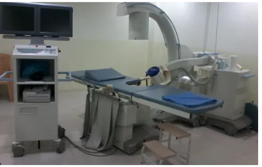
Figure 1. Extracorporeal shock wave lithotripsy in Zakho hospital.
3. Results and Discussion
European treatment guidelines advise active ESWL treatment for all stones larger than 6 - 7 mm [12]. Factors
known to alter the extracorporeal lithotripsy outcome are: stone size, location, chemistry, number as well as pa-tient anatomy. The present work was carried out to study the physical relation between the parameters of a total number of patients 34 male and female. The data were collected between the periods July 2013 to January 2015 from centers of kidney stones breakdown in Zakho hospital after treatment with (ESWL). Age of the patients ranged from 20 to 60 years harboring renal or ureteral stones. Size of stones ranged from 7 mm to 23 mm of compassion (cystine, Struvite, Calcium phosphate, uric acid and calcium oxalate). A number of shock waves were needed to breakdown the stones reached to 18,000 with 6 session and power 5 watt for calcium oxalate stone of size 23 mm. This indication is well agreement with the previous studies states that the stones larger than 15 mm and calcium oxalate monohydrate stones usually require several ESWL procedures for clearance. Uric acid, calcium oxalates dehydrate as well as struvite stones are much easier to be disintegrated. ESWL has poor
results for stones located in the lower calyx (“stone free” rate of 41 - 70) [13]-[16]. In order to explain physically
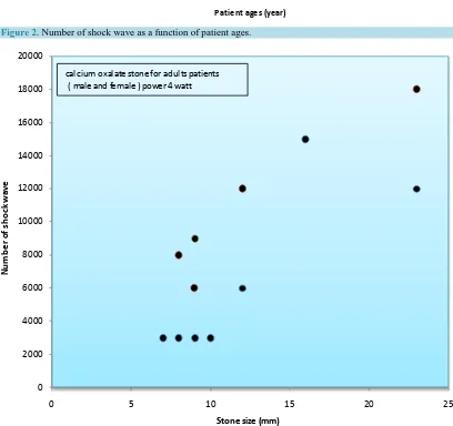
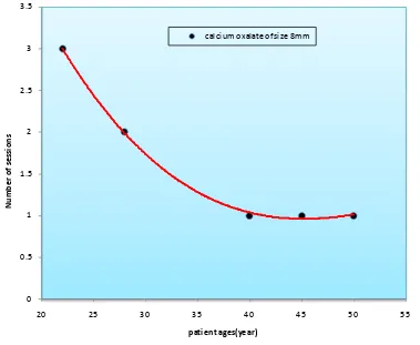
the parameters that affect the extracorporeal lithotripsy outcome such as stone size, location, chemistry, patient’s ages well as patient’s region, the relation represents between them graphically. Figure 2 illustrated the decreas-ing exponential behavior between the number of shock wave and patient’s ages for calcium oxalate in Zakho re-gion under constant power 4 watt. This indicates that the calcium oxalate of adult’s patients is more stiffness than the elders’ patients.
Contrary to that the number of shock wave increases with the size of stone for the same composition for adult’s patients of ages 20 - 30 years (male and female) under same power 4 watt, Figure 3. Because there was a significant positive correlation between stone size and number of treatments in the total sample, respectively, as
the stones was larger needed higher number of treatments [9]. Contrary to that interesting results are presented
by Tarawneh and colleagues, who proved an inverse correlation between the performance of ESWL and size of
the stone [17].
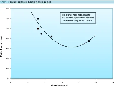
On the other hand, for the same composition and for workers at Zakho region it is found that the size of stones
decrease nearly exponentially with the patient ages as shown inFigure 4. This is attributed to the continuous
work of patients and drinking more water and others liquid during the work, this means that almost of calcium oxalate lost from human body during the live. Also for (calcium, phosphate, oxalate) stone, we obtained the same decreasing exponential behavior between the number of shock wave and the patient age for size stone
8mm and appointed patients due to the same reasons,Figure 5.
Number of shack wave versus type of stones (composition), renal and ureter of size 8 mm for elders patients are plotted in Figure 6. It appears from figure that the stones that are chemically softer required significantly
fewer applications of ESWL, regardless of its size [18]. Recent studies and experience have shown that the
Figure 2. Number of shock wave as a function of patient ages.
Figure 3.Number of shock wave versus stone size.
0 1000 2000 3000 4000 5000 6000 7000 8000 9000 10000
0 10 20 30 40 50 60 70
Num
be
r o
f s
ho
ck
w
av
e
Patient ages (year)
calcium oxalate stone of size = 8 mm zakho region at power = 4 watt
0 2000 4000 6000 8000 10000 12000 14000 16000 18000 20000
0 5 10 15 20 25
N
um
be
r o
f s
ho
ck
w
av
e
Stone size (mm) calcium oxalate stone for adults patients
[image:4.595.109.517.318.702.2]Figure 4. Patient ages as a function of stone size.
Figure 5. Patients ages as a function of stone size.
0 5 10 15 20 25 30 35 40 45 50
0 5 10 15 20 25
P
at
ien
t ag
es(
year
)
Stone size (mm)
calcium oxalate stone in zakho region for worker patient
0 10 20 30 40 50 60 70
0 5 10 15 20 25 30
P
at
ien
t ag
es (
year
)
Stone size (mm)
calcium,phosphate.oxalate stones for appointed patients
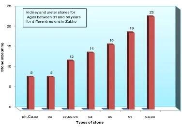
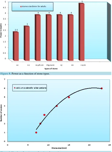
[image:5.595.121.508.405.702.2]As well as type of stones versus stone size for the ages 31 - 60 years (elders), reveals the increasing relation starting from the minimum value for both (phosphate calcium oxalate and oxalate) stones to maximum value for (calcium oxalate) stone as shown in Figure 7.
Another remarkable result of the present research is the (ESWL) power needed for each type of stone to crush
[image:6.595.128.499.159.421.2]in to fragments, Figure 8represent the variation of the (ESWL) power with the type of stone for 8 mm size and
Figure 6. Number of shock waves versus types stone.
Figure 7. Stone size as a function of the stone types.
0 1000 2000 3000 4000 5000 6000 7000 8000 9000
uc ca,mg,ox ca,ph,ox cy ca,ox
2000
3000
6000 6000
9000
N
u
m
b
er
o
f sh
o
ck w
aves
Types of stone kidney and ureterstones of
size 8 mm, for adult (22-30 year)
0 5 10 15 20 25
ph,Ca,ox ox cy,uc,ox ca uc cy ca,ox
8 8
12
14
16
19
23
S
to
n
e si
z
e(
m
m
)
Types of stone
[image:6.595.126.502.445.708.2]different ages of patients. It is clear from the chart diagram that the uric acid stone required the minimum power to break, while the calcium oxalate needs maximum power to fragment due to its hardness composition.
Also another important result of the present study is the number of session of shock wave required for crush-ing each stones size shown in increascrush-ing curve Figure 9. Because large stones which will require several ESWL sessions and consequently increase the number of shock wave are best treated with percutaneous
nephrolithot-omy plus (ESWL) [19], later we plot the number of sessions against the patient’s ages for calcium oxalate of
size 8 mm results nearly a decreasing exponential behavior, Figure 10. Because patient age was a significant
predicting factor affecting the treatment outcome of ESWL for renal, but not ureteric stones [20]. This could be
[image:7.595.130.499.215.713.2]attributed to the fragmentation difficulty of these compositions due to their hardness for adults patients, and eas-ier for elders patients consequently, decreasing number of sessions.
[image:7.595.131.498.215.451.2]Figure 8. Power as a function of stone types.
Figure 9. Number of session as a function of stone size.
0 0.5 1 1.5 2 2.5 3 3.5 4 4.5 5
uc ca ca,ph,ox mg,ca,ox cy ox ca,ox
2.5
3
4 4 4 4
5
Po
w
er
in
(w
at
t)
types of stone stones size 8mm for adults
0 1 2 3 4 5 6 7
0 5 10 15 20 25
Nu
m
be
r o
f se
ssi
on
Figure 10. Number of session versus patient ages (year).
4. Conclusion
ESWL can be safely recommended for patients of urolithiasis irrespective of age and stone size with promising results of stone clearance and patient acceptance. It is a very effective modality for treatment of renal and ure-teric stones which can be performed in outpatient basis. The success rate depends upon size, type and location of stone. As stone size increases, the success rate decreases. Also the success rate is better in ureteric stone
com-pared to renal stone. The factors that influence the treatment outcome of this technique for renal and ureteral
stones as a results of the present work, are patients ages, type, size and location of stones have a significant ef-fect on the results of ESWL. Further studies are needed to identify possible explanations for this observation. Based on the results of this study, it is evidence that almost patients harbor calcium oxalate stones of size 8 mm. This indicates that the patients begin to feel the pain at this size of stones within the ureter due to blocking the urea, as well as smaller size can pass through the ureter without pain. There is a significant positive correlation between the stone size and the number of treatments in the total sample, as well as decreasing exponential beha-vior between the number of shock wave and patients’ ages for calcium oxalate in Zakho region. Uric acid stone requires minimum power to break, while calcium oxalate needs maximum power to fragment due to its hardness composition. The number of sessions against the patient’s ages for calcium oxalate of size 8 mm results in near-ly a decreasing exponential behavior because patient age is a significant predicting factor affecting the treatment outcome of ESWL for renal and ureteric stones.
References
[1] Herdman, R.C. (1984) Effects of Federal “PoliciesonExtracorporealShockWaveLithotripsy” Is Case Study 36 in OTA’S Health Technology Case Study Series. OTA Health and Life Sciences Division.
[2] Lingeman, J.E. (1997) Urologic Clinics of North America, 24, 185-211.
[3] Costa-Bauzá, A., Perelló, J., Isern, B. and Grases, F. (2005) Urological Research, 33, 51-56.
http://dx.doi.org/10.1016/S0094-0143(05)70363-3 0
0.5 1 1.5 2 2.5 3 3.5
20 25 30 35 40 45 50 55
Nu
m
be
r o
f se
ssi
on
s
patient ages(year)
[4] Hossain, M.J., Uddin, M.N. and Islam, M.S. (2009) CMCTA, 20, 45-49.
[5] Weinberg, K. and Ortiz, M. (2009) Biomechanics and Modeling in Mechanobiology, 8, 285-299.
http://dx.doi.org/10.1007/s10237-008-0135-0
[6] Tomescu, P., Pănuş, A., Mitroi, G., Drăgoescu, O., Stoica, L., Dena, S. and Enache, M. (2009) CurrentHealthSci-
encesJournal, 35.
[7] Wang, C.-J. (2012) SurgeryandResearch, 7, 11. http://dx.doi.org/10.1186/1749-799X-7-11
[8] Abid, A.F. (2014) Open Journal of Urology, 4, 26-32. http://dx.doi.org/10.4236/oju.2014.43005
[9] Junuzovic, D., Prstojevic, J.K., Hasanbegovic, M. and Lepara, Z. (2014) Acta Informatica Medica, 22, 309-314.
http://dx.doi.org/10.5455/aim.2014.22.309-314
[10] Sauerbruch, T., Holi, J., Sackmann, M. and Paumgartner, G. (2015) Extracorporeal Shock Wave Lithotripsy of Pan-creatic Stones. http://gut.bmj.com/
[11] Gallagher, H.J. and Tolley, D.A. (2000) CurrentOpinioninUrology, 10, 551-555.
http://dx.doi.org/10.1097/00042307-200011000-00003
[12] Grasso, M., Hsu, J. and Spaliviero, M. (2008) Extracorporeal Shockwave Lithotripsy. eMedicine by WebMD.
[13] Manu, R. (1998) Litotripsia extracorporeală cu unde de şoc (ESWL). în Urologie Clinică, Editura Medicală Amaltea, Bucureşti, 162-164.
[14] Tiselius, H.G., Ackermann, D., Alken, P., Buck, C., Conort, P. and Galluci, M. (2001) European Urology, 40, 362-371.
http://dx.doi.org/10.1159/000049803
[15] Yang, H.S., Park, K.S. and Min, B.K. (1993) Korean Journal of Urology, 34, 109-115. [16] Argyropoulus, A.N. and Tolley, D.A. (2007) European Urology, 52, 344-350.
http://dx.doi.org/10.1016/j.eururo.2007.04.066
[17] Tarawneh, E., Awad, Z., Hani, A., Haroun, A.A., Hadidy, A., Mahafza, W., et al. (2010) Saudi Journal of Kidney
Dis-eases and Transplantation, 21, 660-665.
[18] Kovacevic-Prstojevic, J. (2014) Broj vantjelesnih razbijanja kamenaca u odnosu na veličinu kamenca, njegovu lokali-zaciju u urinarnom traktu, temorfološku strukturu kamenca. Magistarski rad. Medicinski fakultet Univerziteta u Sara-jevu, Sarajevo.