Tinman Regulates NetrinB in the Cardioblasts
of the
Drosophila
Dorsal Vessel
Jamshid Asadzadeh1,2¤, Niamh Neligan1, Sunita G. Kramer3, Juan-Pablo Labrador1,2*
1Smurfit Institute of Genetics, Trinity College Dublin, Dublin, Ireland,2Institute of Neuroscience, Trinity College Dublin, Dublin, Ireland,3Department of Pathology and Laboratory Medicine, Robert Wood Johnson Medical School, Rutgers The State University of New Jersey, Piscataway, New Jersey, United States of America
¤ Current address: Columbia University Medical Center, Department of Pathology and Cell Biology, New York, New York, United States of America
*labradoj@tcd.ie
Abstract
Morphogenesis of theDrosophiladorsal vessel (DV) shares similarities with that of the ver-tebrate heart. Precursors line up at both sides of the embryo, migrate towards the midline and fuse to form a tubular structure. Guidance receptors and their ligands have been impli-cated in this process in vertebrates and invertebrates, as have been a series of evolution-arily conserved cardiogenic transcriptional regulators including Tinman, theDrosophila homolog of the transcription factor Nkx-2.5. NetrinB (NetB), a repulsive ligand for the Unc-5 receptor is required to preserve the dorsal vessel hollow. It localizes to the luminal space of the dorsal vessel but its source and its regulation is unknown. Here, using genetics together with in situ hybridization with single cell resolution, we show howtinis required for NetrinB expression in cardioblasts during DV tubulogenesis and sufficient to promote NetB tran-scription ectopically. We further identify a dorsal vessel-specific NetB enhancer and show that it is also regulated bytinin a similar fashion to NetB.
Introduction
Heart organogenesis begins with the migration of bilaterally paired groups of cardiac precur-sors and the formation of a linear tube after they meet [1,2]. Guidance receptors and their ligands have been implicated at different steps in this process in vertebrates [3–5] and inverte-brates [6–10]. InDrosophilatwo rows of bilaterally lined up cardioblasts (CBs) and pericardial cells (PCs) migrate towards the dorsal midline (Fig 1, Stage 15). After migration, the tubular heart (dorsal vessel, DV) forms as CBs fuse, leaving a central luminal space (Fig 1, Stage 17). This complex process is mediated at least through the actions of two guidance systems; the Slit and its Robo receptors [6–8] and NetB behaving as a repulsive cue for the Unc-5 receptor local-ized at the luminal side of the CBs [9,10] (Fig 1, stage 17). However, several questions remain unanswered. While NetB is present in the DV lumen it is not clear if it is actually produced by cardiac cells [9,11,12]. How these guidance systems are regulated in the DV is also largely unknown. A core network of evolutionary conserved transcription factors (TFs) that regulates heart morphogenesis in both vertebrates and invertebrates, includingNkx2.5and its
OPEN ACCESS
Citation:Asadzadeh J, Neligan N, Kramer SG, Labrador J-P (2016) Tinman Regulates NetrinB in the Cardioblasts of theDrosophilaDorsal Vessel. PLoS ONE 11(2): e0148526. doi:10.1371/journal. pone.0148526
Editor:Edward Giniger, National Institutes of Health (NIH), UNITED STATES
Received:August 6, 2015
Accepted:January 5, 2016
Published:February 3, 2016
Copyright:© 2016 Asadzadeh et al. This is an open access article distributed under the terms of the
Creative Commons Attribution License, which permits unrestricted use, distribution, and reproduction in any medium, provided the original author and source are credited.
Data Availability Statement:All relevant data are within the paper and its Supporting Information files.
Drosophilahomolog,tinman(tin), [13] may coordinate their expression playing a later role after cardiac precursor specification. This situation would be analogous to the neuromuscular system where guidance receptors for Slit and Netrin are regulated by TFs that play an earlier role in motoneuron specification [14–16]. In this respect we have recently shown how the Unc-5 receptor is specifically regulated in cardioblasts bytin[17].
In this work, we show howNetBmRNA specifically localizes in CBs within the DV as does a membrane tethered NetB knocked into theNetBlocus. NetB is regulated in CBs bytinsince its mRNA and protein levels are strongly reduced in tin deficient CBs. Furthermore,tinis suffi-cient to induce ectopic NetB expression when misexpressed in the ectoderm. Finally, we iden-tify a NetB DV enhancer that drives expression specifically in CBs within the DV, show that its activity is strictly dependent on Tinman and that Tinman can bind to phylogenetically con-served sites within the enhancer. Together our data shows thattinregulatesNetBin CBs and strongly suggests thattinmay orchestrate DV assembly, at least partially, through the regula-tion of a NetB autocrine loop in CBs.
Results
NetrinB protein is expressed by Tin-positive cardioblasts in the
Drosophila
dorsal vessel
[image:2.612.201.563.74.282.2]The Unc-5 receptor and its NetB ligand have been shown to play a role late in tubulogenesis during DV morphogenesis inDrosophila[9,10]. While the receptor is expressed by CBs and
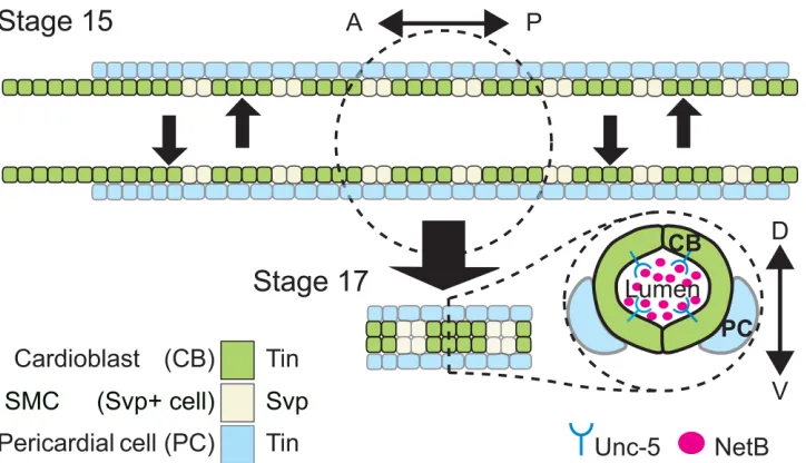
Fig 1. Schematic representation of theDrosophiladorsal vessel’s cellular composition and organization.Top, presentsDrosophilaDV at embryonic stage 15 while cardiac progenitors are migrating towards the dorsal midline. Bottom is an schematic representation of the embryonic heart proper at stage 17 when the DV lumen has formed. Aortic portion (right) is oriented anteriorly and the beating (heart) portion (left), posteriorly. Based on the expressed marker TFs, different cell types are colored. CBs are divided into Tin-(green) or Svp-expressing SMCs (light green) subtypes. SMCs of the last three posterior segments of DV make the future ostial cells (inflow valves). CBs are surrounded by pericardial cells (PCs; blue) on their ventrolateral side. PCs are divided into Tin-positive (blue) or Tin-negative PCs (not pictured). Bottom right, is a schematic representation of cross-section view of the DV lumen at stage 17. CBs on the opposite sides assume a crescent-like shape by contacting each other at the two dorsal and ventral apical sides, avoiding contact at the luminal domains, therefore making a hollow space in between. Unc-5 and NetB have been proposed to play a role in the formation of the hollow space between each CB on each row and its counterpart on the opposite row [9].
doi:10.1371/journal.pone.0148526.g001
Tinman Regulates NetrinB in theDrosophilaDorsal Vessel
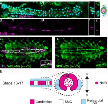
present at their luminal side in the DV, NetB accumulates in the lumen from stage 15 onwards [9]. However,NetBtranscripts were not detected in CBs [9], although other groups have reported its presence in the cardiac mesoderm [11,12]. In order to determine if cardiac cells produce NetB, we took advantage of NetB knock-inDrosophilalines [18]. These lines have been engineered to replace the endogenousNetBgene with either a V5-tagged membrane-bound NetB (NetB-tm) or a myc-tagged secreted one. These lines are very useful tools to deter-mine NetB localization and, in particular, the cells where NetB is produced as NetB-tm labels the membranes of NetB expressing cells. In our experiments, we can specifically detect secreted NetB predominantly within the heart lumen in live dissected embryos at late stage 16 (Fig 2A and 2B) as previously reported [9]. When we analyzed NetB-tm expression pattern we could detect it homogeneously expressed in all Tin+ CB membranes but not in Tin–Svp-expressing cells (SMCs) along the DV (Fig 2C and 2DandS1 Fig). Thus, our results show that NetB is pro-duced in CBs, strongly suggesting that CBs are the source of NetB protein in the DV.
Fig 2. NetB is expressed by CBs and can be detected in the luminal space after dorsal vessel closure. (A and B) Myc staining of embryos expressing a secreted Myc-tagged NetB knock-in [18] (magenta, luminal space) reveals secretion of NetB into the luminal space. CBs and PCs are labeled with Mef2 (green) and a-Zfh1 (cyan), respectively. Two cross sections taken from two points numbered 1 and 2 (places of which indicated in A and B) are shown on the right. The cross section at the point 1 is depicted schematically on the right and also in E. Note that due to secretion from CBs, NetB-Myc is mainly concentrated in the luminal space. (C and D) Co-staining for Tin (magenta) and a transmembrane V5-tagged NetB knock-in [18] (green) reveals NetB expression in Tin-positive cardioblasts. Note the regular gaps in NetB-tm expression
corresponding to Tin-negative SMCs (arrowheads). All panels are dorsal views with anterior to the left. A magnification of the regions delineated by insets is shown for each panel in A and B.
NetB
mRNA expression in cardioblasts is regulated by
tin
To confirm that NetB was transcribed in CBs, we performed in situ hybridization inDrosophila
embryos at late stage 16, which is the onset of DV tube assembly. At this stage,NetBmRNA can be readily detected within CBs in the posterior, beating portion of the DV (heart proper) and also, at lower levels within the aorta (Fig 3A and 3A’), which contrasts with the
homoge-neous expression seen for NetB-tm along the DV (Fig 2). Given that these CBs specifically express the transcription factor Tinman and that we had previously detected NetB in Tin+ CB membranes we wondered iftinwould be controllingNetBexpression.tinmutants are charac-terized by their lack of DV, astinis required for early specification of cardiac precursors [19,
20]. Nevertheless,tinmutants wheretin’sexpression is restored everywhere except in the DV (tin-ABD;tin346/tin346) are able to form a DV and hatch as adults [21]. To determine whether
tinregulatesNetB, we took advantage of these mosaictinmutants to analyze NetB mRNA expression through mRNA in situ hybridization (3B, B’).NetBmRNA was significantly reduced when compared withtinheterozygous DVs (compareFig 3A and 3A’with 3B and 3B’). Thus, not onlyNetBmRNA is transcribed in Tin+ CBs, but also its specific expression pattern matching that of the transcription factor is a consequence of its regulation bytinin these cells during DV tubulogenesis.
NetB protein expression is reduced in
tin
mutants
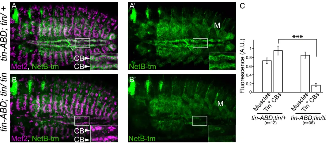
To determine if NetB mRNA reduction intinmutants would be reflected in NetB protein expression, we analyzed NetB-tm levels intinmutants. As readout of mRNA translation we detected NetB intinheterozygous or mutant CBs and compared its expression levels with NetB expression in muscles.tinis not expressed in muscles; therefore muscle levels should
Fig 3.tinregulatesNetBmRNA expressionin vivo.A and A’representNetBmRNA (magenta) expression in CBs (green), labeled with Tau-Myc driven by
TinC-Gal4to label CBs, in heterozygoustinABD;tin346/+embryos (arrowheads).NetBexpression dramatically drops in homozygoustinABD;tin346/tin346
late stage 16 embryos (B and B’; open arrowhead-asterisks in B’point to where CBs are positioned in B). All panels are dorsal views with anterior to the left. A magnification of the regions delineated by insets is shown for each panel. C, quantification of the in situ signal in cardioblast from the anterior, aorta region (A) or the posterior, heart proper (P) region of the dorsal vessel compared to transversal epidermal signal (Ep) intinABD;tin346/+andtinABD;tin346/tin346
embryos (***p<0.003 and**p<0.01).
doi:10.1371/journal.pone.0148526.g003
[image:4.612.33.573.76.312.2]remain constant. As expected, NetB-tm is virtually absent in CBs of tin mutants (compareFig 4A and 4A’with 4B and 4B’) while muscle expression remains unchanged (Fig 4C).
Misexpression of
tin
in the ectoderm can induce NetB ectopically
Given thattinis required for NetB expression in the DV, we investigatedtin’s requirement for its expressionin vivo. We misexpressedtinwith anengrailed-Gal4driver on a striped pattern in the ectoderm, where neither Tin nor NetB are expressed (Fig 5A and 5A’). Subsequently, we
analyzed the induction of NetB and confirmed that ectopic expression oftinis sufficient to induce NetB expression (Fig 5B and 5B’) in the characteristicengrailed(en) striped pattern. These results show that even when Tinman is expressed outside its normal endogenous context it is sufficient to induce expression of NetB. Together, the fact thatNetBmRNA or protein are almost undetectable intinmutant CBs andtin’sability to induce their expression ectopically in the ectoderm, suggest a direct role of Tinman in the regulation of this ligand.
A
NetB
enhancer drives expression in cardioblasts
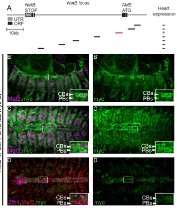
[image:5.612.36.568.75.309.2]In order to understand how is NetB regulated in the developing DV, we analyzed several enhancers within its locus. Screening the available Gal4 lines from the Janelia collection [22], we identified a fragment (~4kb) within the first NetB intron that can drive expression in the DV at late stages of migration and tubulogenesis (NetB-M,Fig 6A). Given the size of the NetB locus and the regions analyzed, this is one potential enhancer element, and we do not exclude the presence of other, potentially more significant, enhancer elements in the locus driving expression in the DV. Nevertheless, the identified enhancer drives expression in mesodermal cells including muscles, pericardial cells and cardioblast at earlier stages (Fig 6B–6C’) and its
Fig 4. NetB protein expression, intinmutants is downregulated in CBs.AsNetBmRNA is almost absent in CBs oftin-ABD;tin346/tin346mutants (Fig 3), we tested the NetB-tm protein expression in this background using the transmembrane V5-tagged NetB knock-in [18]. A and A’represent NetB expression in
tinABD; tin346/+heterozygous embryos (full confocal stack inS1 Fig). There is no significant difference in the NetB-tm levels betweenwild-typeand
heterozygotes. However, intin-ABD;tin346/tin346mutants, NetB-tm expression level is dramatically reduced (B, B’, full confocal stack inS2 Fig) while its expression in the muscles [M] is unaffected (C). CBs are labeled with a-Mef2 (Magenta) and NetB is labeled with a-V5 (green). All panels are dorsal views with anterior to the left. A magnification of the regions delineated by insets is shown for each panel (***p<2.6x10⁻⁵).
expression gets restricted at later stages to CBs within the DV (Fig 6B and 6B’). Our UAS-tau-myc-GFP is driven byNetB-M-Gal4therefore its expression persists to slightly later stages than the endogenous NetB but faithfully replicatesNetBtranscript and protein expression within the DV (Fig 2C and 2DandFig 3A and 3A’).
The
NetB
DV enhancer requires
tin
for its induction in cardioblasts
Given thattinis required for NetB expression in the DV we argued that theNetB-Menhancer may be regulated bytin in vivo. To test iftinwas required to promote transcription through theNetBenhancer, we examined if it was able to promote transcription intin-ABD;tin346/
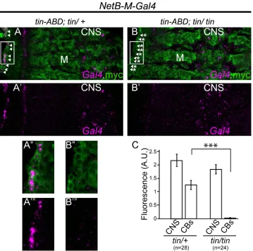
tin346mutant embryos (Fig 7). We analyzed the expression ofGal4mRNA under the control of theNetB-Menhancer. WhileGal4was specifically expressed intinheterozygous CBs, we could not detect any mRNA signal intin-ABD;tin346/tin346CBs (CompareFig 7A and 7A”with 7B– 7B”and 7C). Importantly, reporter expression was not affected intinmutants within the CNS wheretinis not expressed and therefore, not required.
Tinman can bind to the
NetB-M
enhancer
[image:6.612.198.562.77.322.2]Tinman regulates NetB mRNA and protein expression specifically in CBs during tubulogenesis in the DV and it is able to induce its expression ectopically. In addition we have identified a NetB enhancer that drives expression in the DV that is under Tin control in CBs during tubu-logenesis. In order to determine if Tin can bind to the NetB enhancer we performed chromatin immunoprecipitation (ChIP) with followed by qPCR using overlapping primers covering the NetB-M enhancer (Fig 8A). We identified 4 peaks of enrichment in the immunoprecipitate
Fig 5. Ectopic expression of Tin induces NetB ectopic expressionsin vivo.In a wild-type background, staining for NetB protein (magenta), displays little or no NetB expression in the Engrailed stripes (A and A’). However, in anengrailed-Gal4>UAS-tinbackground, NetB is strongly induced in Engrailed stripes which now ectopically express Tin (B and B’, arrowheads). For NetB protein staining, the transmembrane V5-tagged NetB-tm knock-in [18] was used. All panels are lateral views with anterior to the left and dorsal side up. A magnification of the regions delineated by insets is shown for each panel.
doi:10.1371/journal.pone.0148526.g005
indicating that Tin can bind to the NetB enhancer. Further analysis of the sequence revealed phylogenetically conserved Tin consensus binding sequences within each peak that have been preserved across>107years of evolution, strongly suggesting its functional relevance (Fig 8B).
Taken together, our data demonstrates that Tinman regulates transcriptionally theNetB
[image:7.612.207.562.76.493.2]ligand and a DV enhancer present within its regulatory region specifically in CBs during tubu-logenesis. Furthermore, our results also suggest that this regulation may be mediated through a direct interaction of Tin with NetB regulatory regions.
Fig 6. TheNetB-M reporter is expressed in CBs and weakly in muscles.(A) Schematic representation of the positions and the relative sizes of the availableGal4reporters forNetBlocus. We screened Gal4 from the Janelia collection [22] for DV expression. The line expressing Gal4 efficiently in the DV is shown in magenta. (B-C’) Gal4 expression pattern was examined using a UAS-Tau-Myc-GFPconstruct. Co-staining of embryos with a-Myc (green) and a-Mef2 (magenta) revealed Gal4 expression in muscles and the DV at stage 13 (B, B’) and 15 (C, C’). Co-staining of these embryos with a-Myc (green), a-Mef2 (red) and a-Zfh1 (magenta) reveals a reduction in the expression in other in muscles and pericardial cells while expression in CBs persists at late stage 16–17 (D, D’). All panels are dorsal views with anterior to the left. A magnification of the regions delineated by insets is shown for each panel.
Discussion
Early dorsal vessel specification events inDrosophilatake place within the ventral mesoderm. A network of evolutionary conserved cardiogenic transcription factors, including Tinman, integrates their activity to promote DV morphogenesis in the cardiac mesoderm [2,12]. While Tinman is essential for cardiac cell specification, a later role in the embryonic DV morphogen-esis remains largely unexplored. Nevertheless, its function at later stages has been suggested by the defective arrangement of CBs within the DV in cardioblast-specifictinmutants [21]. Dif-ferent guidance systems such as the Slit and its receptors [6–8] and NetB and its Unc-5 receptor
Fig 7.NetB-M enhancer element is regulated by Tinin vivo.In situ hybridization forGal4mRNA driven by theNetB-Menhancer (Gal4, magenta) revealsGal4mRNA expression in some cells in the CNS and in the DV intin-ABD;tin346/+heterozygous background (A and A
’). However, intin-ABD;tin346/tin346mutant
embryosGal4mRNA expression is exclusively absent from the CBs, while CNS cells have the same signal intensity as in heterozygous embryos (B and B’). Reporter driven UAS-tau-myc-GFP is used as a
counterstain as it shows a persistent broad expression pattern within the CNS, muscles and DV (myc, green) at stage 15 (A-B”) while the Gal4 mRNA is clearly absent at this stage. Quantification ofGal4mRNA expression driven byNetB-M element in CBs intin-ABD;tin346/+heterozygous versustin-ABD;tin346/tin346
mutant embryos. Fluorescence ofGal4-expressing CNS cells is used as internal control (C). While in situ signal in the CNS of heterozygous and mutant embryos remains unchanged (2.15±0.22 s.e.m and 1.83±0.164 s.e.m, in heterozygous ortinmutants, respectively), the signal in CBs is significantly dropped from 1.24±0.169 (***p<6.8 x 10⁻⁸) intin-ABD;tin346/+heterozygous embryos to 0.0036±0.032 intin-ABD;
tin346/tin346mutants. All panels are views with dorsal side to the left and anterior to the top. A magnification of the regions delineated by insets in A and A’or B and B’is shown in A”and A”‘or B”and B”‘respectively. doi:10.1371/journal.pone.0148526.g007
[image:8.612.201.560.75.430.2][9,10] are required in late DV morphogenesis for tube assembly. We provide several lines of evidence to demonstrate thattinis a regulator of NetB in CBs during DV tubulogenesis: 1-NetrinB is expressed in the DV by CBs and accumulates in a polarized fashion on their luminal side at late embryonic stages (Fig 2); 2-tinis required forNetBmRNA expression in CBs (Figs
[image:9.612.195.551.71.491.2]3and4); 3-tincan induce NetB expression ectopically (Fig 5); and 4-tinregulates aNetB DV-specific enhancer in CBs (Figs6and7).
Fig 8. Tin binds directly to NetB-M enhancer element.(A) ChIP analysis of theNetB-Mgenomic loci in S2R+ cells transfected with pAct5C-GFP-tinman. The precipitated DNA fragments were amplified by real-time qPCR using overlapping primers (R1-R20) designed over the genomic region covering theNetB-M
enhancer element. ChIP signal is also illustrated as a curve peaking at R1, R10/R11, R14 and R16. A schematic ofNetBlocus is also illustrated below the graph. Note that the schematic view represents only a ~2kb region (chrX:14,744,635–14,746,673) of theNetB-Menhancer that showed significant enrichment as no significant enrichment for the rest of the enhancer was detected above the background. Enrichment is presented as a percentage of total input. (B) Consensus Tin-binding motifs detected with JASPAR within the ChIP enriched region and their conservation in differentDrosophilaspecies (mel,melanogaster; sim,
simulans; sec,sechellia; yak,yakuba; ere,erecta).
NetrinB expression in the DV was not definitely described previously. Its mRNA had been detected in the DV by some groups [11,12] while another failed to detect it [9] leading to the proposition that NetB was produced in non-cardiac cells and actively or passively transported to the DV lumen. We unambiguously show that NetB is specifically expressed by Tin+ CBs in the DV, strongly suggesting that the NetB source in the luminal side is actually the CBs and it may, therefore, work in an autocrine fashion.
Tissue-specific genome-wide ChIP studies of evolutionarily conserved cardiogenic tran-scriptional regulators, including Tin, have previously suggested that they may regulateNetB
through their binding events close to this gene early in cardiac development [12]. Our work analyzesNetBregulation bytinat later stages when CBs and PCs have been fully specified and shows thattinregulatesNetBand aNetBDV enhancer specifically in CBs wheretinis also expressed. Additionally, we show that Tin can bind to the DV enhancer in phylogenetically conserved Tin binding sites. Full confirmation of their functional relevance would require their mutagenesis and analysisin vivo. Thus, our results suggest that Tin specific regulation in CBs may be mediated through a direct binding within theNetBlocus as a vestige of their embryonic origin in the cardiac mesoderm.
Materials and Methods
Genetics
The stocks used in this study were as follows:Tin-ABD;tin346/TM3,eve-lacZ, svp-lacZ[21], en-Gal4, TinC-Gal4 [23], UAS-tau-Myc, Bloomington stocks 49645 (NetB-M-Gal4), 49633, 49643, 49645, 49647,49524, 49521, 49346 [22], NetAΔNetBmyc, NetAΔNetBtm [18].
Immunohistochemistry and mRNA in situ hybridization
Embryo collection, immunohistochemistry and in situ hybridization were performed as previ-ously described [24]. The following antibodies were used for immunohistochemistry: rabbit a-Mef2 (1:2000), guinea pig a-Zfh1 (1:1500) [25], rabbit a-Tin (1:1000) [19], mouse a-V5 (1:1000) (Life technologies; R960-25), chicken a-GFP (1:2000) (Abcam, 13970), mouse a-Myc (1:50) (DSHB). Secondary antibodies: Alexa 555, Alexa 488-conjugated (Invitrogen) and Cy5-conjugated (Jackson ImmunoResearch Laboratories). ForNetBin situ hybridization, digoxigenin-labeled probes were used as previously described [24]. Probe-hybridized embryos were stained with HRP-conjugated anti-digoxigenin antibody (Roche) overnight. The follow-ing day, embryos were incubated with Cy3-conjugated tyramide, as HRP substrate, for 20 (for
NetBmRNA) or 45 (forGal4mRNA) minutes at room temperature. Zeiss Confocal LSM700 Microscope was used for obtaining stacks of images using 20X or 40X objectives. For fluores-cent quantifications, samples were prepared using the same conditions and at the same time, when possible. Image J software was used for image analysis. Background correction was per-formed for all quantifications. The obtained values from the selected regions were then divided by the values in control areas for each embryo. The normalized values were finally averaged for each genotypic group.
The expression pattern of secreted NetB-Myc was revealed in live-dissected embryos follow-ing previously described procedures [18,26] with minor modifications. Live embryos were staged and propped with their ventral side up, on poly L-lysine-coated glass slides (Fisher Sci-entific) and were dissected. Following dissection, embryos were fixed in 4% paraformaldehyde in PBS at RT for 15 min and stained with rabbit anti-Mef2, guinea pig anti-Zfh1, and mouse anti-Myc antibodies.
Chromatin immunoprecipitation
ChIP was performed and analyzed as described previously [17,27]. Briefly, S2R+ cells transfected with either pAct5C-GFP-Tin or pAct5C (as mock control) were fixed at RT in 1% formaldehyde for 10 minutes. Following cell lysis, the fixed nuclei were sonicated to obtain proper size sheared chromatin. Sonicated lysates were then incubated with rabbit anti-GFP (ab290; Abcam) at 4°C for 2 hours. Lysates were then incubated with protein A-sepharose (P9424; Sigma) for an additional 2 hours. Subsequently, sepharose beads were washed to remove unspecific binding. This step was followed by elution of antibody-GFP-Tin-chromatin from beads at 70°C overnight. The next day, DNA fragments were purified using phenol-chloroform method. The immunoprecipitated frag-ments were subsequently amplified by real-time qPCR and quantified by comparison to input.
Statistical analysis
Data are presented as mean values ± s.e.m.. Two-tailed independent samples t-test was used for determining statistical significance for all comparisons between wild-type/heterozygous samples and mutant ones. Histograms were generated using Microsoft Excel 2013.
Supporting Information
S1 Fig. Full stack view of the DV in atin-ABD;tin346/+heterozygousNetB-tmknock-in embryo.NetB-tm is expressed in a broad pattern in muscles, gut and dorsal vessel. Individual slice view within a confocal stack reveals a homogeneous expression of NetB-tm along the DV (green). Note the specific expression of NetB-tm in CBs. Panels 1 and 1’represent the most ventral slice and panels 12 and 12’represent the most dorsal one. Top panels are double stain-ing with a-Mef2 (magenta) and a-V5 (green). Bottom panels only show the NetB-tm pattern from the corresponding top panel. All panels are dorsal views with anterior to left.
(TIF)
S2 Fig. Full stack view of the DV in atin-ABD;tin346/tin346mutantNetB-tmknock-in embryo.NetB-tm signal is almost absent from individual confocal slices of atinmutant. Note the absence of V5 signal (green) in all CBs. Panels 1 and 1’represent the most ventral slice and panels 12 and 12’represent the most dorsal one. Top panels are double staining with a-Mef2 (magenta) and a-V5 (green). Bottom panels only show the NetB-tm pattern from the corre-sponding top panel. All panels are dorsal views with anterior to left.
(TIF)
Acknowledgments
Brian McCabe, Jim Skeath, Hanh Nguyen, Manfred Frasch and Matthew Wolf kindly provided us with stocks, antibodies and other various reagents. Funding sources of this work were as fol-lows: SFI grants 07/IN.1/B913 and 08/ RFP/NSC1617 and a New Foundations Award from the IRC to JPL and a Trinity research award to JA.
Author Contributions
Conceived and designed the experiments: JA JPL SGK. Performed the experiments: JA NN. Analyzed the data: JA JPL SGK. Wrote the paper: JA SGK JPL.
References
2. Bodmer R, Frasch M. Development and aging of the Drosophila heart. In: Rosenthal N, Harvey RP, edi-tors. Heart Development and Regeneration. I. First ed. London: Academic Press; 2010. p. 47–86. 3. Fish JE, Wythe JD, Xiao T, Bruneau BG, Stainier DY, Srivastava D, et al. A Slit/miR-218/Robo
regula-tory loop is required during heart tube formation in zebrafish. Development. 2011; 138(7):1409–19. doi: 10.1242/dev.060046PMID:21385766; PubMed Central PMCID: PMC3050667.
4. Medioni C, Bertrand N, Mesbah K, Hudry B, Dupays L, Wolstein O, et al. Expression of Slit and Robo genes in the developing mouse heart. Dev Dyn. 2010; 239(12):3303–11. doi:10.1002/dvdy.22449 PMID:20941780; PubMed Central PMCID: PMC2996720.
5. Mommersteeg MT, Andrews WD, Ypsilanti AR, Zelina P, Yeh ML, Norden J, et al. Slit-roundabout sig-naling regulates the development of the cardiac systemic venous return and pericardium. Circ Res. 2013; 112(3):465–75. doi:10.1161/CIRCRESAHA.112.277426PMID:23255421.
6. Qian L, Liu J, Bodmer R. Slit and Robo control cardiac cell polarity and morphogenesis. Curr Biol. 2005; 15(24):2271–8. doi:10.1016/j.cub.2005.10.037PMID:16360689.
7. Medioni C, Astier M, Zmojdzian M, Jagla K, Semeriva M. Genetic control of cell morphogenesis during Drosophila melanogaster cardiac tube formation. J Cell Biol. 2008; 182(2):249–61. doi:10.1083/jcb. 200801100PMID:18663140; PubMed Central PMCID: PMC2483531.
8. Santiago-Martinez E, Soplop NH, Patel R, Kramer SG. Repulsion by Slit and Roundabout prevents Shotgun/E-cadherin-mediated cell adhesion during Drosophila heart tube lumen formation. J Cell Biol. 2008; 182(2):241–8. doi:10.1083/jcb.200804120PMID:18663139; PubMed Central PMCID: PMC2483515.
9. Albrecht S, Altenhein B, Paululat A. The transmembrane receptor Uncoordinated5 (Unc5) is essential for heart lumen formation in Drosophila melanogaster. Dev Biol. 2011; 350(1):89–100. doi:10.1016/j. ydbio.2010.11.016PMID:21094637.
10. Macabenta FD, Jensen AG, Cheng YS, Kramer JJ, Kramer SG. Frazzled/DCC facilitates cardiac cell outgrowth and attachment during Drosophila dorsal vessel formation. Dev Biol. 2013; 380(2):233–42. doi:10.1016/j.ydbio.2013.05.007PMID:23685255; PubMed Central PMCID: PMC4137861. 11. Mitchell KJ, Doyle JL, Serafini T, Kennedy TE, Tessier-Lavigne M, Goodman CS, et al. Genetic
analy-sis of Netrin genes in Drosophila: Netrins guide CNS commissural axons and peripheral motor axons. Neuron. 1996; 17(2):203–15. PMID:8780645.
12. Junion G, Spivakov M, Girardot C, Braun M, Gustafson EH, Birney E, et al. A transcription factor collec-tive defines cardiac cell fate and reflects lineage history. Cell. 2012; 148(3):473–86. doi:10.1016/j.cell. 2012.01.030PMID:22304916.
13. Cripps RM, Olson EN. Control of cardiac development by an evolutionarily conserved transcriptional network. Dev Biol. 2002; 246(1):14–28. doi:10.1006/dbio.2002.0666PMID:12027431.
14. Santiago C, Labrador JP, Bashaw GJ. The homeodomain transcription factor Hb9 controls axon guid-ance in Drosophila through the regulation of Robo receptors. Cell reports. 2014; 7(1):153–65. doi:10. 1016/j.celrep.2014.02.037PMID:24685136; PubMed Central PMCID: PMC4128229.
15. Zarin AA, Asadzadeh J, Hokamp K, McCartney D, Yang L, Bashaw GJ, et al. A transcription factor net-work coordinates attraction, repulsion, and adhesion combinatorially to control motor axon pathway selection. Neuron. 2014; 81(6):1297–311. Epub Feb 20th. doi:10.1016/j.neuron.2014.01.038PMID: 24560702; PubMed Central PMCID: PMC4128230.
16. Zarin AA, Asadzadeh J, Labrador JP. Transcriptional regulation of guidance at the midline and in motor circuits. Cell Mol Life Sci. 2014; 71(3):419–32. doi:10.1007/s00018-013-1434-xPMID:23917723. 17. Asadzadeh J, Neligan N, Canabal-Alvear JJ, Daly AC, Kramer SG, Labrador JP. The Unc-5 Receptor
Is Directly Regulated by Tinman in the Developing Drosophila Dorsal Vessel. PloS one. 2015; 10(9): e0137688. doi:10.1371/journal.pone.0137688PMID:26356221; PubMed Central PMCID: PMC4565662.
18. Brankatschk M, Dickson BJ. Netrins guide Drosophila commissural axons at short range. Nat Neurosci. 2006; 9(2):188–94. doi:10.1038/nn1625PMID:16429137.
19. Azpiazu N, Frasch M. tinman and bagpipe: two homeo box genes that determine cell fates in the dorsal mesoderm of Drosophila. Genes Dev. 1993; 7(7B):1325–40. PMID:8101173.
20. Bodmer R. The gene tinman is required for specification of the heart and visceral muscles in Drosoph-ila. Development. 1993; 118(3):719–29. PMID:7915669.
21. Zaffran S, Reim I, Qian L, Lo PC, Bodmer R, Frasch M. Cardioblast-intrinsic Tinman activity controls proper diversification and differentiation of myocardial cells in Drosophila. Development. 2006; 133 (20):4073–83. doi:10.1242/dev.02586PMID:16987868.
22. Jenett A, Rubin GM, Ngo TT, Shepherd D, Murphy C, Dionne H, et al. A GAL4-driver line resource for Drosophila neurobiology. Cell reports. 2012; 2(4):991–1001. doi:10.1016/j.celrep.2012.09.011PMID: 23063364; PubMed Central PMCID: PMC3515021.
23. Lo PCH, Frasch M. A role for the COUP-TF-related gene seven-up in the diversification of cardioblast identities in the dorsal vessel of Drosophila. Mechanisms of Development. 2001; 104:49–60. doi:10. 1016/S0925-4773(01)00361-6PMID:11404079
24. Zarin AA, Daly AC, Hulsmeier J, Asadzadeh J, Labrador JP. A GATA/homeodomain transcriptional code regulates axon guidance through the Unc-5 receptor. Development. 2012; 139(10):1798–805. doi:10.1242/dev.070656PMID:22461564.
25. Ward EJ, Skeath JB. Characterization of a novel subset of cardiac cells and their progenitors in the Dro-sophila embryo. Development. 2000; 127(22):4959–69. PMID:11044409.
26. Lee HK, Wright AP, Zinn K. Live dissection of Drosophila embryos: streamlined methods for screening mutant collections by antibody staining. Journal of visualized experiments: JoVE. 2009;(34: ). doi:10. 3791/1647PMID:20040910; PubMed Central PMCID: PMC3149970.