Journal of Chemical and Pharmaceutical Research, 2015, 7(2):62-69
Research Article
ISSN : 0975-7384
CODEN(USA) : JCPRC5
Segmentation of brain MR images for tumor area and size detection by using
of clustering algorithm
Shinu Sadeyone
1and S. Freeda
21
EIE, Sathyabama University, Chennai
2EIE, A. C. T. Engineering College, Chngalpattu
_____________________________________________________________________________________________
ABSTRACT
There are different types of tumors are available. Astrocytoma is the most common type of tumor (30% of all brain tumor) and is usually a malignant one. Astrocytoma can be subdivided into four grades. Each grade has its own characteristics and unique treatment. If any wrong treatment is given to these grades that leads to death. So finding the position and shape of tumor is very important for the further treatment. The proposed system of this paper is to find the exact position and shape of the tumor cells. That helps the physician for further treatment. In the proposed system, it consists of four modules (i) Pre-processing, (ii) Segmentation of brain in MR Images,(iii) Quality extraction and (iv) Approximate reasoning. Pre processing is done by filtering. Segmentation is done by advanced K-means and Fuzzy C-means algorithms. Quality extraction is by thresholding. Finally, Approximate reasoning method to recognize the tumor shape and position in MRI image. If the tumor is a mass in shape then k-means algorithm is enough to extract it from brain cells. Suppose if it is a malignant (spread over the brain) one then the Fuzzy C-means algorithm will be used for accurate tumor diagnosis, since the Fuzzy method is used for floating point prediction of the tumor cells. At the end of the process the tumor shape, position, area and its stage will be determined. In this project the two strong algorithms areused for segmentation. So, the entire system for tumor segmentation is more accurate than other methods.
Key words: pre-processor, k-means clustering, fuzzy c-means clustering, grouping.
_____________________________________________________________________________________________
INTRODUCTION
we went for combination of two algorithms that will segment the malignant tumor such as astrocytoma tumor. The proposed method consists of five modules.
They are Pre-processing
Segmentation using K-means Segmentation using FCM Feature extraction Tumour Area calculation
I. System model
enhanced. But the possibilities for the noise in MRI images are very less. Here we are using the medial filter for the noise removal.
a)Removing Noise by means of Median Filtering:
Median filtering is similar to using an averaging filter, in that each output pixel is set to an average of the pixel values in the neighbourhood of the corresponding input pixel. However, with median filtering, the value of an output pixel is determined by the median of the neighbourhood pixels, rather than the mean. The median is much less sensitive than the mean to extreme values (called outliers). Median filtering is therefore better able to remove these outliers without reducing the sharpness of the image. The medfilt22 function implements median filtering.
Since there is no possibility for noise in the image. So the output of pre-processed image same as that of the original image. This image given as a input to the next level that is segmentation level done by both advanced k-means clustering and fuzzy c-means clustering. But for the whole system we artificially add and remove the noise.
III. K-means segmentation
K-means is one of the simplest unsupervised learning algorithms that solve the well known clustering problem [3][4].
Algorithm:
1. At first it decide on a value for k.
2. Initialize the k cluster centres (randomly, if necessary).
3. Decide the class memberships of the M objects by assigning them to the nearest cluster centre. 4. Re-estimate the k cluster centres, by assuming the memberships found above are correct. 5. If none of the M objects changed membership in the last iteration, exit. Otherwise go to step 3.
For a given assignment d, compute the cluster means α
=∑: ,k=1,….,K. (1)
For a current set of cluster means, assign each observation as,
d(i) = arg min|| αk || , i=1,…….,N (2)
Iterate above two steps until convergence.
Advantages of using k-means algorithm Technique:
• With a large number of variables, K-Means may be computationally faster than hierarchical clustering (if K is small).
Fig2. Flowchart for k-means algorithm
μ
∑
(3)
3.Compute the fuzzy centres 'vj' using
=∑∑ (4)
4.Repeat step 2 and 3 until the minimum 'j' value is achieved or
||U(l+1) - U(l)|| < ƞ (5)
where,
‘l’ is the iteration step.
‘ƞ’ is the termination criterion between [0, 1]. ‘U’ = (µij)n*c’ is the fuzzy membership matrix.
‘j’ is the objective function.
After each iteration membership and cluster centres are updated based on the above conditions.
Advantages of using this Technique:
• Gives best result for overlapped data set and comparatively better then k-means algorithm.
• Unlike k-means where data point must exclusively belong to one cluster centre here data point is assigned membership to each cluster centre as a result of which data point may belong to more than one cluster centre.
V. Tumor area calculation
The tumour detected image is converted to binary image by thresholding (binarisation).By using the tumour detection formula the area is calculated. Based on the area the various stages of tumours are identified [7] - [10]. Tumor Calculation formula:
Binary image = ∑ ∑ [f 0 + f 1 ]/00
1 2 /00
3 2 Where,
Pixels = Length (L)x Breadth (B) = 256x 256. f(0) = white pixel (digit 0)
f(1) = black pixel (digit 1)
Number of white pixels, W=∑/003 2∑ [f 0 ]/001 2 Size of tumor,
S = [(W)*0.264] mm2
Where,
W = number of white pixels(width*height) 1Pixel = 0.264 mm
Different stages of tumour:
• If the detected white pixels< 50, then no tumor was detected. The result gives ‘activity found normal’.
• If the detected white pixels>50, and detected <=300, then tumor was in initial stage. The result gives ‘initial stage of brain tumor detected’.
• If the detected white pixels>300, and detected <=500, then tumor was in intermediate stage. The result gives ‘intermediate stage of brain tumor detected’.
• If the detected white pixels>=500, then tumor was in critical stage. The result gives ‘critical stage of brain tumor detected’.
RESULTS
filter, the output gives an image without noise. The advantage of median filter is, it can reduce small amount of noise present in an image.
Fig5. Combined output for pre-processing and K-means with K=5
In this section, fig 5 gives the clustered images of MR Images. In which the pixels are get grouped on the basis of k-means clustering algorithm when the tumor is in mass level.
Fig6. Output image of for Fuzzy c-means algorithm
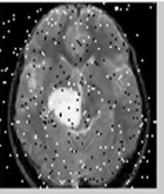
Fig7. Size detection (Output image detected white pixels > 50 & detected <= 300, Initial Stage of Brain Tumor)
In this section, it gives the output of stages of tumor level. Fig 7 gives the output of tumor level based on the quantity of white pixels.
CONCLUSION
There are different types of tumors are available. They may be as mass in brain or malignant over the brain. This project deals with the mass tumor detection and extraction. This project is used to extract the mass tumor from Brain MRI scan image and it can find out the area. This will help the physician for further treatment. In future, mass tumor volume can be calculated by using some morphological operation and 3D slicer.
REFERENCES
[1]R. Verma, E. I. Zacharaki, Y. Ou, H. Cai, S. Chawla, S.-K. Lee, E. R. Melhem, R.Wolf, and C. Davatzikos, Acad. Radiol., vol. 15, no. 8, pp. 966–977, Aug. 2008.
[2]C. Hogea, G. Biros, F. Abraham, and C. Davatzikos, Phys.Med. Biol., vol. 52, no. 23, pp. 6893–6908, Dec. 2007. [3]K.S. Ravichandran and 2B. Ananthi(2009), International Journal of Computational and Applied Mathematics ISSN 1819-4966 Volume 4 Number 2, pp. 153 –157.
[4]Anil Z Chitade(2010) , International Journal of Engineering Science and Technology Vol. 2(10), 5319-5325. [5]Tse-Wei Chen , Yi-Ling Chen , Shao-Yi Chien (2010), Journal of Scientific Research ISSN 1452-216X Vol.44 No.2, pp.337-351.
[6]S. Zulaikha Beevi M. Mohamed Sathik(2010), European Journal of Scientific Research, ISSN 1450-216X Vol.41 No.3 pp.437-451.