Copyright © 1999, American Society for Microbiology. All Rights Reserved.
Subtyping of
Haemophilus influenzae
Strains by Pulsed-Field
Gel Electrophoresis
MITSUMASA SAITO,* AKIKO UMEDA,ANDSHIN-ICHI YOSHIDA
Department of Bacteriology, Faculty of Medicine, Kyushu University, Fukuoka 812-8582, Japan
Received 23 November 1998/Returned for modification 11 January 1999/Accepted 29 March 1999
A total of 200 isolates ofHaemophilus influenzaewere analyzed by serotyping, biotyping, and pulsed-field gel
electrophoresis (PFGE). A total of 178 epidemiologically unrelated strains ofH. influenzaedemonstrated a
variety of genome patterns by PFGE, and 165 genotypes were thus obtained in this study. PFGE typing proved to have a much stronger discriminatory power than either serotyping or biotyping. Six serotype b strains were all classified into discrete genotypes. A PFGE analysis of 18 strains obtained from the nasopharynx, blood, and
cerebrospinal fluid of patients with meningitis also supported the hypothesis that invasive H. influenzae
disseminates from the nasopharynx to the bloodstream and then subsequently to other body sites. PFGE typing
of 10 other strains isolated from household contacts of patients withH. influenzaeinfection revealed that the
strain that caused theH. influenzaeinfection often colonized the nasopharynges of household contacts. Our
findings suggest that PFGE analysis is useful for the epidemiological study ofH. influenzaeinfection, even when
the invasive disease is caused by serotype b strains.
Haemophilus influenzae is a pathogen exclusively found in humans, and it causes infections that range from asymptomatic colonization of the upper respiratory tract to serious invasive diseases, such as meningitis. Strains ofH. influenzaeare usually classified according to the antigenicities and compositions of their polysaccharide capsules. There are six structurally and antigenically distinct capsular types (serotypes), designated se-rotypes a to f (17). Among them, serotype b is a common cause of serious infections in younger children.
H. influenzaecan also be divided into eight biotypes on the basis of three biochemical tests (13). However, this biotyping system is of limited use for epidemiological studies since the majority of clinical isolates are distributed into three biotypes (biotypes I, II, and III). More than 90% of invasive type b strains are of biotype I. The majority of strains isolated from the nasopharynges of healthy people are nonencapsulated and of biotypes II and III.
More useful subtyping procedures that use the outer mem-brane proteins (1, 15, 21), lipopolysaccharides (12), or isozymes (16) ofH. influenzaehave been developed. These methods dem-onstrated considerable heterogeneity among nontypeable strains; however, type b strains showed much less variation.
Typing based on the bacterial genomic DNA fingerprinting pattern obtained by pulsed-field gel electrophoresis (PFGE) has been reported to be a convenient tool for the epidemio-logical investigation of bacterial infections (18). In this study, we analyzed 200 clinical isolates ofH. influenzaeby PFGE and then compared the findings obtained by genome typing with those obtained by other typing methods (serotyping, biotyping).
MATERIALS AND METHODS
Bacterial strains.From 1994 to 1996, 200H. influenzaestrains were collected: 160 from Fukuoka Children’s Hospital and Medical Center for Infectious Dis-ease, Fukuoka, Japan; 36 from Kyushu University Hospital, Fukuoka, Japan; 3 from Kyushu Kousei-nenkin Hospital, Fukuoka, Japan; and 1 from Saga Prefec-tual Kouseikan Hospital, Saga, Japan. They were isolated from various sources
including the nasopharynx (148 strains), sputum (19 strains), eye mucus from patients with conjunctivitis (4 strains), middle-ear mucus from patients with otitis media (8 strains), urine from patients with urinary tract infection (7 strains), vaginal mucus from a patient with vaginitis (1 strain), blood from patients with sepsis (5 strains), and cerebrospinal fluid (CSF) from patients with meningitis (8 strains) (see Table 1). Among the isolates, 178 strains were isolated from epi-demiologically unrelated patients who happened to be admitted to the hospitals mentioned above. In six of eight patients with meningitis, cultures of nasopha-ryngeal or blood specimens were performed. In total, 12H. influenzaestrains were obtained from cultures of their nasopharyngeal or blood specimens. Ten other strains were isolated from household contacts of patients with an H. influenzaeinfection.
Bacteriological methods.The isolates were identified asH. influenzaeon the basis of the following biological characteristics: they were gram-negative small rods and required X and V factors for growth. The growth factor requirement was tested for with a small disc containing either X or V factor. All of the strains were serotyped by a slide agglutination assay with type a- to f-specific antiserum (Denka Seiken Co. Ltd., Tokyo, Japan) and were then biotyped on the basis of the findings of three biochemical tests (indole production, urease activity, orni-thine decarboxylase activity) (13).
Preparation of chromosomal DNA.The bacteria were suspended in 100 mM EDTA buffer (pH 8.0) at a concentration of 109cells/ml. The suspension was
mixed with melted low-melting-point agarose L (Wako Pure Chemical Industries Ltd., Osaka, Japan). After solidification the plugs were incubated in 100 mM EDTA buffer containing lysozyme (1 mg/ml) for 5 h at 37°C. Then the plugs were incubated in 250 mM EDTA containing 0.5 mg of proteinase K per ml and 1% sodium dodecyl sulfate overnight at 50°C (5, 8).
PFGE.The DNA extracted from each agarose block prepared as described above was digested with the enzymeSmaI (Toyobo Co. Ltd., Osaka, Japan) (3, 4). Pulsed-field gel electrophoresis (PFGE) was performed in 0.53TBE (Tris-borate-EDTA) buffer in a contour-clamped homogeneous electric field appara-tus (CHEF-DR II apparaappara-tus; Bio-Rad, Richmond, Calif.). Portions of the aga-rose plugs containingSmaI-digested DNA were loaded directly into the wells of a 1% agarose gel. Electrophoresis was performed for 30 h at 12°C at 5.0 V/cm with a ramped pulse time of 1 to 26 s. After electrophoresis, the gels were stained for 50 min with 0.5 mg of ethidium bromide per liter. DNA bands were visualized on a UV transilluminator and were photographed through a red filter (3–5, 8). ID.A numerical index of discrimination (ID) for serotyping, biotyping, and PFGE typing was calculated by the method of Hunter and Gaston (9, 10), which is based on the probability that two unrelated strains sampled from the test population will be placed into different groups by the typing method. An ID of 1.0 indicates that the typing method is able to distinguish each strain from the test population. Conversely, an ID of 0.0 indicates that all strains of the test population have identical types.
RESULTS
Analysis of isolates by serotyping and biotyping.A total of
200 isolates were serotyped and biotyped. The results are shown in Table 1. All of the serotype b and c strains were * Corresponding author. Mailing address: Department of
Bacteriol-ogy, Faculty of Medicine, Kyushu University, 3-1-1 Maidashi, Higashi-ku, Fukuoka 812-8582, Japan. Phone: 6130. Fax: 81-92-642-6133. E-mail: msaito@bact.med.kyushu-u.ac.jp.
2142
on May 15, 2020 by guest
http://jcm.asm.org/
isolated from patients with meningitis. Serotype a and non-typeable strains were from various sources including one pa-tient with meningitis. Serotype a strains were biotyped as bio-type I or V. Serobio-type b strains were biobio-typed as bio-type I or II. Serotype c strains belonged to biotype I. Nontypeable strains were biotyped into all eight types, and the majority of them belonged to biotype II (44%) or III (30%).
PFGE analysis. We compared the fingerprints of the
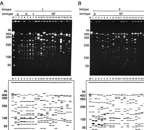
genomic DNAs of all the strains which were obtained in this study. The strains belonging to the same biotype showed var-ious distinctive PFGE patterns (Fig. 1). Twenty-seven distinc-tive restriction patterns were identified among the 28 epide-miologically unrelated strains which were biotyped as biotype I. Figure 1A shows examples of the PFGE patterns of the 20 strains which belonged to biotype I. It is noteworthy that these two strains, which belonged to different serotypes (lane 5, serotype b; lane 7, serotype c), had the same PFGE patterns. Sixty-eight restriction patterns were identified among 76 bio-type II strains. Examples of the PFGE patterns for 20 strains are demonstrated in Fig. 1B. Forty-eight restriction patterns were identified among 52 biotype III strains. Figure 1C shows examples of the PFGE patterns of the 20 strains that belonged to biotype III. The strains which belonged to biotypes IV to VIII showed different genome patterns, as shown in Fig. 1D. The variations in the PFGE patterns for the H. influenzae
strains examined in this study are summarized in Table 2. The genome patterns of the 178 epidemiologically unrelated iso-lates were classified into 165 groups. In our study, no group obtained by genotyping contained strains of more than one group obtained by biotyping.
PFGE analysis of isolates from patients with meningitis.We
obtained 12 isolates from either nasopharyngeal or blood sam-ples, in addition to CSF, from six of eight patients with men-ingitis. These isolates were also analyzed by PFGE, and their PFGE patterns were then compared with the PFGE patterns of the strains isolated from the CSF of the individual patients. The results are shown in Fig. 2. For patients 1 (serotype b, biotype I), 2 (serotype b, biotype I), 3 (serotype b, biotype II), and 5 (serotype c, biotype I), H. influenzaestrains were also obtained from both nasopharyngeal and blood samples, and PFGE analysis demonstrated that the genome patterns of the strains were the same as those of the isolates from CSF. We obtained an isolate (serotype b, biotype II) from a blood sam-ple from patient 4 but not from his nasopharyngeal samsam-ple.
H. influenzae strains which were isolated from CSF and pharynx of patient 6 were nontypeable by serotyping and also had the same genome patterns. We could not isolateH. influ-enzaefrom a blood sample from this patient. After a detailed
examination, we confirmed that this patient with meningitis had CSF leakage into his nasopharynx.
PFGE analysis of isolates from household contacts. We
obtainedH. influenzaestrains isolated from the nasopharynges of the family members of seven patients. The PFGE patterns of these isolates are shown in Fig. 3. For patients 1 (serotype b, biotype I), 8 (serotype NT [nontypeable], biotype III), 9 (se-rotype NT, biotype III), and 10 (se(se-rotype NT, biotype II), the strains from the patients had the same PFGE patterns as iso-lates that colonized the nasopharynges of their household con-tacts. For patients 3 (serotype b, biotype II), 7 (serotype NT, biotype III), and 11 (serotype b, biotype I), the strains obtained from the household contacts had PFGE patterns different from those of the patients with meningitis. For patient 11, strains of the same genotype colonized the nasopharynges of two house-hold contacts, although the genotype of the isolates from the patient with meningitis was different from that of the strains colonizing the household contacts.
IDs for various typing methods. The IDs for three typing
methods (serotyping, biotyping, and PFGE typing) are shown in Table 3. An extremely high level of discrimination was observed by PFGE typing. Serotyping and biotyping alone al-lowed less discrimination than PFGE typing. The indices for various combinations of these typing methods are also shown in Table 3. The combination of serotyping and PFGE typing had the highest level of discrimination.
DISCUSSION
H. influenzae type b strains cause invasive disease in chil-dren, including sepsis and meningitis. However, the nonencap-sulated strains of H. influenzaecommonly colonize the upper respiratory tracts of healthy persons and are also implicated in such mucosal infections as otitis media, sinusitis, bronchitis, and conjunctivitis. Although various methods for the typing of clinicalH. influenzaeisolates have been reported in the past, no method has proven to be significantly useful in epidemiological investigations because of their poor discriminatory ability, par-ticularly regarding type b strains.
In recent years, PFGE has become a useful tool for the typing and differentiation of strains for epidemiological studies with multiple bacterial species (5, 11, 14, 20). PFGE has been used for epidemiological studies of H. influenzae infections. Heath et al. (7) analyzedH. influenzaeserotype b strains iso-lated from cultures of blood from two elderly nursing home residents by PFGE. The isolates from those patients were suggested to be related because their PFGE patterns were identical. Gazagne et al. (6) used PFGE to compare clinical ampicillin-resistant, non-beta-lactamase-producingH. influen-zae strains. PFGE enabled the identification of 20 different patterns among 31 strains. They also studied the strains using other molecular biology tools, ribotyping and arbitrarily primed PCR with two primers, and each technique gave nearly the same number of different patterns as PFGE. However, ribotyping is more fastidious and time-consuming than arbi-trarily primed PCR and PFGE (6).
The apparatuses for PFGE have recently come into wide-spread use in clinical laboratories. PFGE is considered a pop-ular tool for epidemiological studies with various bacterial species. This study was carried out in order to determine whether the PFGE typing system could be useful as a tool for epidemiological studies ofH. influenzaeon the basis of the use of IDs.
The epidemiologically unrelated strains of H. influenzae
[image:2.612.53.294.91.222.2]which were collected in this study showed a variety of genome patterns by PFGE and also had high IDs. A total of 165 TABLE 1. Distribution into serotypes and biotypes of
the 200H. influenzaestrains in this study
Source No. ofstrains
No. of strains with the following serotype and biotype: a b
c, I Nontypeable I V I II I II III IV V VI VII VIII Nasopharynx 148 2 1 4 1 2 16 63 42 5 1 1 10
Sputum 19 1 8 7 2 1
Eye mucus 4 1 2 1
Middle ear mucus 8 2 1 3 1 1
Urine 7 3 3 1
Vaginal mucus 1 1
Blood 5 2 2 1
CSF 8 4 2 1 1
on May 15, 2020 by guest
http://jcm.asm.org/
genotypes were obtained in this study, and a larger number of different genome patterns are also expected to be found if more strains are collected and analyzed by PFGE. It was note-worthy that sixH. influenzaetype b strains had separate PFGE patterns. These data confirm the fact that PFGE analysis is indeed useful even for epidemiological studies of invasive dis-ease caused byH. influenzaetype b. All serotype b strains were epidemiologically unrelated; however, the number of fragment
[image:3.612.59.541.72.510.2]differences among them was only three to nine. Serotype b strains are thus considered to be genetically related to each other. This phenomenon may explain why serotype b strains have been shown to have very few variations when they are typed by other subtyping methods. The guidelines for PFGE typing (19) indicated that variations in some bands were ob-served for strains of some species when they were cultured repeatedly over time. We subcultured three different strains of FIG. 1. Examples of PFGE patterns of chromosomal DNAs extracted from clinicalH. influenzaeisolates. Chromosomal DNA was digested with theSmaI restriction endonuclease. Lanes M, bacteriophage lambda concatamer molecular size marker. (A) Patterns of 20 strains of biotype I. Nineteen different patterns were observed, and a pair with the same pattern (lanes 5 and 7) was found. Lanes: 1 and 2, serotype a; 3 to 6, serotype b; 7, serotype c; 8 to 20, nontypeable (NT) strains (lanes 8 to 14, strains isolated from the nasopharynx; lane 15, strain isolated from sputum; lane 16, strain isolated from eye mucus; lanes 17 and 18, strains isolated from middle ear mucus; lanes 19 and 20, strains isolated from urine). (B) Patterns of 20 strains of biotype II. The strains in lanes 7 and 13 showed the same genome patterns, and the strains in lanes 9, 10, and 12 and in lanes 11 and 15 also showed the same genome patterns. Therefore, 16 different patterns are shown in panel B. Lanes: 1 and 2, serotype b; 3 to 20, nontypeable strains (lanes 3 to 10, strains isolated from the nasopharynx; lanes 11 to 14, strains isolated from sputum; lane 15, strain isolated from eye mucus; lane 16, strain isolated from middle-ear mucus; lanes 17 and 18, strain isolated from urine; lane 19, strain isolated from vaginal mucus; lane 20, strain isolated from CSF). (C) Patterns of 20 strains of biotype III. The strains in lanes 17 and 20 had the same genome patterns; therefore, 19 different patterns are shown in panel C. All strains were nontypeable by serotyping. Lanes: 1 to 11, strains isolated from nasopharynx; lanes 12 to 15, strains isolated from sputum; lane 16, strain isolated from eye mucus; 17 to 19, strains isolated from middle-ear mucus; 20, strain isolated from urine. (D) Patterns of 19 strains of biotypes IV to VIII. All strains except the one in lane 5 (serotype A) were nontypeable by serotyping. Lanes: 1 to 4, biotype IV strains isolated from the nasopharynx; 5 to 9, biotype V strains (lanes 5 and 6, strains isolated from the nasopharynx; lanes 7 and 8, strains isolated from sputum; 9, strains isolated from middle-ear mucus); 10, biotype VI strain isolated from nasopharynx; 11 to 18, biotype VII strains isolated from nasopharynx; 19, biotype VIII strain isolated from sputum.
on May 15, 2020 by guest
http://jcm.asm.org/
H. influenzaeseven times and compared their PFGE types with those obtained before passage. The genome patterns of no strains changed after seven passages (data not shown). This result demonstrates the reproducibility of the PFGE typing technique withH. influenzae.
The present study also showed that strains of two different serotypes could be classified into the same group by genotyp-ing. The degree of correlation between serotyping and PFGE typing was not significantly high. On the other hand, in our study no group delineated by genotyping contained strains of more than one group delineated by biotyping.
For five of eight patients with meningitis, H. influenzae
strains were isolated from their blood samples, in addition to CSF, and the genome patterns of the strains were the same as those of isolates from CSF. It is generally known that invasive
H. influenzaeinfections including meningitis are caused by the dissemination of bacteria, which are almost always of serotype b, from the nasopharynx to the bloodstream and subsequently to other body sites (22). All of the isolates simultaneously obtained from the nasopharynx, blood, and CSF of five pa-tients with meningitis were thus considered to be the same strain. As expected, the isolates from each patient were clas-sified into a single genotype. These results indicate that al-though PFGE demonstrated various genome patterns, identi-TABLE 2. Variations in PFGE patterns of the 178
epidemiologically unrelated strains ofH. influenzaein this study Biotype No. of strains No. of different PFGE patterns
I 28 27
II 76 68
III 52 48
IV 4 4
V 5 5
VI 1 1
VII 11 11
VIII 1 1
[image:4.612.66.536.73.513.2]Total 178 165
FIG. 1—Continued.
on May 15, 2020 by guest
http://jcm.asm.org/
cal strains were placed into the same group by genotyping. For the patient with meningitis caused by a nontypeableH. influ-enzaestrain and with CSF leakage, no bacteria were isolated from his blood sample. This is probably because the bacteria disseminated from his nasopharynx to his spinal cord directly through CSF leakage. A previous study reported that anatomic disorders, including skull fractures, craniotomy, and CSF leak-age, are the major predisposing factors for invasive disease
caused by nontypeableH. influenzaestrains in older children (2).
H. influenzae strains from four of seven patients had the same PFGE pattern as the isolates from the nasopharynges of household contacts. This finding strongly suggests that the same strain that caused H. influenzae serotype b infections (meningitis) also often colonized the nasopharynges of house-hold contacts.
Our data therefore suggest that genotyping is a powerful tool for the epidemiological typing ofH. influenzaestrains.
ACKNOWLEDGMENTS
We gratefully acknowledge the following persons for supplying the strains used in this study: K. Takemori, Kyushu University Hospital, Fukuoka, Japan; T. Murao, K. Nakamura, T. Sakamoto, and K. Tsu-zuki, Fukuoka Children’s Hospital and Medical Center for Infectious Disease; T. Ohno and H. Kariyazono, Kyushu Kousei-nenkin Hospital, Fukuoka, Japan; and E. Ishii, Saga Prefectual Kouseikan Hospital, Saga, Japan. We also thank K. Okada and K. Amako for helpful discussions and advice. We also express our appreciation to B. Quinn for valuable editorial advice on the manuscript.
REFERENCES
1.Barenkamp, S. J., R. S. J. Munson, and D. M. Granoff.1981. Subtyping isolates ofHaemophilus influenzaetype b by outer-membrane protein pro-files. J. Infect. Dis.143:668–676.
2.Bol, P., L. Spanjaard, L. van Alphen, and H. C. Zanen.1987. Epidemiology ofH. influenzaemeningitis in patients more than 6 years of age. J. Infect. 15:81–94.
3.Butler, P. D., and E. R. Moxon.1990. A physical map of the genome of
Haemophilus influenzaetype b. J. Gen. Microbiol.136:2333–2342. 4.Curran, R., K. R. Hardie, and K. J. Towner.1994. Analysis by pulsed-field
gel electrophoresis of insertion mutation in the transferrin-binding system of
Haemophilus influenzaetype b. J. Med. Microbiol.41:120–126.
5.Fujita, M., S. Fujimoto, T. Morooka, and K. Amako.1995. Analysis of strains ofCampylobacter fetusby pulsed-field gel electrophoresis. J. Clin. Microbiol. 33:1676–1678.
6.Gazagne, L., C. Delmas, E. Bingen, and H. Dabernat.1998. Molecular epidemiology of ampicillin-resistant non-beta-lactamase-producing Hae-mophilus influenzae. J. Clin. Microbiol.36:3629–3635.
7.Heath, T. C., M. C. Hewitt, B. Jalaludin, C. Roberts, A. G. Capon, P. Jelfs, and G. L. Gilbert.1997. InvasiveHaemophilus influenzaetype b disease in elderly nursing home residents: two related cases. Emerg. Infect. Dis.3:179– 181.
8.Hu, L., A. Umeda, S. Kondo, and K. Amako.1995. Typing ofStaphylococcus aureuscolonising human nasal carriers by pulsed-field gel electrophoresis. J. Med. Microbiol.42:127–132.
9.Hunter, P. R.1990. Reproducibility and indices of discriminatory power of microbial typing methods. J. Clin. Microbiol.28:1903–1905.
10. Hunter, P. R., and M. A. Gaston.1988. Numerical index of the discrimina-tory ability of typing system: an application of Simpson’s index of diversity. J. Clin. Microbiol.26:2465–2466.
11. Ichiyama, S., M. Ohta, K. Shimokata, N. Kato, and J. Takeuchi.1991. Genomic DNA fingerprinting by pulsed-field gel electrophoresis as an epi-demiological marker for study of nosocomial infections caused by methicil-lin-resistantStaphylococcus aureus. J. Clin. Microbiol.29:2690–2695. 12. Inzana, T.1983. Electrophoretic heterogeneity and interstrain variation of
the lipopolysaccharide ofHaemophilus influenzae. J. Infect. Dis.148:492– 499.
[image:5.612.58.290.72.285.2]13. Kilian, M.1976. A taxonomic study of the genusHaemophiluswith the proposal of a new species. J. Gen. Microbiol.93:9–62.
FIG. 2. PFGE patterns of isolates from six patients (patients 1 to 6) with meningitis. The strains were isolated from CSF (lanes C), pharynx (lanes P), nasal cavity (lanes N), and blood (lanes B). Lane M, molecular size marker. Patients 1 and 2 had meningitis caused by serotype b, biotype I strains, patients 3 and 4 had meningitis caused by serotype b, biotype II strains, patient 5 had meningitis caused by a serotype c, biotype I strain, and patient 6 had meningitis caused by a nonencapsulated biotype II strain.
FIG. 3. PFGE patterns of isolates from seven patients withH. influenzae
[image:5.612.311.552.92.177.2]infection (p) and their household contacts (no mark). Patients 1, 3, and 11 had meningitis; patients 7, 8, 9, and 10 had respiratory tract infections. Patients 1 and 3 are the same as patients 1 and 3 in Fig. 2.
TABLE 3. IDs of three typing methods for 178 epidemiologically unrelated strains ofH. influenzae
Method No. of types ID
Serotyping 4 0.108360
Biotyping 8 0.706405
PFGE typing 165 0.999047
Serotyping and biotyping 13 0.726274
Serotyping and PFGE typing 166 0.999111
Biotyping and PFGE typing 165 0.999047
Serotyping, biotyping, and PFGE typing 166 0.999111
on May 15, 2020 by guest
http://jcm.asm.org/
[image:5.612.63.283.470.692.2]14.Kristjansson, M., M. H. Samore, D. N. Gerding, P. C. Degirolami, K. M. Bettin, A. W. Karchmer, and R. D. Arbeit.1994. Comparison of restriction endonuclease analysis, ribotyping, and pulsed-field gel electrophoresis for molecular differentiation ofClostridium difficilestrains. J. Clin. Microbiol. 32:1963–1969.
15.Loeb, M. R., and D. H. Smith.1980. Outer membrane protein composition in disease isolates ofHaemophilus influenzae: pathogenic and epidemiolog-ical implications. Infect. Immun.30:709–717.
16. Musser, J. M., D. M. Granoff, P. E. Pattison, and R. K. Sellander.1985. A population genetic framework for the study of invasive disease caused by serotype b strains ofHaemophilus influenzae. Proc. Natl. Acad. Sci. USA 82:5078–5082.
17. Pittman, M.1931. Variation and type specificity in the bacterial species
Haemophilus influenzae. J. Exp. Med.53:471–493.
18.Smith, C. L., and C. R. Canter.1987. Purification, specific fragmentation,
and separation of large DNA molecules. Methods Enzymol.155:449–467. 19. Tenover, F. C., R. D. Arbeit, R. V. Goering, P. A. Mickelsen, B. A. Murray,
D. H. Persing, and B. Swaminathan.1995. Interpreting chromosomal DNA restriction patterns produced by pulsed-field gel electrophoresis: criteria for bacterial strain typing. J. Clin. Microbiol.33:2233–2239.
20. Thong, K., Y. Ngeow, M. Altwegg, P. Navaratnam, and T. Pang.1995. Molecular analysis ofSalmonella enteritidisby pulsed-field gel electrophore-sis and ribotyping. J. Clin. Microbiol.33:1070–1074.
21. van Alphen, L., T. Riemens, J. Poolman, C. Hopman, and H. C. Zanen.1983. Homogeneity of cell envelope protein subtypes, lipopolysaccharide sero-types, and biotypes amongHaemophilus influenzaetype b from patients with meningitis in The Netherlands. J. Infect. Dis.148:75–81.
22. Ward, J. I., J. M. Lieberman, and S. L. Cochi.1994.Haemophilus influenzae
vaccines, p. 337–386.InS. A. Plotkin and E. A. Mortimer (ed.), Vaccines. The W. B. Saunders Co., Philadelphia, Pa.