Copyright © 2001, American Society for Microbiology. All Rights Reserved.
Detection of
Mycobacterium bovis
in Bovine Clinical Specimens
Using Real-Time Fluorescence and Fluorescence Resonance
Energy Transfer Probe Rapid-Cycle PCR
MALCOLM JAMES TAYLOR,* MARY SIOBHAN HUGHES, ROBIN ALFRED SKUCE,
ANDSYDNEY DONNELLY NEILL
Department of Agriculture and Rural Development, Veterinary Sciences Division, Stormont, Belfast BT4 3SD, Northern Ireland
Received 25 May 2000/Returned for modification 29 August 2000/Accepted 31 December 2000
Nucleic acid sequence capture extraction was coupled with LightCycler PCR amplification and product
detection using real-time fluorescence for rapid, definitive detection ofMycobacterium bovis in lymph node
specimens from 38 cattle with bovine tuberculosis lesions. PCR amplification of sequence-captured DNA using both a conventional heating block thermocycler and a LightCycler thermocycler was compared with culture and histopathological analyses. Conventional PCR enabled detection of 26 of 28 culture-positive specimens (93%) in approximately 9 h, and the LightCycler PCR detected 20 of 28 culture-positive specimens (71%) in
only 30 min. Specific confirmation ofMycobacterium tuberculosiscomplex DNA was achieved by LightCycler
PCR amplification using Syb Green 1 and anM. tuberculosiscomplex-specific Cy5-labeled fluorescence
reso-nance energy transfer probe. The system described here enabled rapid and specific laboratory confirmation of
bovine tuberculosis, and this is the first report of the detection ofM. bovisin tissues using LightCycler PCR.
The fluorescence technology used in the study has potential to allow development of a high-throughput molecular diagnostic test for bovine tuberculosis.
Mycobacterium bovis, a member of theMycobacterium tuber-culosiscomplex, is the causative agent of bovine tuberculosis. This zoonotic disease continues to have considerable economic and public health implications (18, 19). National eradication programs employ tuberculin testing and slaughter strategies (17). Culture is used commonly to confirm infection in post-mortem specimens from cattle slaughtered following a positive skin test reaction. Although culture is considered to be the “gold standard” for confirming tuberculosis, this procedure may take several weeks (15).
Consequently, rapid nucleic acid amplification techniques, including the PCR- and transcription-mediated amplification, have been applied to detectM. bovisdirectly in clinical speci-mens (1, 2, 15, 28; S. Roring et al., unpublished data). While PCR amplifications have enabled detection of nonviable my-cobacteria (15, 16, 28), they are not as sensitive as culture. In comparison to M. tuberculosis in human sputa, which often contain large numbers of bacilli (28), bovine tuberculous tis-sues are associated with few bacilli. Extraction of mycobacte-rial DNA from sputa was considered to be less difficult than extraction from tissues, and consequently, improvements in nucleic acid extraction were recommended to increase sensi-tivity of PCR detection ofM. bovisin tissue (6).
Mycobacterial DNA extraction efficiency has been improved with the development of a nucleic acid sequence capture pro-cedure, which has enabled detection of mycobacteria in pauc-ibacillary forms of tuberculosis (3, 16). Sequence capture PCR
has been used successfully for simultaneous detection and strain typing ofM. bovisfrom BACTEC cultures (22) and more recently from bovine lymph node tissue (23). These studies were performed in conventional heating block thermocyclers (HBTC) with end point detection of PCR products by agarose gel electrophoresis. Automation of the sequence capture PCR procedure should facilitate its use in routine diagnosis of
M. tuberculosisin clinical specimens (3, 16).
Rapid-cycle PCR amplifications, using an air thermocycler (ATC), have increased the rapidity ofM. tuberculosisdetection by decreasing the amplification time (6, 13) and thus may assist automation. Additionally, alternative PCR product detection systems have the potential to improve automation, as tradi-tional methods of detection may be laborious and time-con-suming (12, 20).
Fluorimeter-based closed-tube PCR assays permit the con-tinual monitoring of accumulating fluorescently labeled PCR products, termed real-time fluorescence (11). Rapid-cycle PCR in conjunction with fluorimeter-based closed-tube PCR assays have provided a rapid and sensitive method for identification and quantification of PCR products (21, 29). Real-time fluo-rescence has been used to detect M. tuberculosis in sputum using the TaqMan system (8). In this study, sequence capture is combined with rapid-cycle PCR and real-time fluorescence involving the use of anM. tuberculosis complex-specific fluo-rescence resonance energy transfer (FRET) probe in a Light-Cycler LC32 (Biogene Ltd., Kimbolton, United Kingdom) to detectM. bovisin bovine tissues.
MATERIALS AND METHODS
Biological material.A bovine field isolate ofM. bovis, characterized by re-striction fragment length polymorphism analysis (24), was cultured in
Middle-brook 7H9 broth (Difco Laboratories). Serial dilutions of culture broth (10⫺3to
* Corresponding author. Mailing address: Department of Agricul-ture and Rural Development, Veterinary Sciences Division, Stoney Rd., Stormont, Belfast BT4 3SD, Northern Ireland. Phone: 44 (0) 2890 525719. Fax: 44 (0) 2890 525745. E-mail: malcolm.taylor@dardni .gov.uk.
1272
on May 15, 2020 by guest
http://jcm.asm.org/
10⫺8) were made in sterile saline and subcultured on 7H9 agar plates. Aliquots
of the culture dilution series (400l) were stored at⫺20°C and used for the
determination of sequence capture PCR efficiency at a later date.
M. bovisgenomic DNA was extracted from a second, similarly characterized
bovine field isolate, and the IS6110gene copy number was estimated from DNA
concentrations determined byA260s. Serial dilutions ofM. bovisgenomic DNA
(2.5⫻104to 2.5⫻10⫺1IS6110genome equivalents) were used as positive PCR
controls.
Lymph nodes were collected from 38 cattle exhibiting lesions at slaughter. These cattle were either intradermal skin test positive or identified as having lesions during routine meat inspection. Specimens were subjected to histopathol-gical examination (5) and to microbiolohistopathol-gical decontamination prior to culture (26). Aliquots of the latter specimens were cultured both on slopes of Lowen-stein-Jensen (L-J) solid medium (Media for Mycobacteria; Sully, South Glam-organ, United Kingdom) and in BACTEC 12B liquid medium (Becton Dickin-son, Oxford, United Kingdom). Residual decontaminated tissue was stored at
⫺20°C for sequence capture PCR. L-J slope cultures were monitored for
bac-terial growth after 28 days, and BACTEC cultures were examined up to 84 days postinoculation. The presence of acid-fast bacilli in BACTEC cultures was con-firmed by Ziehl-Neelsen staining and microscopic examination (25). Isolates
from L-J slopes were confirmed asM. tuberculosiscomplex using the Accuprobe
(Gen-Probe, San Diego, Calif.).
DNA extractions.DNA sequence capture PCR, as previously described (16) and modified (23), was applied to homogenates of the 38 decontaminated bovine lymph nodes with the following additional modifications. In brief, suspensions of
decontaminated tissue homogenate (500l) were transferred to screw-cap
mi-crocentrifuge tubes containing 500l of 0.1-mm-diameter zirconium beads
(Bio-spec Products Inc., Bartlesville, Okla.). Samples were centrifuged and washed as
described previously (23). The pellet and beads were resuspended in 500l of
TES [N-tris(hydroxymethyl)methyl-2-aminoethanesulfonic acid]–100 mM
Tris-HCl (pH 7.4)–50 mM EDTA–150 mM NaCl and incubated in a sonicating water bath for 15 min. Samples were agitated in a FastPrep Bio 101 bead shaker (Savant Instruments Inc., Holbrook, N.Y.) at 6 m/s for 45 s. Proteinase K (Sigma, Poole, Dorset, United Kingdom) was added to a final concentration of 3 mg/ml, and the mixture was incubated at 50°C for 18 h. Samples were shaken in the FastPrep Bio 101 as before. Aliquots of proteinase K-treated homogenates (500
l) were denatured at 100°C for 15 min and immediately transferred to ice for a
further 5 min. The biotinylated capture oligonucleotides CapDRa (5⬘biotin-AA
AAAGGTTTTGGGTCTGACGAC) and CapDRa (5⬘biotin-AAAAACCGAG
AGGGGACGGAAAC) [Genosys Biotechnologies (Europe), Pampisford,
Cambs., United Kingdom] were used to capture the DR region of theM.
tuber-culosiscomplex (16). Capture oligonucleotides (2.5 pmol of each) in 3.75 M NaCl solution were added to homogenates to a final concentration of 1 M NaCl, mixed well, and hybridized at 42°C with gentle agitation for 3 h. Following
hybridiza-tion, 50g of streptavidin M-280 Dynal beads (Dynal, Oslo, Norway) was added,
mixed well, and incubated at 36°C with gentle agitation for 2 h. The streptavidin M-280 Dynal beads were separated from the supernatant and washed using a
magnetic bead separator (6 by 1.5 ml) (Stratagene, La Jolla, Calif.) in 750l of
wash buffer (10 mM Tris-HCl [pH 7.4], 1 mM EDTA) and then in sterile
deionized H2O. Magnetic beads were resuspended in 25l of sterile deionized
H2O and stored at⫺20°C prior to PCR amplification. Immediately prior to
amplification, captured mycobacterial DNA was released from magnetic beads by heat treatment at 100°C for 5 min. Following a brief centrifugation, the resulting supernatant was subjected to the appropriate amplification.
PCR amplification.Sequence-captured mycobacterial nucleic acid was sub-jected to rapid thermal cycling and continuous monitoring of PCR products in a LightCycler LC32 (Biogene Ltd.) and to conventional PCR amplification using an HBTC (model 480; Perkin-Elmer, Warrington, United Kingdom)
(HBTC-PCR). Oligonucleotides specific for theM. tuberculosiscomplex IS6110sequence
(9) (Genosys) were used for the amplifications. A standard PCR protocol (LC-PCR) utilizing the LC32 instrumentation was adopted only after extensive mod-ification and optimization of previously reported PCR conditions (9). Optimal magnesium ion and oligonucleotide concentrations were determined along with cycling parameters such as denaturation, annealing and elongation temperatures, incubation periods, and temperature transition rates. LC32 instrumentation vari-ables were optimized as directed by the manufacturers.
Standard LightCycler protocol.After optimization, the following standard LC-PCR protocol was applied to all specimens. A commercial PCR master
mixture (Bio/Gene Ltd.), containing 2.5 U of Taqpolymerase, 250M
de-oxynucleoside triphosphates, and 3 mM MgCl2, was pretreated withTaq-Start
antibody (7l) (Sigma). The PCR mixture consisted of pretreated PCR master
mixture supplemented with IS6110-specific oligonucleotides (500 nM), Syb
Green 1 (1/60,000 dilution) (Biogene Ltd.), lambda DNA (5 pg/l), and Cy5
3⬘-labeled FRET probe LCP (100 nM) together with 2.5l of target DNA in a
final reaction volume of 10l. The FRET probe LCP (5⬘GCCCAGGTCGAC
ACATAGG3⬘-Cy5), specific for theM. tuberculosiscomplex, was designed using
OLIGO 5 primer design software (Biogene Ltd.) and to the specifications de-scribed by Biogene Ltd.
The PCR mixture (3-l aliquot) was applied to the top of a glass capillary
reaction vessel (part 1720; Biogene Ltd.) which was filled by pulse centrifugation in a microcentrifuge. Conditions for cycling were 94°C for 45 s, followed by 50 cycles of 94°C for 0 s, 62°C for 0 s, and 74°C for 10 s; fluorescence was monitored at the end of every 74°C step. The amplification program was followed by a melting program of 45°C for 10 s and then 45 to 95°C at a transition rate of 0.2°C/s with continuous monitoring of fluorescence. The temperature transition
rate for all cycling steps was 20°C per s except for those between 62 and 74°C,
where a transition rate of 1°C per s was used. The gain on the F1 channel photometric detector was routinely set at 64.
HBTC protocol. HBTC-PCR included either 2.5l of sequence-captured
magnetic beads or a 5-l inoculum of either genomic DNA or heat-inactivated
M. bovis. A PCR master mix (50-l final volume) contained 2.5 U ofTaq
poly-merase (Sigma), a 500 nM concentration of each of the IS6110primers, 250M
deoxynucleoside triphosphates, and 1.75 mM MgCl2, with a mineral oil overlay
(Sigma). Conditions for cycling were 94°C for 5 min, followed by 30 cycles of 94°C for 1 min, 68°C for 2 min, and 74°C for 1 min. Amplification mixtures were incubated for a further 7 min at 74°C, with a final 4°C soak.
For reamplification, 5l of first-round PCR product was subjected to a second
round of PCR amplification using the same PCR conditions. The 123-bp PCR
product of IS6110was identified by 2% (wt/vol) agarose gel electrophoresis with
TAE buffer (40 mM Tris-acetate, 1 mM EDTA).
RESULTS
Optimization of light cycler reactions. (i) Preliminary
ex-periments.PCR amplifications and monitoring of fluorescence
emission during PCR were performed using a LightCycler LC32. An amplification program, recommended by the LC32 manufacturer, was modified so that annealing temperatures similar to those previously published for this primer set were used (9). Preliminary experimentation using positive controls of heat-killed mycobacterial suspension together with negative controls of water established suboptimal reaction conditions of 3 mM magnesium (range of 2 to 5 mM tested in 1 mM incre-ments), annealing temperature of 60°C (range of 60 to 66oC tested in 1°C increments), 500M primer (100 and 500 nM tested), and 100 nM LCP (10, 50, and 100 nM tested).
Run profile software graphically presented relative fluores-cence versus time during the amplification program. Fluo-rescence signal acquisition, graphically displayed as a curve, increased in value with time as product was synthesized. Re-action mixtures containingM. bovisgenomic DNA exhibited an exponential increase in signal after 20 cycles, while negative controls demonstrated a slow signal acquisition only after 40 cycles (Fig. 1).
Linking a melt cycle program to the amplification program facilitated the identification of amplification products. The continuous monitoring of fluorescence emissions during the slow denaturation step in the melt program permitted a precise calculation of the melting temperatures (Tms) of all PCR
prod-ucts. Melting-curve software (Idaho Technologies Inc.) con-verted fluorescence versus temperature to the rate of change of fluorescence emissions versus temperature (⫺dF/dT versus temperature), termed peak analysis. PCR product purity was demonstrated by determining theTms of amplified products.
Adjustment of points to average to 7 was found to smooth curves, aiding interpretation by reducing noise without com-promising resolution. Fluorescence channel F1 detected emis-sions from the intercalation of the dye Syb Green 1 with all
on May 15, 2020 by guest
http://jcm.asm.org/
double-stranded nucleic acid PCR products. PCRs with M. bovisgenomic DNA exhibited a peak at 89°C, while negative controls were characterized by a peak at 86°C. The identities of PCR products were confirmed by gel electrophoresis and ethidium bromide staining of LC32 reaction vessel contents. A 123-bp IS6110-specific PCR product was associated with the 89°C peak, while a smaller primer artifact was associated with the 86°C peak. LC-PCR amplifications of serial dilutions of
M. bovisgenomic DNA, over 4 log units (104to 101 IS6110 gene copies), demonstrated a sensitivity of 101 gene copies (Fig. 2).
Fluorescence channel F2 detected emissions from Cy5-la-beled FRET LCP stimulated by resonance energy donated by intercalated Syb Green 1. Fluorescent emissions from the melt program, monitored by both F1 and F2 optics, (F2/F1) after peak analysis exhibited a peak between 60 and 65°C for
reac-tion mixtures containingM. bovisgenomic DNA, while nega-tive controls displayed no such peak (Fig. 3).
(ii) Improved reproducibility and sensitivity.In preliminary
experiments, amplifications performed on consecutive days were found to lack reproducibility (data not shown). Positive control run profiles demonstrated inconsistencies in signal ac-quisition. This lack of reproducibility of the LC-PCR was at-tributed to Syb Green 1 instability. Revised protocols from the manufacturer (Biogene Ltd.) required that Syb Green 1 be stored at⫺20°C, in an undiluted form, for storage of up to 1 year without loss of activity. Once diluted 1/1,000 in the dilu-tion buffer supplied by the manufacturer, Syb Green 1 must be stored in the dark at 4°C for no more than 1 month and shaken vigorously for 1 min immediately prior to use.
Several modifications were made to the PCR master mixture recommended by the manufacturer (Biogene Ltd.) to improve sensitivity. Commercial master reaction mixtures (500-l ali-quots) were pretreated withTaq-Start antibody (7l) (Sigma) incubated at room temperature for 20 min prior to storage at
⫺20°C. Pretreatment, while reducing background in the neg-ative control, delayed the onset of exponential signal in posi-tive controls. Lambda DNA was added to the master mixture to a final concentration of 47.5 pg/l in order to improve the specificity of PCRs. Storage of positive control reaction mix in the dark at room temperature for prolonged periods (24 h) was not detrimental to either specificity or sensitivity. The FRET LCP was stored in aliquots at 10M at⫺20°C and diluted to 1M immediately prior to use. The temperature transition rate of the cycle program, between annealing and extension temperatures, was modified to 1°C/s. The melt program was modified to include a 15-s hold at 45°C prior to the commence-ment of the melt program with a temperature transition rate of 0.2°C/s.
[image:3.612.55.290.71.227.2]The preliminary protocol was optimized using positive con-trols of heat-killed mycobacterial suspension and purified ge-nomic mycobacterial DNA and negative controls of water, establishing reaction conditions of 3 mM magnesium (2 to 5 FIG. 1. Screen capture image (CorelDRAW; Corel Corporation,
Ottawa, Ontario, Canada) of run profile analysis demonstrating the accumulation of F1 fluorescence during PCR of 10-fold serial dilutions ofM. bovisgenomic DNA representing 2.5⫻104to 2.5⫻10⫺1IS6110
gene copies using Syb Green 1 only.
FIG. 2. Screen capture (CorelDRAW) of F1 melting-peak analysis of 10-fold serial dilutions ofM. bovisgenomic DNA representing 2.5⫻ 104to 2.5⫻10⫺1IS6110gene copies using Syb Green 1. Each trace
graphically displays the rate of change of F1 fluorescence emissions versus temperature; a peak represents theTmof the PCR product. The
IS6110123-bp species displays aTmof 89°C, while a primer artifact
displays a peak at 86°C.
FIG. 3. Screen capture (CorelDRAW) of F2 melting-peak analysis of anM. bovisgenomic DNA dilution series representing 2.5⫻104to
2.5⫻10⫺1IS6110gene copies using FRET probe LCP and Syb Green
1. TheTmof each PCR product is identified as a peak; each trace
represents the rate of change of F2/F1 fluorescence emissions versus temperature. The IS6110123-bp species displays aTmof 60 to 65°C,
while the primer artifact displays none. The F1 emissions are respon-sible for the F1 peaks at the right side of the graph.
on May 15, 2020 by guest
http://jcm.asm.org/
mM tested), 62°C annealing temperature (60 to 68°C tested), 500M primer concentration (100 to 500 nM tested), and 100 nM LCP (10 to 100 nM tested) using the cycling conditions stated in Materials and Methods.
Sensitivity.LightCycler PCR amplifications using the
stan-dard protocol reproducibly detected 25 copies of theM. bovis
IS6110gene DNA with melt peak analysis (dF/dTversus tem-perature) of both F1 and F2 emissions (Fig. 2 and 3). LC-PCR amplification of sequence-capturedM. bovis culture dilution series detected 36 bacilli using the F1 channel and 360 bacilli using the F2 channel. A single round of HBTC-PCR amplifi-cation of the same dilution series ofM. bovisIS6110gene DNA as used for the LC-PCR amplifications reproducibly detected 625 gene copies. A second round of HBTC amplification, how-ever, was required in order to detect 62 gene copies. Two rounds of HBTC amplifications were also required to detect 36 sequence-capturedM. bovisbacilli.
Clinical specimens.Lymph node tissues were cultured and
examined histopathologically. Of the 38 lymph node specimens
from animals with lesions, 13 were from cattle which were skin test positive (Table 1), and 11 of these 13 specimens were cul-ture positive. Twenty-seven of the 38 specimens were culcul-ture positive using both L-J slope and BACTEC media (Table 1). All culture isolates were confirmed asM. tuberculosiscomplex by Accuprobe (Gen-Probe) (data not shown). Three were BACTEC cultured positive after 7 days, and the remaining 24 were pos-itive after 14 days. Histopathological examination identified 26 bovine tuberculosis-positive specimens. Five of the 28 culture-positive specimens were negative by histopathology, and 3 of the 10 culture-negative specimens (Table 1) were positive by histopathology. One specimen, identified with morphology typ-ical of actinomycetes by histopathology, was BACTEC culture negative; however, this specimen was L-J culture positive after 28 days.
Sequence-captured nucleic acid derived from decontami-nated homogenates was subjected to PCR amplification utiliz-ing both LC32 and HBTC methodologies. DNA representutiliz-ing 10% of that sequence captured was amplified in either system. Using HBTC,M. bovis was detected in 26 (93%) of the 28 culture-positive tissue specimens (Table 2 and Fig. 4a). LC-PCR detectedM. bovisin 20 (71%) of the 28 culture-positive tissue specimens (Table 3 and Fig. 4b). All of the culture-negative specimens were PCR culture-negative in both the HBTC- and LC-PCRs (Table 1). There was also good agreement between the LC- and HBTC-PCR results; 13 of 14 (93%) of the HBTC strong positives, 4 of 5 (80%) of the HBTC moderate positives, and 3 of 7 (43%) of the HBTC weak positives were positive by LC-PCR. In addition, the two culture-positive HBTC-PCR-negative specimens were also HBTC-PCR-negative by LC-PCR.
DISCUSSION
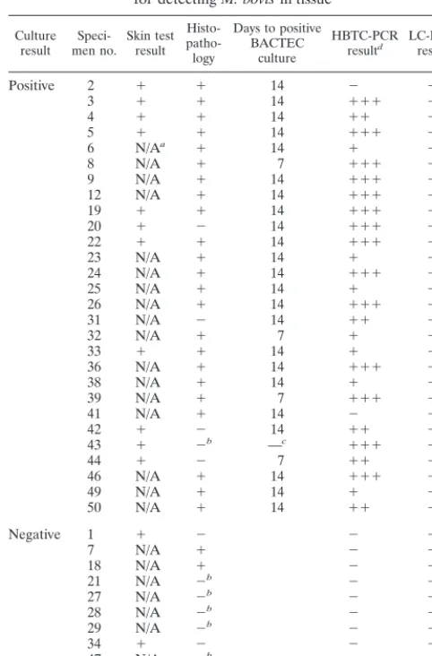
[image:4.612.51.292.98.464.2]This work is the first reported application of sequence cap-ture PCR and real-time fluorescence detection ofM. bovisin clinical tissue. The benefits of target enrichment and removal of inhibitory substances attributed to sequence capture were combined successfully with the alternative PCR product detec-tion technology offered by real-time fluorescence. Several nu-cleic acid amplification-based techniques have become acces-sible to clinical mycobacteriological laboratories in recent years. However, poor performance in paucibacillary situations and high costs have limited their widespread application (20). Requirement of a high degree of operator skill to perform and interpret molecular analyses has also hindered adoption of molecular methods in diagnostic laboratories (10). The appli-cation of PCR to disease diagnosis is dependent on specificity, sensitivity, ease of use, and high sample throughput (3, 16, 20). In this study improved DNA extraction and fluorescent detec-tion methodologies were used to examine these criteria and to TABLE 1. Histopathological and bacteriological status of tissue
specimens and performance of HBTC-PCR and LC-PCR for detectingM. bovisin tissue
Culture
result men no.Speci- Skin testresult
Histo- patho-logy
Days to positive BACTEC
culture
HBTC-PCR
resultd LC-PCRresult
Positive 2 ⫹ ⫹ 14 ⫺ ⫺
3 ⫹ ⫹ 14 ⫹⫹⫹ ⫺
4 ⫹ ⫹ 14 ⫹⫹ ⫺
5 ⫹ ⫹ 14 ⫹⫹⫹ ⫹
6 N/Aa ⫹ 14 ⫹ ⫹
8 N/A ⫹ 7 ⫹⫹⫹ ⫹
9 N/A ⫹ 14 ⫹⫹⫹ ⫹
12 N/A ⫹ 14 ⫹⫹⫹ ⫹
19 ⫹ ⫹ 14 ⫹⫹⫹ ⫹
20 ⫹ ⫺ 14 ⫹⫹⫹ ⫹
22 ⫹ ⫹ 14 ⫹⫹⫹ ⫹
23 N/A ⫹ 14 ⫹ ⫹
24 N/A ⫹ 14 ⫹⫹⫹ ⫹
25 N/A ⫹ 14 ⫹ ⫺
26 N/A ⫹ 14 ⫹⫹⫹ ⫹
31 N/A ⫺ 14 ⫹⫹ ⫹
32 N/A ⫹ 7 ⫹ ⫺
33 ⫹ ⫹ 14 ⫹ ⫹
36 N/A ⫹ 14 ⫹⫹⫹ ⫹
38 N/A ⫹ 14 ⫹ ⫺
39 N/A ⫹ 7 ⫹⫹⫹ ⫹
41 N/A ⫹ 14 ⫺ ⫺
42 ⫹ ⫺ 14 ⫹⫹ ⫹
43 ⫹ ⫺b —c ⫹⫹⫹ ⫹
44 ⫹ ⫺ 7 ⫹⫹ ⫹
46 N/A ⫹ 14 ⫹⫹⫹ ⫹
49 N/A ⫹ 14 ⫹ ⫺
50 N/A ⫹ 14 ⫹⫹ ⫹
Negative 1 ⫹ ⫺ ⫺ ⫺
7 N/A ⫹ ⫺ ⫺
18 N/A ⫹ ⫺ ⫺
21 N/A ⫺b ⫺ ⫺
27 N/A ⫺b ⫺ ⫺
28 N/A ⫺b ⫺ ⫺
29 N/A ⫺b ⫺ ⫺
34 ⫹ ⫺ ⫺ ⫺
47 N/A ⫺b ⫺ ⫺
48 N/A ⫹ ⫺ ⫺
aN/A, animals were not skin tested prior to analysis.
bActinomycesinfection.
cA few colonies grew on L-J slope media.
d⫹⫹⫹, strong band;⫹⫹, medium band;⫹, weak band.
TABLE 2. Comparison of HBTC-PCR with culture results for the detection ofM. bovisin tissue
Culture result No. of specimens HBTC-PCR:
Positive Negative
Positive 26 2
Negative 0 10
on May 15, 2020 by guest
http://jcm.asm.org/
[image:4.612.310.551.673.728.2]facilitate PCR diagnosis of M. bovis in bovine tuberculosis specimens.
Culture of M. bovis from clinical specimens has been re-garded as the standard against which otherM. bovisdiagnostic methodologies have been measured (15). However, culture of
M. bovisis slow, and incubation periods of several weeks may be required (15, 28). Radiometric culture (BACTEC) permits more rapid detection of mycobacteria; however, minimum incubation periods of several days are still required (14). In this study, only 4 of the 28 culture-positive specimens were BACTEC culture positive within 7 days. A further 23 were culture positive within 14 days. The remaining specimen was BACTEC culture negative and exhibited only poor growth on L-J slopes. The inability to culture mycobacteria from tissue specimens has previously been associated with sample autolysis or microbial contamination (15, 28), which may account for the latter result.
Histopathological examination, used commonly for routine diagnosis of bovine tuberculosis, permits rapid identification of lesions but may not differentiate between those caused byM. bovisand by other mycobacterial or closely related agents (7). In addition to this lack of specificity, the sensitivity of micro-scopic examination is limited (31). The results of this study indicated the limitations of histopathological analysis, as only 82% (23 of 28) of the culture-positive specimens were positive by histopathological analysis. The five histopathologically neg-ative specimens were both culture and PCR positive. Three
histopathologically positive specimens were negative by both culture and PCR, suggesting that these histopathology results may represent false positives.
Limited sensitivity has also been associated with molecular technologies for mycobacterial diagnosis (16). PCR has been applied widely to the detection ofM. tuberculosisin sputa and bronchiolar lavages from human tuberculosis patients with suc-cess but not so for bovine tuberculosis, where the bacterial load is substantially less (8, 28).
The application of sequence capture and conventional PCR amplification methodologies in this study demonstrated
M. bovisdetection sensitivities comparable to those of culture. This is in agreement with other sequence capture studies (3, 16, 23). Ninety-three percent of culture-positive specimens were detected using two rounds of PCR amplification in the HBTC followed by analysis of the PCR products by agarose gel electrophoresis. This was comparable to a sensitivity of 91% achieved in earlier PCR studies (1, 28). PCR amplification and product detection, using HBTC, took approximately 9 h, which was much shorter than the period required for detection by culture.
Further reductions in the PCR detection time forM. tuber-culosis have been achieved using an ATC (6, 13). In these studies, air thermal cycling used small-volume glass capillaries and high-velocity heated air to generate temperature transition rates in excess of 10°C/s and culminated in amplification times of less than 30 min. In addition, the use of reduced reaction volumes associated with an ATC resulted in a substantial re-duction in reagent costs (6). Until recently, PCR amplification and product analysis have been sequential procedures. The LC32 used in this study measures real-time fluorescence, com-bining the rapid-cycle capabilities of the ATC with continual fluorimetric detection of accumulating PCR products.
Interpretation of fluorimetric emissions generated by Syb Green 1 was confirmed initially by agarose gel electrophoresis analysis of PCR products evacuated from the capillary reaction vessels and resolved on agarose gels, as was the case in earlier studies utilizing fluorescent detection systems (27). The pres-ence of the expected 123-bp PCR product along with PCR artifacts established that the molecular enzymology of the LC32 had resulted in the synthesis of the predicted IS6110
PCR product. The sensitivity achieved in this report with the IS6110target was 25 IS6110gene copies, which was compara-ble to that of previous studies (16). Accumulation of fluores-cence may not equate to specific PCR product synthesis, as Syb Green 1 detects all double-stranded DNA, including PCR ar-tifacts (21, 27).
In this present study PCR product differentiation was achieved, in most cases, by analyzing the melting curve char-acteristics of the PCR products. Melting curves have previously FIG. 4. (a) Agarose gel electrophoresis of HBTC-PCR-amplified
[image:5.612.52.294.74.337.2]DNAs from lymph nodes from animals with bovine tuberculosis le-sions. Amplification products from five specimens listed in Table 1 are displayed. The size of the amplified target DNA is shown. (b) Screen capture (CorelDRAW) of F2 melting peak analysis of LC-PCR am-plification of DNAs from lymph nodes from five animals with bovine tuberculosis lesions (Table 1).
TABLE 3. Comparison of LC-PCR with culture results for the detection ofM. bovisin tissue
Culture result No. of specimens LC-PCR:
Positive Negative
Positive 20 8
Negative 0 10
on May 15, 2020 by guest
http://jcm.asm.org/
[image:5.612.311.551.673.727.2]differentiated multiple PCR products in concordance with those products identified by gel electrophoresis. PCR products with
Tms differing by only 2°C have been distinguished (21). Again
in the present study, the 123-bp IS6110-specific product exhib-ited aTmof 89°C, while the PCR primer artifact displayed aTm
of 86°C. Primer artifacts occasionally generated shoulder ef-fects on F1 signal melt peaks of specific PCR products, making their interpretation difficult. Primer artifacts withTms similar
to those of specific products have been reported in other stud-ies (27, 30). These reports suggested that melt curve analysis alone is not definitive in identifying specific PCR products.
Sequence-specific detection using FRET hybridization probes (specific oligonucleotide probes labeled with fluores-cent dyes) has been reported. Several FRET formats have been developed (12). One format, hybridization probes, which employs two single fluorescence-labeled oligonucleotides that hybridize to adjacent regions of target DNA, has been used in conjunction with LightCycler instrumentation (4). In the pres-ent study, because the target was small and GC rich, only a single-oligonucleotide FRET probe was used as a hybridiza-tion probe. Resonance energy, received from Syb Green 1 intercalated between the oligonucleotide and PCR product, was transferred to the Cy5 fluorophore attached to the oligo-nucleotide’s 3⬘terminus. The broad emission spectrum of the fluorescence of Syb Green 1 overlaps that of Cy5, but the melt curve analysis compensates by dividing the F2 signal by the F1 signal. This FRET probe enabled detection of anM. tubercu-losis complex target which exhibited aTmof 60 to 65°C. A
sensitivity of 25 IS6110gene copies, similar to that achieved using the DNA dilution series and Syb Green 1 only, was demonstrated. The FRET probe circumvented the previously mentioned difficulties with interpretation of the F1 signal melt peak. Few studies have used this single-probe approach, but those that have report a greater sensitivity than with the dou-ble-probe format (4).
The Cy5-labeled FRET hybridization probe LCP described in this study detectedM. bovisgenomic DNA in decontami-nated tissue homogenates of 20 of the 28 culture-positive spec-imens. This sensitivity of detection was lower than the 26 of 28 culture-positive specimens detected by PCR in the HBTC. The reduced sensitivity with the FRET probe was attributed in part to the perturbation of fluorescent signals by the red iron oxide present in the sequence capture magnetic beads. This pertur-bation was alleviated by detachment of captured mycobacterial DNA from magnetic beads by heat treatment at 100°C for 5 min and centrifugation prior to amplification. This treatment resulted in significant improvements in signal interpretation. PCR in the HBTC was probably more sensitive than that in the LC32 due to the second round of amplification employed in the HBTC. It is clear that the benefits of PCR reamplification need to be balanced against the associated increased risk of sample cross contamination and increased assay time. Further optimization of the LC32 system is expected to elevate the sensitivity to that of HBTC. This study demonstrated the ra-pidity of PCR diagnosis compared to culture. Rapid-cycle PCR and fluorimetric detection technologies have reduced detec-tion time further to several minutes and would allow rapid screening of tissue specimens for the presence ofM. bovis. The same fluorescent technology should facilitate development of a high-throughput molecular diagnostic assay and provide a
more practical approach for confirmation of tuberculosis di-rectly from clinical specimens.
ACKNOWLEDGMENTS
We are grateful to D. Brittain for the provision ofM. bovisgenomic DNA and to all of the staff in the tuberculosis diagnostic laboratory and histopathology laboratory for their technical assistance.
REFERENCES
1.Aranaz, A., E. Lie´bana, A. Mateos, D. Vidal, M. Domingo, and L. Domin-guez.1995. Direct detection ofM. bovisfrom tissue samples. Improvement of
a DNA extraction method for PCR amplification, p. 60–63.InF. Griffin and
G. de Lisle (ed.), Tuberculosis in wildlife and domestic animals. University
of Otago Press, Dunedin, New Zealand.
2.Bollo, E., F. Guarda, M. T. Capucchio, and F. Galietti.1998. Direct
detec-tion ofMycobacterium tuberculosiscomplex in tissue specimens from cattle
through identification of specific rRNA sequences. J. Vet. Med. Ser. B
45:395–400.
3.Brugie`re, O., M. Vokurka, D. Lecossier, G. Mangiapan, A. Amrane, B. Milleron, C. Mayaud, J. Cadranel, and A. J. Hance.1997. Diagnosis of smear-negative pulmonary tuberculosis using sequence capture polymerase
chain reaction. Am. J. Respir. Crit. Care Med.155:1478–1481.
4.Cane, P. A., P. Cook, D. Ratcliffe, D. Mutimer, and D. Pillay.1999. Use of real-time PCR and fluorimetry to detect lamivudine resistance-associated
mutations in hepatitis B virus. Antimicrob. Agents Chemother.43:1600–
1608.
5.Cassidy, J. P., D. G. Bryson, and S. D. Neill.1999. Tonsillar lesions in cattle
naturally infected withMycobacterium bovis. Vet. Rec.144:139–142.
6.Chapin, K., and T.-L. Lauderdale.1997. Evaluation of a rapid air thermal
cycler for detection ofMycobacterium tuberculosis.J. Clin. Microbiol.35:
2157–2159.
7.de Lisle, G. W., G. F. Yates, and D. M. Collins.1993. Paratuberculosis in farmed deer: case reports and DNA characterisation of isolates of
Myco-bacterium paratuberculosis. J. Vet. Diagn. Investig.5:567–571.
8.Desjardin, L. E., Y. Chen, M. D. Perkins, L. Teixeira, M. D. Cave, and K. D. Eisenach.1998. Comparison of the ABI 7700 system (TaqMan) and
com-petitive PCR for quantification of IS6110DNA in sputum during treatment
of tuberculosis. J. Clin. Microbiol.36:1964–1968.
9.Eisenach, K. D., M. D. Cave, J. H. Bates, and J. T. Crawford.1990. Poly-merase chain reaction amplification of a repetitive DNA sequence specific forMycobacterium tuberculosis. J. Infect. Dis.161:977–981.
10. Friedman, C. R., M. Y. Stoeckle, W. D. Johnston, and L. W. Riley.1995. Double-repetitive-element PCR method for subtyping mycobacteria
tuber-culosis clinical isolates. J. Clin. Microbiol.33:1382–1384.
11. Higuchi, R., G. Dollinger, S. Walsh, and R. Griffith.1992. Simultaneous
amplification and detection of specific-DNA sequences. Bio/Technology10:
413–417.
12. Isaksson, A., and U. Landegren.1999. Accessing genomic information:
al-ternatives to PCR. Curr. Opin. Biotechnol.10:11–15.
13. Kearns, A. M., R. Freeman, M. Steward, and J. G. Magee.1998. A. rapid
polymerase chain reaction technique for detectingM. tuberculosisin a variety
of clinical specimens. J. Clin. Pathol.51:922–924.
14. Kirihara, J. M., S. L. Hillier, and M. B. Coyle.1985. Improved detection
times forMycobacterium aviumandMycobacterium tuberculosis with the
BACTEC radiometric system. J. Clin. Microbiol.22:841–845.
15. Lie´bana, E., A. Aranaz, A. Mateos, M. Vilafranca, E. Gomez-Mampaso, J. C. Tercero, J. Alemany, G. Suarez, M. Domingo, and L. Dominguez.1995.
Simple and rapid detection ofMycobacterium tuberculosiscomplex organisms
in bovine tissue samples by PCR. J. Clin. Microbiol.33:33–36.
16. Mangiapan, G., M. Vokurka, L. Schouls, J. Cadranel, D. Lecossier, J. van Embden, and A. J. Hance.1996. Sequence capture-PCR improves detection
of mycobacterial DNA in clinical specimens. J. Clin. Microbiol.34:1209–
1215.
17. Neill, S. D.1995.Mycobacterium bovisinfection in cattle and its control in
developed countries, p. 183–186.InF. Griffin and G. de Lisle (ed.),
Tuber-culosis in wildlife and domestic animals. University of Otago Press, Dunedin, New Zealand.
18. Neill, S. D., J. M. Pollock, D. B. Bryson, and J. Hanna.1994. Pathogenesis ofMycobacterium bovisinfection in cattle. Vet. Microbiol.40:41–52. 19. O’Reilly, L. M., and C. J. Daborn.1995. The epidemiology ofMycobacterium
bovisinfections in animals and man: a review. Tuberc. Lung Dis.76(Suppl.
1):1–46.
20. Pfyffer, G. E.1999. Nucleic acid amplification for mycobacterial diagnosis.
J. Infect.39:21–26.
21. Ririe, K. M., R. P. Rasmussen, and C. T. Wittwer.1997. Product differen-tiation by analysis of DNA melting curves during the polymerase chain
reaction. Anal. Biochem.245:154–160.
22. Roring, S., M. S. Hughes, L.-A. Beck, R. A. Skuce, and S. D. Neill.1998.
Rapid diagnosis and strain differentiation ofMycobacterium bovisin
on May 15, 2020 by guest
http://jcm.asm.org/
metric culture by spoligotyping. J. Clin. Microbiol.61:71–80.
23.Roring, S., M. S. Hughes, R. A. Skuce, and S. D. Neill.2000. Simultaneous
detection and strain differentiation ofMycobacterium bovisdirectly from
bovine tissue specimens by spoligotyping. Vet. Microbiol.74:227–236.
24. Skuce, R. A., D. Brittain, M. S. Hughes, and S. D. Neill.1996. Differentiation ofMycobacterium bovisisolates from animals by DNA typing. J. Clin.
Mi-crobiol.34:2469–2474.
25. Steven, A., and R. J. Frances.1996. Micro-organisms, p. 291–308.InJ. D.
Bancroft and A. Stevens (ed.), Theory and practice of histological
tech-niques, 4th ed. Churchill and Livingston, New York, N.Y.
26. Strong, B. E., and G. P. Kubica.1987. Isolation and identification of Myco-bacterium tuberculosis, a guide for the level II laboratory, p. 43–88. Centers for Disease Control, Atlanta, Ga.
27. Stu¨rzenbaum, S. R.1999. Transfer RNA reduces the formation of primer
artefacts during quantitative PCR. BioTechniques27:50–52.
28. Wards, B. J., D. M. Collins, and G. W. de Lisle.1995. Detection of Myco-bacterium bovisin tissues by polymerase chain reaction. Vet. Microbiol.
43:227–240.
29. Wittwer, C. T., K. M. Ririe, R. V. Andrew, D. A. David, R. A. Gundry, and U. J. Balis.1997. The LightCycler™: a microvolume multisample fluorimeter
with rapid temperature control. BioTechniques22:176–181.
30. Woo, T. H. S., B. K. C. Patel, L. D. Smythe, M. L. Symonds, M. A. Norris, R. S. Weyant, and M. F. Dohnt.1998. Identification ofLeptospira inadiaby continuous monitoring of fluorescence during rapid cycle PCR. Syst. Appl.
Microbiol.21:89–96.
31. Yeager, H., Jr., J. Lacy, L. R. Smith, and C. A. LeMaistre.1967. Quantitative studies of mycobacterial populations in sputum and saliva. Am. Rev. Respir.
Dis.95:998–1004.