Chemistry Dissertations Department of Chemistry
Spring 5-9-2016
Novel Anticancer Agents That Upregulate p53 and
A New Type of Neighbouring Group Assisted
Click Reactions
Alexander B. Draganov
Follow this and additional works at:https://scholarworks.gsu.edu/chemistry_diss
This Dissertation is brought to you for free and open access by the Department of Chemistry at ScholarWorks @ Georgia State University. It has been accepted for inclusion in Chemistry Dissertations by an authorized administrator of ScholarWorks @ Georgia State University. For more information, please contactscholarworks@gsu.edu.
Recommended Citation
Draganov, Alexander B., "Novel Anticancer Agents That Upregulate p53 and A New Type of Neighbouring Group Assisted Click Reactions." Dissertation, Georgia State University, 2016.
NEIGHBOURING GROUP ASSISTED CLICK REACTIONS
by
ALEXANDER DRAGANOV
Under the Direction of Binghe Wang, PhD
ABSTRACT
In the everlasting battle against cancer the development of drugs targeting new therapeutic
pathways is of crucial importance. In the attempt to develop new anticancer agents we have
synthesized a library of anthraquinone compounds that show selectivity against leukemia.
Mechanistic evaluation of the lead compound reveal that this class of compounds achieve their
effects through inhibition of MDM2-MDM4 heterodimer and upregulation of the tumor suppressor
p53. Computer aided rational design resulted in the development of a number of compounds with
activities in the nanomolar range against various cancer cells. Analysis of the physicochemical
properties of selected compounds allowed for their evaluation as potential drug candidates. The
successful development of non-toxic formulations permits for the further in vivo investigation of
bioconjugation, and biolabeling. A number of very useful click reactions have been discovered,
which allow for various applications. In bioconjugation applications, the ability to conduct a
secondary conjugation will be very useful in, e.g., protein pull down and binding site identification.
Along this line, we describe a neighboring group-assisted facile condensation between an aldehyde
and a vicinal aminothiol moiety, leading to the formation of benzothiazoles. The conversion is
completed within 5 minutes at low micromolar concentrations at ambient temperature. The facile
reaction was attributed to the presence of a neighboring boronic acid, which functions as an
intramolecular Lewis Acid in catalyzing the reaction. The boronic acid group is compatible with
most functional groups in biomolecules and yet can also be used for further functionalization via
a large number of well-known coupling reactions.
INDEX WORDS: Rhein, MDM2, MDM4, p53, anticancer, Click chemistry, Boronic acids,
NEIGHBOURING GROUP ASSISTED CLICK REACTIONS
by
ALEXANDER DRAGANOV
A Dissertation Submitted in Partial Fulfillment of the Requirements for the Degree of
Doctor of Philosophy
in the College of Arts and Sciences
Georgia State University
Copyright by
NEIGHBOURING GROUP ASSISTED CLICK REACTIONS
by
ALEXANDER DRAGANOV
Committee Chair: Binghe Wang
Committee: David Wilson
Suri Iyer
Electronic Version Approved:
Office of Graduate Studies
College of Arts and Sciences
Georgia State University
DEDICATION
I would like to dedicate this work to my grandmother Dr. Danka Pavlova who has been an
inspiration and a role model for me my whole life. I only wish you could share this moment with
us; may you rest in peace.
I would also like to dedicate this dissertation to my parents Boryan Draganov and
Temenuzhka Draganova. Without their encouragement and endless support this would have never
been possible. I love you both and I thank you with all my heart!
My wife, Elizabeth Bennett Draganova, has been right next to me in this intellectual
journey. I would like to dedicate my work to her as well. You have been my rock, you brought
ACKNOWLEDGEMENTS
I would like to express my gratitude to my advisor Dr. Binghe Wang. His guidance and
support throughout the years shaped me as a scientist and the professional that I am today. I would
like to thank Dr. David Wilson and Dr. Suri Iyer for their help, support and advice throughout this
journey. I would like to especially thank Dr. Chaofeng Dai who answered every question that I
had and was a significant contribution to my training in the skill of organic synthesis. I would like
to thank Dr. Nanting Ni, Dr. Yunfeng (Jerry) Cheng, Dr. Weixuan Chen, Dr. Hanjing Peng, Dr.
Ke Wang, Dr. Danzhu Wang, Dr. Sarah Zingales, Dr. Krishna Damera, Dr. Lifang Wang, Dr.
Bowen Ke and Dr. Siming Wang for the educational and inspiring discussions and collaborations
throughout the years. I would like to thank Jalisa Holmes, Zeus De Los Santos, Yueqin Zheng,
Sarah Laughlin-Toth, Mengyuan Zhu, Dr. Arpana Sagwal, and Dr. Mei Cui for being such a great
and supportive group of people that made the days in graduate school seem to pass so quickly.
Last, but not least I would like to thank all other, present and past group members. Every single
TABLE OF CONTENTS
ACKNOWLEDGEMENTS ... v
LIST OF TABLES ... x
LIST OF FIGURES ... xi
List of Schemes ... xiii
1 Novel anticancer agents that upregulate p53 ... 1
1.1 Background and introduction ... 1
1.1.1 p53... 1
1.1.2 MDM2/MDM4 ... 3
1.1.3 Compounds targeting MDM2-p53 interactions as anticancer therapeutic strategy 6 1.1.4 Anthraquinone compounds used as anticancer agents ... 14
1.1.5 References ... 16
1.2 Mechanistic studies of the lead compound BW-AQ-101 ... 23
1.2.1 Experimental section... 30
1.2.2 References ... 34
1.3 Design and synthesis of novel potent anthraquinone compounds as anticancer agents 34 1.3.2 Synthesis of 1,8-diethoxyanthracene-9,10-dione compounds ... 40
1.3.4 Synthesis of 1,8-bis(benzyloxy)anthracene-9,10-dione compounds ... 52
1.3.5 Synthesis of 1,8-bis(2-azidoethoxy)anthracene-9,10-dione compounds ... 55
1.3.6 Synthesis of 1,8-bis(vinyloxy)anthracene-9,10-dione compounds ... 58
1.3.7 Synthesis of 1,8-diisobutoxyanthracene-9,10-dione compounds ... 61
1.3.8 Synthesis of 1,8-bis(isopentyloxy)anthracene-9,10-dione compounds ... 65
1.3.9 Synthesis of additional anthracene-9,10-dione analogs ... 70
1.3.10 Computational evaluation of the designed compounds ... 73
1.3.11 References ... 80
1.4 Biological evaluation of novel potent anthraquinone compounds as anticancer agents 81 1.4.1 Evaluation of the 1,8-dimethoxyanthracene-9,10-dione compounds ... 81
1.4.2 Evaluation of the 1,8-diethoxyanthracene-9,10-dione compounds ... 82
1.4.3 Evaluation of 1,8-dipropoxyanthracene-9,10-dione compounds ... 83
1.4.4 Evaluation of 1,8-bis(benzyloxy)anthracene-9,10-dione compounds ... 84
1.4.5 Evaluation of 1,8-bis(2-azidoethoxy)anthracene-9,10-dione compounds .. 85
1.4.6 Evaluation of 1,8-bis(allyloxy)anthracene-9,10-dione compounds ... 86
1.4.7 Evaluation of 1,8-diisobutoxyanthracene-9,10-dione compounds ... 87
1.4.8 Evaluation of 1,8-bis(isopentyloxy)anthracene-9,10-dione compounds... 88
1.4.9 Evaluation of additional anthracene-9,10-dione analogs ... 88
1.4.11 Experimental section... 90
1.5 Solubility, formulation, and stability studies of selected anthraquinone analogs 91 1.5.1 Solubility evaluation of potent anthraquinone analogs ... 92
1.5.2 Formulation of selected anthraquinone analogs ... 97
1.5.3 Stability studies of selected anthraquinone analogs ... 104
1.5.4 References ... 107
1.6 Conclusions ... 107
2 A new type of neighboring group assisted click reactions ... 108
2.1 Introduction ... 108
2.1.1 Click and click: biorthogonal reactions allowing for secondary functionalization ... 109
2.1.2 Dual orthogonal conjugations ... 112
2.1.3 Multiple orthogonal conjugations ... 113
2.2 Neighbouring boronic acid promoted click reaction ... 115
2.3 Conclusion ... 121
2.4 Experimental section ... 122
2.5 References ... 128
APPENDICES ... 137
Appendix B HPLC data for the stability studies described in Chapter 1 ... 239
LIST OF TABLES
Table 1.1 IC50 values of synthesized 1,8-dimethoxyanthracene-9,10-dione analogs ... 82
Table 1.2 IC50 values of synthesized 1,8-diethoxyanthracene-9,10-dione analogs ... 83
Table 1.3 IC50 values of synthesized 1,8-dipropoxyanthracene-9,10-dione analogs ... 84
Table 1.4 IC50 values of synthesized 1,8-bis(benzyloxy)anthracene-9,10-dione analogs ... 85
Table 1.5 IC50 values of synthesized 1,8-bis(2-azidoethoxy)anthracene-9,10-dione analogs ... 86
Table 1.6 IC50 values of synthesized 1,8-bis(allyloxy)anthracene-9,10-dione analogs ... 87
Table 1.7 IC50 values of synthesized 1,8-diisobutoxyanthracene-9,10-dione analogs ... 87
Table 1.8 IC50 values of synthesized 1,8-bis(isopentyloxy)anthracene-9,10-dione analogs ... 88
Table 1.9 IC50 values of synthesized anthracene-9,10-dione analogs ... 89
Table 1.10 Physicochemical characteristics of selected rhein analogs ... 95
Table 1.11 Formulation of BW-AQ-101 ... 98
Table 1.12 Formulation of BW-AQ-112 ... 99
Table 1.13 Formulation of BW-AQ-113 ... 100
Table 1.14 Formulation of BW-AQ-124 ... 101
Table 1.15 Formulation of BW-AQ-126 ... 102
Table 1.16 Formulation of BW-AQ-131 ... 103
Table 1.17 Stability studies in solution at 37 ˚C ... 105
Table 2.1 Biorthogonal reactants used in single molecule for sequential “click” reactions/ conjugations. ... 111
Table 2.2 Design of boronic acid facilitated “click” reaction ... 116
LIST OF FIGURES
Figure 1.1.1 A cartoon representation of the p53’s response to cellular stress. ... 3
Figure 1.1.2 A cartoon representation of the MDM2 and MDM4 primary structures. ... 5
Figure 1.1.3 A cartoon representation of the MDM2-p53 regulatory loop. ... 6
Figure 1.1.4 Selected examples of potent MDM2-p53 PPI inhibitors. ... 11
Figure 1.1.5 Selected MDM2 ligase inhibitors. ... 14
Figure 1.1.6 Rhein and selected clinically used anthaquinone anticancer agents. ... 16
Figure 1.2.1 Structure and activity (based on MTT assay) of BW-AQ-101. ... 24
Figure 1.2.2 Effects of BW-AQ-101 on p53, MDM2 and MDM4; ... 26
Figure 1.2.3 The binding pocket of MDM2:... 27
Figure 1.2.4 ITC binding assay of BW-AQ-101 against: A) MDM2 and B) MDM4 ... 29
Figure 1.2.5 Comet assay of EU-1 cells ... 30
Figure 1.3.11,8-dimethoxyanthracene-9,10-dione analogs ... 38
Figure 1.3.2 Synthesis of 1,8-diethoxyanthracene-9,10-dione analogs ... 42
Figure 1.3.3 Synthesis of 1,8-dipropoxyanthracene-9,10-dione analogs... 47
Figure 1.3.4 Synthesized of 1,8-bis(benzyloxy)anthracene-9,10-dione analogs ... 53
Figure 1.3.5 Synthesized of 1,8-bis(2-azidoethoxy)anthracene-9,10-dione analogs ... 57
Figure 1.3.6 Synthesized of 1,8-bis(vinyloxy)anthracene-9,10-dione analogs. ... 60
Figure 1.3.7 Synthesized of 1,8-diisobutoxyanthracene-9,10-dione analogs ... 62
Figure 1.3.8 Synthesized of 1,8-bis(isopentyloxy)anthracene-9,10-dione analogs ... 66
Figure 1.3.10 Structures of the MDM2 RING homodimer and the MDM2-MDM4 RING
heterodimer ... 74
Figure 1.3.11: The Residues forming the binding cavity of the MDM2 RING homodimer ... 75
Figure 1.3.12 Superimposed MDM2 RING domain extracted from the heterodimer and the homodimer ... 76
Figure 1.3.13 View of successfully bound anthraquinone analogues ... 77
Figure 1.3.14: Relative docking score of selected compounds ... 77
Figure 1.3.15 Various athraquinone analogs docked in the binding pocket ... 78
Figure 1.3.16 The binding hypothesis... 79
LIST OF SCHEMES
Scheme 1.1 General synthetic route to various active anthraquinones ... 36
Scheme 1.2: General synthetic route to compounds 8a-8d ... 37
Scheme 1.3 Synthesis of compound 11 through Sonogashira coupling ... 37
Scheme 1.4 General synthetic route to compounds 14a-14g ... 41
Scheme 1.5 Synthesis of compound 14h ... 41
Scheme 1.6 General synthetic route to compounds 16a-16i... 46
Scheme 1.7 Synthesis of compound 16j ... 46
Scheme 1.8 General synthetic routes to compounds 18a-18d ... 52
Scheme 1.9 Synthesis of compound 18e... 52
Scheme 1.10 Synthesis of compound 21a and 21b ... 56
Scheme 1.11 General synthetic route to compounds 23a and 23b ... 59
Scheme 1.12 Synthesis of compound 23c... 59
Scheme 1.13 General synthetic route to compounds 25a-25e ... 62
Scheme 1.14 General synthetic route to compounds 27a-27f ... 65
Scheme 1.15 Synthesis of compound 27g ... 66
Scheme 1.16 Synthesis of compound 29, 31, 33 ... 71
Scheme 2.1 Proposed mechanism (R1 = H, Me, OMe, OBn, OCF3, F; R2 = H, Cl, CF3) ... 120
1 NOVEL ANTICANCER AGENTS THAT UPREGULATE P53
1.1 Background and introduction
Cancer is one of the leading causes of death in modern society and the medical and scientific
communities throughout the world have been engaged in an ongoing battle with this deadly disease
for decades. Although, incremental progress in controlling this modern-day “plague” has been
achieved and the grand statistics for 2015 have shown a decrease in mortality, the number of global
cancer incidence steadily keeps rising.1 Furthermore, the incidence in adolescent acute solid tumor
and leukemia have been increasing by approximately 0.6 % per year since 1975.1 Thus, new and
more efficient therapeutic strategies against cancer are of immediate need. Herein, we disclose the
development of series of anthraquinone-based compounds that show potency against a number of
cancer cell lines. These compounds show preferential inhibition against leukemia cell lines as well
as efficacy against drug resistant cancer cells. The developed lead compound induces cellular death
through the inhibition of MDM2/MDM4 interactions, consequently elevating p53 levels in the
cell, leading to apoptosis.
1.1.1 p53
One of the key factors in the mammalian system responsible for the cellular viability in a
“stress” situations is the transcription factor p53. This so-called “genome guardian” is a tumor
suppressor protein that is 43.7 kDa in size and its levels in normal cells are tightly regulated.2 In
addition to protein suppression through transcription, p53 possesses regulatory cytoplasmic
activity.3,4 p53 has been shown to interact with proteins from the Bcl-2 family, thus activating, in
a non-transcriptional manner, the pro-apoptotic BH3 proteins.5 There are also reports indicating
the importance of p53 in double-stranded RNA degradation, autophagy inhibition, and improved
mammalian cell is of extreme importance. There is a number of ways to regulate p53 activity;
through translocation and degradation via ubiquitin ligases, post-translational modifications,
transcriptional co-activator, etc.7 However, the key player in the p53 regulatory process is MDM2;
an active E3 ubiquitin ligase. The importance of this ligase has been exemplified in a number of
studies where deletion of Mdm2 gene resulted in early embryonic death due to p53-induced
apoptosis.8 Although tightly regulated under physiological cellular environment, in stress
conditions p53 regulation is swiftly abolished and activated p53 establishes a stress response
(Figure 1). Activation of p53 has been shown in response to DNA damage, genotoxic disturbances,
abnormal cell proliferation, hypoxia, and nutrient depravation.4,9-12 There is a number of ways that
p53 suppresses tumor development: apoptosis, autophagy, senescence, and promotion of growth
arrest.13,14 In the case of DNA damage, for example due to radiation, two major kinases ATM
(ataxia telangiectasia mutated) and Chk2 activate p53 through subsequent phosphorylation
events.15-17 Overexpression of p53 in this case will directly signal for either programmed cell death
through transcription of the Bax gene or stimulation of other pro-apoptotic proteins from the
Bcl-2 family.13 Abnormal cell proliferation and growth activate p53 through the p14ARF protein.18 This
protein inhibits the negative regulation of p53 and is a product of increased mitotic signaling,
normally due to the production of the Ras and Myc oncogenes during the process of cellular
division.18 These events result in cell cycle arrest through p53 dependent expression of p21, a
cyclin-dependent kinase (CDK) inhibitor. Unfortunately, about 50% of the cancer cells have
developed p53 mutations, thus keeping it dormant.2,19 Despite that, the majority of cancers control
p53 levels by overexpressing its lead negative regulator; MDM2. 2,20 Overexpression of the MDM2
ubiquitin ligase and its homolog MDM4 are benchmark source of inactivation/regulation of the
short half-life (10 – 15 min).2 There is a constant cycle of proteosomal-dependent degradation and
production of p53.2 There are numbers of E3 ubiquitin ligases that play part in this process,
however, the major contributor to the p53 degradation is MDM2.21-23 The inhibition of MDM2
allows the buildup of p53 to be quickly achieved under stress conditions in normal cells.
Furthermore, this process is clearly observed under stress conditions such as, hypoxia, pH shock,
heat stress, genotoxic stress, and ribosomal stress.24,25 Cancer cells that possess wild-type p53 have
developed a mechanism where MDM2 levels are kept high, resulting in down regulation of p53
[image:19.612.252.402.296.452.2]and inhibition of pro-apoptotic response. This makes MDM2 an attractive therapeutic target.
Figure 1.1.1 A cartoon representation of the p53’s response to cellular stress.
1.1.2 MDM2/MDM4
The MDM2 (murine double minute 2) protein was first isolated from 3T3DM
mouse cell lines in the 1980s.2 Shortly after the protein was isolated it was found that it can bind
and inhibit p53 tumor suppressor.26 MDM2 is an E3-ubiquitin ligase from the RING (Really
Interesting New Gene) family that exists as a homodimer under physiological conditions. MDM2
consists of 491 amino acids and has three major domains: the C-terminal RING domain (the E3
inhibitor, the regulation of MDM2 is very strictly controlled. MDM2 undergoes post-translational
modifications under cellular stress, so the p53 protein concentrations can be stabilized or
increased.2 On the other hand when cells are under physiological conditions and there is lack of
external cellular stress, MDM2 inactivates p53 in two major ways. The first and most obvious way
of p53 regulation by the MDM2 is through the E3 ubiquitin ligase activity; meaning that p53 is
being ubiquitinated and proteosomally degraded.27,28 The second method of p53 inactivation by
MDM2 is shown to be through binding to the transactivation domain of p53 and blocking its
transcriptional activity.26,29 Studies have shown that MDM2 itself is oncogenic and can cause
tumor formations in nude mice when overexpressed.30 MDM2 has been found to be overexpressed
in a number of cancer cells containing wild-type p53 including, but not limited to, leukemia cells,
osteosarcomas and sarcomas.31,32 Direct regulation of MDM2 can be achieved via
prost-translational modifications such as inactivation through phosphorylation by ATM or Chk2
kinases.17 Another major regulatory way of this ligase is through a negative feedback loop that
uses overexpressed p53 to abolish the MDM2 gene transcription (Figure 3).33 An interesting fact
is that MDM2 can regulate itself through self-ubiquitination as well.24 This can be achieved via
post-translational modifications (phosphorylation, SUMOlation, etc.) independent from MDM2’s
activity against p53.34,35 Weissman and co-workers showed that the self-ubiquitination activity of
MDM2 is RING independent.35 The RING domain is a common structural feature of the RING E3
ubiquitin ligase family. In an experiment where the RING region of MDM2 was replaced with
PRAJA1 domain, the p53 ubiquitination was abolished but the self-ubiquitination activity was
Figure 1.1.2 A cartoon representation of the MDM2 and MDM4 primary structures.
A number of p53 and MDM2 knockout studies has given proof that MDM2 is in fact a
direct negative regulator of p53.36-38 As previously mentioned, a crucial structural feature of
MDM2 is the RING domain.39 The transfer of ubiquity from E2 ligases to a target protein is not
the only essential function of this domain, it is also utilized as a handle for protein-protein
interactions; MDM2 uses the RING region to form stable heterodimeric structures with its
homolog MDM4.2 Although MDM4 is a structural homolog of MDM2, it does not carry E3 ligase
activity. However, it has been proposed that through the heterodimer of the two homologs, MDM2
and MDM4, has enhanced E3 ubiquitin ligase activity compared to the MDM2 homodimer.40,41 In
fact, evidence has shown that the MDM2-MDM4 complex is responsible for polyubiquitination of
p53 compared to monoubiquotination by sole MDM2 homodimer.2,42 In addition, MDM2 serves
as negative regulator of MDM4 through direct ubiquitination.42,43 MDM4 is a 90% homolog of
MDM2 and has been found to be overexpressed in a number of cancer cells that contain the p53
wild-type protein.2,44,45 This fact suggests that MDM4 plays an important role in the regulation of
p53. In fact MDM4 binds to p53 and directly regulates its activity.24,46 There are two major theories
that place MDM4 in the regulation pathway of p53.2 The first theory deals with the idea that
MDM2 and MDM4 play distinct roles in the regulation of p53 and the second theory is that the
experimental results and the question of what is the exact mechanism of p53 regulation regarding
MDM2 and MDM4 stays open. Various in vivo tests show that MDM2 inhibits the apoptotic
activity of p53 and MDM4 inhibits the cell arrest activity of p53.47-49 On the other hand a number
of structural studies have shown that MDM2 and MDM4 depend on each other not only for
improvement of the E3 ligase activity, but also the fact that they must function as a unit and control
p53 during embryotic development.19,50-52 It has also been reported that MDM2 and MDM4 form
energetically much more stable heterodimer as compared to the MDM2 homodimer.53 As the
question for the exact regulation of p53 stays open, it is clear that the disruption of the
[image:22.612.220.432.344.526.2]p53-MDM2-MDM4 regulatory cycle is an attractive therapeutic strategy.
Figure 1.1.3 A cartoon representation of the MDM2-p53 regulatory loop.
1.1.3 Compounds targeting MDM2-p53 interactions as anticancer therapeutic strategy
In addition to the fact that MDM2 and MDM4 are overexpressed in over 50% of cancers,
their importance in the negative regulation of the vital tumor suppressor p53 makes these two
proteins very critical anticancer targets. In fact there is a number of academic and industrial groups
clinical trials.54 The development of small molecules to successfully block protein-protein
interactions (PPIs) is one of the biggest challenges in medicinal chemistry. Generally speaking
protein-protein interactions are dynamic processes and occur on a relatively large and hydrophobic
surface area of the proteins. Thus, it is understandable that in order to be efficient, a small molecule
inhibitor of PPIs must be an extremely specific and potent binder. This section of the chapter
briefly describes some of the most potent MDM2-p53 inhibitors.
1.1.3.1 Nutlin and nutlin-based analogs as MDM2 binders
The breakthrough discovery in the field of MDM2-p53 inhibition was done by Hoffman
La Roche with the development of nutlin.54 This compound has an IC50 = 90 nM (enzymatic
inhibition) and was the first small molecule to be co-crystalized with MDM2. The crystal structure
of this binding truly opened the door for the development of a number of active compounds
targeting this binding pocket. Nutlin and its analogs target the N-terminal residues of MDM2 that
form a hydrophobic pocket (Phe19, Trp23, Leu26), which has been shown to be the key interface
in the MDM2-p53 interactions. Although, nutlin showed cellular growth inhibition in the range of
1-2 µM IC50 and lack of obvious toxicity at 200 mg/kg twice a day for 20 days in mouse models,
it needed improvement in its pharmacokinetic properties.54,55 An optimized version of nutlin
(RO5045337) with a lower molecular weight reached phase I clinical trials after showing
improvement in MDM2 binding affinity (IC50 = 18 nM), pharmacokinetic properties, improved
metabolic profile, and improved redox stability (Figure 4).55,56 Despite the fact that the compound
showed potency, the phase I results indicated that the drug candidate showed some hematological
1.1.3.2 Pyrrolidine-containing MDM2 inhibitors
La Roche developed a second compound (RO5503781) that reached phase I clinical trials
(Figure 4).57,58 This compound was developed based on the crystal structure of nutlin bound to
MDM2; however, the compound is structurally very different. RO5503781 retains the general
three aromatic rings peripheral groups responsible for pi-pi interactions in the binding pocket.
However, the five membered core is pyrrolidine rather than being an imidazole ring. This of course
is a basis for a completely new class of compounds targeting the inhibition of MDM2-p53
interactions. RO5503781 has an IC50 = 6 nM, 80 % oral bioavailability, and good tolerance in
mice. The reported maximum tolerated dose is 500 mg, in a 5 day schedule, twice daily.58 The
compound did exhibit side effects, such as neutropenia, and diarrhea at the maximum tolerated
dose. The disclosed side effects were well within the acceptable limits, and as of 2015, the
compound was scheduled to progress to phase II clinical trials.
1.1.3.3 Spirooxindole-containing MDM2 inhibitors
An MDM2 inhibitor (SAR405838) developed by Shaomeng Wang’s group at University
of Michigan was advanced to clinical trials by Sanofi S.A. in 2012 (Figure 4).54,59 This compound
contains a central pyrolidine ring; however, a spirooxindole structural feature significantly
distinguishes this compound from the previously discussed MDM2 inhibitors. The compound was
developed through computational design and docking studies, in an attempt to obtain a strong fit
in the Phe19, Trp23, Leu26 binding pocket of MDM2. Co-crystal structure of compound
SAR405838 and MDM2 showed that in addition to the mimicked interactions of the p53 binding
residues to Phe19, Trp23, Leu26 the compound is stabilized in the pocket through additional
interactions.59 For example, the 2-fluoro-3-chlorophenyl is actively involved in a pi-pi stacking
interactions. These structural features allow for the compound to have MDM2 binding constant of
about 0.88 nM with cellular IC50 range of 100-200 nM in various cancer cell lines.59 The phase I
clinical trial results have not been disclosed at this point, however, the compound has shown
significant tumor regression at 100 mg/kg daily dose through oral administration in animal
models.60 SAR405838 is the most successful example among a number of spirooxindole MDM2
inhibitors that have been developed thus far.
1.1.3.4 Piperidinone-containing MDM2 inhibitors
Virtual exploration of the Phe19, Trp23, Leu26 binding pocket of MDM2 led to the
discovery of another class of active p53-MDM2 interaction inhibitors. This class of compounds
and in particular AMG 232 developed by Amgen is structurally different not only by the central
core ring, but also with the branched chains (Figure 4).61 The only resemblance of this compound
with the active analogs from the spirooxindole, pyrrolidine, and nutlin based MDM2 inhibitors is
the two aromatic side chains containing Cl on positions 2 and 3 respectively. AMG 232 and
inhibitors from its class contain a piperidinone core ring, a six membered cyclic amide. This
structural feature introduces a hydrogen bond acceptor in the core structure. In addition, a free
carboxylic acid and a sulfonyl group were introduced as side chains. These structural features not
only allow for additional hydrogen bond and salt bridge formations in the MDM2 binding pocket,
but also contribute to the bioavailability and pharmacokinetic properties of the compound. AMG
232 has enzymatic IC50 of 0.6 nM and cellular growth inhibition of 9.1 nM in the p53 wild-type
SJSA-1 cell line.61 The compound has shown complete tumor regression in in vivo models through
oral administration of 60 mg/kg daily and 16 mg/kg twice daily dose in SJSA-1 and HCT-116
effects of this compound have been reported, and its pharmacokinetic and pharmacodynamics
properties are being further investigated.
1.1.3.5 Dihydroisoquinolinone-based MDM2 inhibitors
Another very potent class of MDM2 inhibitors developed from virtual screening and
computational design based on the Phe19, Trp23, Leu26 binding pocket came from Novartis with
their discovery of CGM097 (Figure 4).63 This is a compound that has a dihyrdoisoquinolinone core
structure. The cyclic amide fragment resembles the piperidinone cores of the class of compounds
described in section 1.3.4; however, CGM097 and the analogs from its class are much more
constrained as per the presence of a fused aromatic system. The presence of 4-chloropehyl ring is
another reoccurring structural feature that is of importance in making the hydrophobic contact in
the MDM2 binding pocket. These functionalities resulted in excellent binding with an affinity of
CGM097 to MDM2 of about 1.7 nM. These results were supported by promising proliferation
inhibition values in the range of 200-300 nM for various p53 wild-type cancer cell lines.64 The
developed compound did not show significant toxicity. However, a minor fraction in animals
(mouse and monkey) studies indicated perturbations in the organs with high cellular turnover (i.e.
bone marrow, the lymphoid organs, the GI tract, etc.).64 The results were considered sufficiently
promising that the compound was advanced to phase I clinical trials.
1.1.3.6 Additional potent MDM2 inhibitors and compounds in clinical trial
Two additional inhibitors with undisclosed chemical structures SCH 900242 and MK-8242
have been reported in clinical trials by Daiichi Sankyo Inc. and Merk respectively.54 These
compounds are being tested in advanced leukemia and solid-state tumor patience in combination
with cytarabine. There is a large number of other potent agents that are designed to mimic the p53
potential to be considered drug candidates. The importance of this therapeutic target is clear and
there is a number of outstanding reviews that go into details describing each class of
compounds.54,65,66 Some of the most noteworthy classes that may lead to compounds entering
clinical trials are chalcones, benzodiazepine, isoindolinone, chromenotriazolopyrimidine, and
dihydroimidazothiazol-containing compounds. In addition, a number of the reported compounds
with the following structures have shown dual inhibition of MDM2 and MDM4 binding to p53:
cis-imidazolines, indolyl-hydantoins, pyrrolopyrimidines, and oxalopiperidinone lactams.54
Although highly potent, none of the compounds that belong to the listed classes have yet reached
[image:27.612.112.534.319.593.2]clinical trials.
1.1.3.7 Peptides as MDM2 inhibitors.
As it was discussed earlier in this chapter, generally speaking disruption of PPI by small
molecules is very difficult. In the case of MDM2 inhibitor development, a vast majority of small
molecules have been designed based on the structure of the Phe19, Trp23, Leu26 binding pocket.
It has been shown that this pocket binds an α-helical region of p53, thus substantial effort has been
applied towards the development of successful molecular mimics of this helical interaction with
MDM2. Since the natural substrate of this binding pocket is a helical peptide, the development of
synthetic peptides as MDM2 binders has been attempted as well. Thus, employing the strategy of
synthetically binding or “stapling” peptides, so that they retain specific structure (i.e. α-helix),as
MDM2 binders has shown promise. The downside of this strategy is that synthetic linear peptides
can exist in various conformations under physiological conditions. The idea of structurally
constraining small peptides as active agents has been extensivelly explored in the field of
antibiotics development.67-69 Two examples of stapled peptides targeting MDM2-p53 interactions
stand out in the literature: SAH-p53-8 and ATSP-7041. After a number of modifications in an
attempt to develop stabilized α-helix (SHA) of p53 as a potent PPI inhibitor Walensky and
co-workers developed SAH-p53-8.70 The developed peptides used olefin metathesis to staple
strategically incorporated hydrocarbons together and stabilize the helical structure. SAH-p53-8 is
a compound that actively permeates cellular membrane and has proliferation inhibition of 8.5 µM
against SJSA-1cell line. The in vitro data clearly shows p53 dependent apoptosis of the cancer
cells after treatment with the designed peptide.70 In addition, the peptide was reported to be more
potent MDM4 binder (KD = 2.3 nM) than MDM2 (KD = 55 nM).71In vivo experiments showed the
significant inhibition of tumor growth in a JEG-3 xenograft mouse model and lack of significant
data for SAH-p53-8 does not allow for this compound to be advanced to clinical trials at this point.
Another very potent stapled peptide as dual MDM2, MDMX inhibitor was developed by Aileron
Therapeutics, Inc.72 The peptide successfully induces apoptosis through the p53 signaling network.
ATSP-7041 was developed through a similar olefin metathesis as utilized by Walensky’s lab. In
vitro and in vivo studies showed the efficacy of this peptide in reducing tumor growth.72 The
compound was tested in 3 animal models, mouse, rat, and monkey and exhibited promising PK
and PD profiles.72 These peptides serve as examples that the dual inhibition of MDM2 and MDM4
interactions with p53 might be an alternative in the development of new anticancer strategy.
1.1.3.8 Inhibitors of the MDM2 E3 Ligase
In addition to MDM2-p53 PPI inhibition and the dual MDM2/MDM4-p53 PPI inhibition,
a third strategy to activate the p53 pathway has emerged in the recent years; selective inhibition of
the MDM2 activity. This strategy aims to develop selective binders to the E3 active center (the
RING domain) of MDM2, thus inactivating its ligase activity towards p53 upon binding. To our
best knowledge, there have only been two reports of MDM2 inhibitors that activate the p53
apoptotic pathway upon binding to the E3 ligase RING domain.42,73,74 Both groups came across
these two classes of compound after high-throughput screening of libraries of compounds against
RING E3 ligases. In 2005 Weissman and co-worker reported the first pyrimido-dione-quinoline
as MDM2 RING domain binder.74 This compound opened a new class of inhibitors, which possess
a flat aromatic core structure. The first generation of compounds (HLI98) described by Weissman
generally suffered from poor water solubility, hindering the ability to devleop the compounds as
potent therapeutic agents (Figure 5). In terms of proof of principle, these compounds showed
selective inhibition of MDM2 in the 20-50 µM (PRE cells) range and mechanistic studies clearly
possessing pyrimido-dione-quinoline and improved IC50 value of 3 µM against PRE cells.73 The
compound, however, showed off-target effects that did not allow for their further development as
[image:30.612.105.480.153.255.2]therapeutic agent.
Figure 1.1.5 Selected MDM2 ligase inhibitors.
Utilizing Foster resonance energy transfer (FRET)-based high-throughput assay, a
quinoline-based MDM2-MDM4 RING-RING PPI inhibitors were reported by Wang and
co-workers in 2015 (Figure 5).42 These inhibitors showed significant upregulation of p53 and
downregulation of MDM2 in leukemia cell lines. In addition to the reported IC50 = 0.5 µM against
lymphoma cell lines (wt-p53); interestingly the self-ubiquitination activity of MDM2 was retained.
The retention of self-ubiquitination activity is evidence for the specific blockage of
MDM4 RING-RING interactions, but not MDM2 interactions. The inhibition of
MDM2-MDM4 PPIs with the purpose of activation of p53 is truly a novel anticancer therapeutic strategy.
The therapeutic evaluation of the reported compounds, MMRi6 and MMri64, needs to be further
investigated; however, the mechanistic evidence of their action truly opens a new therapeutic
ground for exploration.
1.1.4 Anthraquinone compounds used as anticancer agents
The development of novel (in terms of mechanism of action and structural scaffold)
nontoxic, target-specific anticancer agents is of great need. In attempt to solve this issue, a number
development, nature has found a way to control abnormalities in earth’s eco-system. One of the
ways that nature does that is through the enzymatic/evolutionary development of natural products,
which have served as active agents against a number health-threatening conditions. With this in
mind and in the search of new anticancer therapies we looked to nature and into the
anthraquione-based natural product rhein.
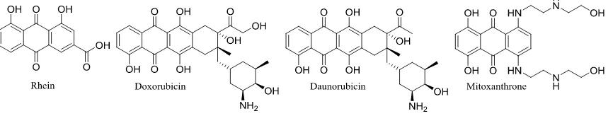
Rhein is a natural product isolated from the ground plant Rhubarb, which belongs to the
Rheum family. Compounds that have structural similarities to rhein, such as doxorubicin,
daunorubicin and mitoxanthrone, have been successfully marketed as anticancer drugs (Figure
5).75-77 These compounds belong to the anthracycline family and have been extensively used in
clinical settings for the treatment of various cancers. Despite the excellent anticancer activity,
however, they have shown serious side effects such as cardiotoxicity.75,77 Generally speaking the
compounds from the anthracycline family have been reported to follow one or more of the
following mechanisms of action: a) DNA intercalation and inhibition of its synthesis, b) DNA
damage through free radical generation, c) binding of DNA, d) DNA alkylation/cross-linking, e)
DNA damage through topoisomerase II inhibition, and f) direct topoisomerase II inhibition.78 A
common belief is that the cardiotoxicity of doxorubicin and daunorubicin derives from their redox
chemistry leading to free radical formation and the subsequent lipid peroxidation.78 It is well
known that single electron addition to the dienone ring of the anthracycline core generates reactive
oxygen species (ROS), which lead to cellular damage. The generation of ROS and oxidative DNA
damage has been observed at supraclinical concentrations of doxorubicin; however, at low
micromolar concentration the effects of DNA damage through topoisomerase inhibition is
predominant. The generation of ROS at high treatment concentrations is of major concern and
family of drugs. Recent findings that carbon monoxide can ameliorate doxorubicin’s cardiotoxicity
through mitochondrial biogenesis also lends insight into the mechanism of the cardiotoxic actions
of doxorubicin.79,80 Clinical studies have shown that 36% of patients treated with multiple doses
[image:32.612.111.544.189.271.2]of 500 mg/m2 doxorubicin develop cardiomyopathy.81
Figure 1.1.6 Rhein and selected clinically used anthaquinone anticancer agents.
There is no question of the efficiency of this class of drugs against various cancers;
however, the observed lethal side effects are a serious issue that needs to be addressed. Studies
have shown that rhein is well tolerated by the human body when used as a laxative, but it has
relatively low anticancer activity with IC50 in the range of 12~120 µM.82-86 The low, but promising
anticancer activity of rhein and its lack of toxicity make this molecule an excellent starting point
for the development of chemical analogs, which can potentially become drug candidates. The
development of a new anticancer agent based on the clinically used anthraquinone scaffold, yet
lacking intrinsic toxicity to normal cells, is a plausible strategy. The following sections of this
chapter describe the utilization of this strategy to develop novel, active anticancer compounds
based on rhein.
1.1.5 References
(1) http://www.cancer.org/research/cancerfactsstatistics/cancerfactsfigures2015/
2015.
(3) Chipuk, J. E.; Bouchier-Hayes, L.; Kuwana, T.; Newmeyer, D. D.; Green, D. R.
Science2005, 309, 1732.
(4) Vousden, K. H.; Lane, D. P. Nature Rev. Mol. Cell Biol.2007, 8, 275.
(5) Yee, K. S.; Vousden, K. H. Carcinogenesis2005, 26, 1317.
(6) Green, D. R.; Kroemer, G. Nature2009, 458, 1127.
(7) Laptenko, O.; Prives, C. Cell Death Differ.2006, 13, 951.
(8) Marine, J. C.; Francoz, S.; Maetens, M.; Wahl, G.; Toledo, F.; Lozano, G. Cell
Death Differ.2006, 13, 927.
(9) Levine, A. J.; Hu, W.; Feng, Z. Cell Death Differ.2006, 13, 1027.
(10) Levine, A. J.; Oren, M. Nat. Rev. Cancer2009, 9, 749.
(11) Nordstrom, W.; Abrams, J. M. Cell Death Differ.2000, 7, 1035.
(12) Derry, W. B.; Putzke, A. P.; Rothman, J. H. Science2001, 294, 591.
(13) Vousden, K. H.; Prives, C. Cell2009, 137, 413.
(14) Donehower, L. A.; Lozano, G. Nat. Rev. Cancer2009, 9, 830.
(15) Vogelstein, B.; Lane, D.; Levine, A. J. Nature2000, 408, 307.
(16) Carr, A. M. Science2000, 287, 1765.
(17) Cheng, Q.; Chen, J. Cell Cycle2010, 9, 472.
(18) Sherr, C. J.; Weber, J. D. Curr. Opin. Genet. Dev.2000, 10, 94.
(19) Huang, L.; Yan, Z.; Liao, X.; Li, Y.; Yang, J.; Wang, Z.-G.; Zuo, Y.; Kawai, H.;
Shadfan, M.; Ganapathy, S.; Yuan, Z.-M. P. Natl. Acad. Sci.2011, 108, 12001.
(20) Hollstein, M.; Sidransky, D.; Vogelstein, B.; Harris, C. C. Science1991, 253, 49.
(21) Wang, X.; Jiang, X. Febs Lett.2012, 586, 1390.
(23) Brooks, C. L.; Gu, W. Mol. Cell2006, 21, 307.
(24) Wade, M.; Wang, Y. V.; Wahl, G. M. Trends Cell Biol.2010, 20, 299.
(25) Zhang, Y.; Lu, H. Cancer Cell2009, 16, 369.
(26) Momand, J.; Zambetti, G. P.; Olson, D. C.; George, D.; Levine, A. J. Cell1992,
69, 1237.
(27) Haupt, Y.; Maya, R.; Kazaz, A.; Oren, M. Nature1997, 387, 296.
(28) Honda, R.; Tanaka, H.; Yasuda, H. Febs Lett.1997, 420, 25.
(29) Oliner, J. D.; Pietenpol, J. A.; Thiagalingam, S.; Gvuris, J.; Kinzler, K. W.;
Vogelstein, B. Nature1993, 362, 857.
(30) Fakharzadeh, S. S.; Trusko, S. P.; George, D. L. EMBO1991, 10, 1565.
(31) Cordoncardo, C.; Latres, E.; Drobnjak, M.; Oliva, M. R.; Pollack, D.; Woodruff,
J. M.; Marechal, V.; Chen, J. D.; Brennan, M. F.; Levine, A. J. Cancer Res.1994, 54, 794.
(32) Leach, F. S.; Tokino, T.; Meltzer, P.; Burrell, M.; Oliner, J. D.; Smith, S.; Hill, D.
E.; Sidransky, D.; Kinzler, K. W.; Vogelstein, B. Cancer Res.1993, 53, 2231.
(33) Piette, J.; Neel, H.; Marechal, V. Oncogene1997, 15, 1001.
(34) de Bie, P.; Ciechanover, A. Cell Death Differ. 2011, 18, 1393.
(35) Fang, S. Y.; Jensen, J. P.; Ludwig, R. L.; Vousden, K. H.; Weissman, A. M. J.
Biol. Chem.2000, 275, 8945.
(36) Montes De Oca Luna, R.; Wagner, D. S.; Lozano, G. Nature 1995, 378, 203.
(37) Jones, S. N.; Roe, A. E.; Donehower, L. A.; Bradley, A. Nature1995, 378, 206.
(38) Ringshausen, I.; O'Shea, C. C.; Finch, A. J.; Swigart, L. B.; Evan, G. I. Cancer
Cell2006, 10, 501.
(40) Stad, R.; Ramos, Y. F. M.; Little, N.; Grivell, S.; Attema, J.; van der Eb, A. J.;
Jochemsen, A. G. J. Biol. Chem.2000, 275, 28039.
(41) Sharp, D. A.; Kratowicz, S. A.; Sank, M. J.; George, D. L. J. Biol. Chem.1999,
274, 38189.
(42) Wu, W.; Xu, C.; Ling, X.; Fan, C.; Buckley, B. P.; Chernov, M. V.; Ellis, L.; Li,
F.; Munoz, I. G.; Wang, X. Cell Death Dis2015, 6, e2035.
(43) Pan, Y.; Chen, J. D. Mol. Cell. Biol.2003, 23, 5113.
(44) Danovi, D.; Meulmeester, E.; Pasini, D.; Migliorini, D.; Capra, M.; Frenk, R.; de
Graaf, P.; Francoz, S.; Gasparini, P.; Gobbi, A.; Helin, K.; Pelicci, P. G.; Jochemsen, A. G.;
Marine, J. C. Mol. Cell. Biol.2004, 24, 5835.
(45) Riemenschneider, M. J.; Buschges, R.; Wolter, M.; Reifenberger, J.; Bostrom, J.;
Kraus, J. A.; Schlegel, U.; Reifenberger, G. Cancer Res.1999, 59, 6091.
(46) Toledo, F.; Wahl, G. M. Int. J. Bio. Cell Biol.2007, 39, 1476.
(47) Parant, J.; Chavez-Reyes, A.; Little, N. A.; Yan, W.; Reinke, V.; Jochemsen, A.
G.; Lozano, G. Nature Gen.2001, 29, 92.
(48) Grier, J. D.; Xiong, S. B.; Elizondo-Fraire, A. C.; Parant, J. M.; Lozano, G. Mol.
Cell. Biol.2006, 26, 192.
(49) Chavez-Reyes, A.; Parant, J. M.; Amelse, L. L.; Luna, R. M. D.; Korsmeyer, S. J.;
Lozano, G. Cancer Res.2003, 63, 8664.
(50) Priest, C.; Prives, C.; Poyurovsky, M. V. Nucleic Acids Res.2010, 38, 7587.
(51) Gu, J. J.; Kawai, H.; Nie, L. G.; Kitao, H.; Wiederschain, D.; Jochemsen, A. G.;
(52) Pant, V.; Xiong, S.; Iwakuma, T.; Quintas-Cardama, A.; Lozano, G. PNAS2011,
108, 11995.
(53) Liu, T.; Zhang, H. L.; Xiong, J.; Yi, S.; Gu, L. B.; Zhou, M. X. Mol. Cancer2015,
14, 12.
(54) Zhao, Y.; Aguilar, A.; Bernard, D.; Wang, S. J. Med. Chem.2015, 58, 1038.
(55) Vu, B.; Wovkulich, P.; Pizzolato, G.; Lovey, A.; Ding, Q.; Jiang, N.; Liu, J.-J.;
Zhao, C.; Glenn, K.; Wen, Y.; Tovar, C.; Packman, K.; Vassilev, L.; Graves, B. Med. Chem.
Lett.2013, 4, 466.
(56) ClinicalTrials.gov identifiers for RG7112: NCT00559533, NCT00623870,
NCT01677780, NCT01164033, NCT01605526, NCT01143740, and NCT01635296.
(57) Ding, Q.; Zhang, Z.; Liu, J.-J.; Jiang, N.; Zhang, J.; Ross, T. M.; Chu, X.-J.;
Bartkovitz, D.; Podlaski, F.; Janson, C.; Tovar, C.; Filipovic, Z. M.; Higgins, B.; Glenn, K.;
Packman, K.; Vassilev, L. T.; Graves, B. J. Med. Chem.2013, 56, 5979.
(58) Siu, L. L.; Italiano, A.; Miller, W. H.; Blay, J.-Y.; Gietema, J. A.; Bang, Y.-J.;
Mileshkin, L. R.; Hirte, H. W.; Reckner, M.; Higgins, B.; Jukofsky, L.; Blotner, S.; Zhi, J.;
Middleton, S.; Nichols, G. L.; Chen, L. C. J. Clin. Oncol.2014, 32.
(59) Wang, S.; Sun, W.; Zhao, Y.; McEachern, D.; Meaux, I.; Barriere, C.; Stuckey, J.
A.; Meagher, J. L.; Bai, L.; Liu, L.; Hoffman-Luca, C. G.; Lu, J.; Shangary, S.; Yu, S.; Bernard,
D.; Aguilar, A.; Dos-Santos, O.; Besret, L.; Guerif, S.; Pannier, P.; Gorge-Bernat, D.;
Debussche, L. Cancer Res.2014, 74, 5855.
(60) ClinicalTrials.gov identifiers for MI-77301/SAR405838: NCT01636479 and
(61) Sun, D.; Li, Z.; Rew, Y.; Gribble, M.; Bartberger, M. D.; Beck, H. P.; Canon, J.;
Chen, A.; Chen, X.; Chow, D.; Deignan, J.; Duquette, J.; Eksterowicz, J.; Fisher, B.; Fox, B. M.;
Fu, J.; Gonzalez, A. Z.; De Turiso, F. G.-L.; Houze, J. B.; Huang, X.; Jiang, M.; Jin, L.; Kayser,
F.; Liu, J.; Lo, M.-C.; Long, A. M.; Lucas, B.; McGee, L. R.; McIntosh, J.; Mihalic, J.; Oliner, J.
D.; Osgood, T.; Peterson, M. L.; Roveto, P.; Saiki, A. Y.; Shaffer, P.; Toteva, M.; Wang, Y.;
Wang, Y. C.; Wortman, S.; Yakowec, P.; Yan, X.; Ye, Q.; Yu, D.; Yu, M.; Zhao, X.; Zhou, J.;
Zhu, J.; Olson, S. H.; Medina, J. C. J. Med. Chem.2014, 57, 1454.
(62) ClinicalTrials.gov identifiers for AMG 232: NCT01723020 and NCT02016729.
(63) Holzer, P.; Masuya, K.; Furet, P.; Kallen, J.; Valat-Stachyra, T.; Ferretti, S.;
Berghausen, J.; Bouisset-Leonard, M.; Buschmann, N.; Pissot-Soldermann, C.; Rynn, C.; Ruetz,
S.; Stutz, S.; Chene, P.; Jeay, S.; Gessier, F. J. Med.Chem.2015, 58, 6348.
(64) Holzer, P.; Masuya, K.; Furet, P.; Kallen, J.; Valat-Stachyra, T.; Ferretti, S.;
Berghausen, J.; Bouisset-Leonard, M.; Buschmann, N.; Pissot-Soldermann, C.; Rynn, C.; Ruetz,
S.; Stutz, S.; Chene, P.; Jeay, S.; Gessier, F. J. Med. Chem.2015, 58, 6348.
(65) Wang, W.; Hu, Y. Med. Res. Rev.2012, 32, 1159.
(66) Vassilev, L. T. Trends Mol. Med.2007, 13, 23.
(67) Boman, H. G. Annu. Rev. Immunol.1995, 13, 61.
(68) Fischbach, M. A.; Walsh, C. T. Chem. Rev.2006, 106, 3468.
(69) Hancock, R. E. W.; Chapple, D. S. Antimicrob. Agents Ch.1999, 43, 1317.
(70) Bernal, F.; Tyler, A. F.; Korsmeyer, S. J.; Walensky, L. D.; Verdine, G. L. J. Am.
Chem. Soc.2007, 129, 5298.
(71) Bernal, F.; Wade, M.; Godes, M.; Davis, T. N.; Whitehead, D. G.; Kung, A. L.;
(72) Chang, Y. S.; Graves, B.; Guerlavais, V.; Tovar, C.; Packman, K.; To, K.-H.;
Olson, K. A.; Kesavan, K.; Gangurde, P.; Mukherjee, A.; Baker, T.; Darlak, K.; Elkin, C.;
Filipovic, Z.; Qureshi, F. Z.; Cai, H.; Berry, P.; Feyfant, E.; Shi, X. E.; Horstick, J.; Annis, D. A.;
Manning, A. M.; Fotouhi, N.; Nash, H.; Vassilev, L. T.; Sawyer, T. K. P. Natl. Acad. Sci.2013,
110, E3445.
(73) Kitagaki, J.; Agama, K. K.; Pommier, Y.; Yang, Y.; Weissman, A. M. Mol.
Cancer Ther.2008, 7, 2445.
(74) Yang, Y. L.; Ludwig, R. L.; Jensen, J. P.; Pierre, S. A.; Medaglia, M. V.;
Davydov, I. V.; Safiran, Y. J.; Oberoi, P.; Kenten, J. H.; Phillips, A. C.; Weissman, A. M.;
Vousden, K. H. Cancer Cell2005, 7, 547.
(75) Tan, J. H.; Zhang, Q. X.; Huang, Z. S.; Chen, Y.; Wang, X. D.; Gu, L. Q.; Wu, J.
Y. Eur. J. Med. Chem.2006, 41, 1041.
(76) Katzhendler, J.; Gean, K.-F.; Bar-ad, G.; Tashma, Z.; Ben-shoshan, R.; Ringel, I.;
Bachrach, U.; Ramu, A. Eur. J. Med. Chem.1989, 24, 23.
(77) Huang, H. S.; Chin, H. F.; Yeh, P. F.; Yuan, C. L. Helv Chim Acta2004, 87, 999.
(78) Minotti, G.; Menna, P.; Salvatorelli, E.; Cairo, G.; Gianni, L. Pharmacol. Rev.
2004, 56, 185.
(79) Bartz, R. R.; Suliman, H. B.; Piantadosi, C. A. Front. Physiol.2015, 6, 8.
(80) Ji, X.; Damera, K.; Zheng, Y.; Yu, B.; Otterbein, L. E.; Wang, B. J. Pharm. Sci.,
105, 406.
(81) Lefrak, E. A.; Pitha, J.; Rosenheim, S.; Gottlieb, J. A. Cancer1973, 32, 302.
(82) Ip, S. W.; Weng, Y. S.; Lin, S. Y.; Mei-Dueyang; Tang, N. Y.; Su, C. C.; Chung,
(83) Cui, X. R.; Tsukada, M.; Suzuki, N.; Shimamura, T.; Gao, L.; Koyanagi, J.;
Komada, F.; Saito, S. Eur. J. Med. Chem.2008, 43, 1206.
(84) van Gorkom, B. A. P.; Timmer-Bosscha, H.; de Jong, S.; van der Kolk, D. M.;
Kleibeuker, J. H.; de Vries, E. G. E. Brit J Cancer2002, 86, 1494.
(85) Cichewicz, R. H.; Zhang, Y. J.; Seeram, N. P.; Nair, M. G. Life Sci2004, 74,
1791.
(86) Shi, P.; Huang, Z. W.; Chen, G. C. Am J Chinese Med2008, 36, 805.
1.2 Mechanistic studies of the lead compound BW-AQ-101
*All biological and mechanistic work described in this section was performed exclusively
by Dr. Muxiang Zhou’s lab at Emory University. The author of the chapter contributed with
synthetic route optimization, preliminary MTT screening against six cell lines, and computational
analysis.
In the pursuit of novel active agents form the anthraquinone family our initial efforts
resulted in the development of a few active compounds.1 One in particular (BW-AQ-101) stood
out as a potent agent against leukemia cell lines; IC50 = 0.69 µM against Molt4 cells. These
preliminary findings gave us the stimulus to develop a more efficient synthetic route to
BW-AQ-101 and further examine its biological activity and mechanism of action. The synthetic
development of a more efficient route was previously disclosed by the author and will not be
covered in this section.2 Extensive examination against number solid and leukemia cell lines
showed that compound BW-AQ-101 has selectivity over leukemia cell lines (Figure 1.2.1). The
compound showed low micromolar IC50 values against solid tumor cell lines (Hela, KB, Cos 7,
K562, EU-1). Understanding the mechanism of action of a given compound is of extreme
importance in the drug development process. Although there is a number of examples of active
agents that have reached clinical trials and even market, the targeted therapeutics strategy has been
proven to be the key to the development of safe and efficient drug candidates.3,4 Therefore, in
collaboration with Dr. Zhou (Emory University) we explored the possible mechanism of action of
this compound. As previously described p53 is the key player in cellular response upon cellular
stress. Our effort was focused on the examination of the p53 signaling network upon cellular
treatment with BW-AQ-101.
Compound Hela (µM) KB (µM) Cos7 (µM) T98G (µM) Molt4 (µM) K562 (µM) EU1 (µM)
BW-AQ-101 2.2 16 3.0 >13 0.69 0.93 0.83
Figure 1.2.1 Structure and activity (based on MTT assay) of BW-AQ-101.
Because BW-AQ-101 showed potency against leukemia cell lines, the investigation of its
mechanism of action was done by utilizing a number of leukemia cell lines. Five
previously-characterized acute lymphoblastic leukemia (ALL) cell lines were selected; Sup-B13, EU-1 and
EU-3 cells that have the wt-p53; EU-6 that has a p53 mutation; and EU-8 that has a p53-null
phenotype. All cell lines except EU-8 express MDM2 and MDM4. Interestingly BW-AQ-101
showed strong cytotoxic effect in all cell lines expressing wt-p53 and MDM2 and considerably
less effect on the other two cell lines (data not shown). This was the first indication that the active
that BW-AQ-101 interrupts the p53-MDM2-MDM4 regulatory cycle, causing apoptosis. Western
blot assays indicated significant downregulation of MDM2 in the p53 wild type cell lines treated
with the compound, which supported the established hypothesis (Figure 1.2.2 A). In addition,
BW-AQ-101 did not show significant effect on the expression of MDM4. As previously discussed, p53
and MDM2 exist in a “love-hate” regulatory cycle, where p53 expresses MDM2 when needed and
on the other hand MDM2 downregulates p53 through ubiquitination. Thus, the evaluation of the
transcriptional effect of BW-AQ-101 on MDM2 was curtail. RT-PCR experiments demonstrated
that the MDM2 mRNA level was significantly elevated by 101. Furthermore,
BW-AQ-101 showed no direct inhibitory effect on MDM2 mRNA expression (Figure 1.2.2 B). These
results lead to the conclusion that the transcriptional activity of p53 was not abolished. On the
contrary, the enhanced MDM2 mRNA expression upon BW-AQ-101 treatment was a result of p53
Figure 1.2.2 Effects of BW-AQ-101 on p53, MDM2 and MDM4;
A) Western blot assays for expression of proteins, as indicated in four MDM2-positive ALL cell lines, treated with 1 µM BW-AQ-101 for 24 h. C, control; T, treatment. B) The mRNA levels of MDM2 and p53 at different time intervals upon treatment with 1 µM BW-AQ-101. C) IP-western blot assays of EU-1 cells upon treatment with 1 µM BW-AQ-101
Since it was indicated that BW-AQ-101 appears to induce MDM2 protein degradation, and as
discussed in the introductory chapter, the regulation of MDM2 protein stabilization is mainly
controlled by its own C-terminal RING domain. Computational tools were utilized to exploit
potential interactions between the developed compound and the RING domain of MDM2. One of
the key aspects that should be kept in mind is that MDM2 forms a homodimer or a heterodimer
with MDM4 through direct RING-RING domain interactions. The purpose of the molecular
modeling analysis was to see whether there was a potential binding pocket for BW-AQ-101 in the
MDM2 RING domain. The molecular docking was examined using a simulated aqueous
binding pockets formed by the two MDM2 RING domains’ interactions were revealed (Figure
1.2.3A). However, the heterodimeric structure of MDM2-MDM4 showed absence of any cavities
(Figure 1.2.3B). It has been reported that the MDM2 RING domains are very unstable in solution
and that they easily form dimeric or other types of aggregate structures; therefore, it is safe to
assume that the binding pockets seen in the homodimer exist under physiological conditions. The
BW-AQ-101 docking study was in good agreement with the observed activity, and did not show
any sites for possible covalent modifications. The computational results gave insight into the
possible mechanism of action of BW-AQ-101. The docking studies revealed clear binding pocket
and potential for stable interactions of the compound with the MDM2 RING domain. However,
the residues involved in these interactions do not play part in the MDM2-MDM4 heterodimer
formation (Figure 1.2.3 C, D). The fact that the MDM2 RING domains predominantly exist in
dimeric or more complex aggregate forms entails for constant, dynamic conformational changes
that allow for the formation of complexes and their stabilizations. The conformational changes
open large surface areas that can serve as interfaces for the formation of stable MDM2-MDM4
heterodimer. We hypothesize that upon binding of BW-AQ-101 to the MDM2 RING homodimer,
the MDM2 RING structure “locks” into a folded conformation, which prevents unfolding and thus
inhibits MDM2-MDM4 heterodimer formation.
A) MDM2 homodimer showing the relative cavity depth of the binding pocket (blue: no cavity; yellow: cavity). B) MDM2-MDM4 heterodimer, showing the lack of a cavity by coloring the surface of the MDM2 RING residues involved in cavity formation in the homodimeric form.C) MDM2 homodimer (orange) and the binding pocket forming amino acid residues (blue). D) MDM2 (orange)-MDM4 (cyan) heterodimer and the MDM2 amino acid residues (blue) participating in pocket formation in the homodimeric form. The image shows the absence of a binding cavity as well as lack of direct interactions between the pocket-forming amino acid residues of MDM2 (blue) with the MDM4 monomer (cyan).
In order to validate the computational results, two types of binding assays were performed;
fluorescent titration and ITC assays. After titration of BW-AQ-101 to the GST-MDM2 RING
protein a Kd = 0.31 µM was obtained from the fluorescence assay (data not shown). The control
titration of BW-AQ-101 to the GST protein itself, did not result in any binding. The ITC assays,
showed similar binding affinity of Kd = 0.29 µM and confirmed that BW-AQ-101 binds to the
MDM2 RING domain. ITC experiment with MDM4 did not detect binding of BW-AQ-101
(Figure 1.2.4). Furthermore, both the fluorescent titration and ITC assays for detection of possible
binding between BW-AQ-101 and an MDM2 fragment (residues 1 - 424) with deletion of the
RING domain, were performed and no binding activity was detected (data not shown). In order
to validate the theory that BW-AQ-101 binds to the MDM2 homodimer and blocks MDM4
interaction, co-immunoprecipitation (Co-IP) experiments were performed. Previous studies had
demonstrated that the MDM2 protein becomes stabilized in the heterodimer form
(MDM2-MDM4).5 Binding of BW-AQ-101 to MDM2 may disrupt the formation of the MDM2-MDM4
heterodimer, resulting in MDM2 destabilization. Co-IP and Western blot assays, showed that
101 dissociated MDM2 from the MDM2-MDM4 complex (Figure 1.2.2C). Because
BW-AQ-101 has an anthraquinone core, these experiments were performed against doxorubicin. MDM2
and MDM4 were unaffected in a co-ip experiment with doxorubicin (Figure 1.2.2C). Thus, the
Naturally another comparison between the two compounds has to be made to verify that the
mechanism of action of BW-AQ-101 is truly as proposed above. Flat aromatic, polycyclic
compounds are known DNA binders and it is well know that doxorubicin induces apoptosis
through DNA damage. In order to verify that BW-AQ-101 does not interact with DNA in the
utilized concentrations, a comet assay was performed. The assay indicated that at 1 µM
concentration, doxorubicin induced clear DNA damage after 4 hours, while BW-AQ-101 exhibited
only DNA fragmentation after 8 hour treatment (Figure 1.2.5).
Figure 1.2.4 ITC binding assay of BW-AQ-101 against: A) MDM2 and B) MDM4
The computational results as well as the mechanistic experiments described above show
strong evidence that the lead compound BW-AQ-101 is indeed a MDM2 RING binder. The
compound does not show significant DNA damage and correlation between molecular modeling
and binding assays agree with the hypothesis that the compound induces apoptosis through
disruption of the p53-MDM2 regulatory mechanism. These observations give an indication that a
compound BW-AQ-101. The correlation between in vitro and in silico binding studies allows us
[image:46.612.235.410.162.276.2]to develop a computational model and design other active rhein-based anticancer agents.
Figure 1.2.5 Comet assay of EU-1 cells
Cells were treated with 1 µM of either AQ-101 or doxorubicin, for 4 h and 8 h. Red arrow indicates DNA damage, blue arrow indicates nuclear fragmentation/apoptosis.
1.2.1 Experimental section
Compound-protein binding assay
The binding properties between AQ-101 and the MDM2/MDM4 RING domains were
examined by both fluorescent titration (data not shown) and isothermal titration calorimetry (ITC)
assays. The expression and purification of the MDM2 and MDM4 RING domain proteins was
performed as described previously. 6
ITC assay
The MDM2 RING protein in a Hepes buffer (10 mM Hepes, pH 7.2 and 150 mM NaCl)
solution was placed in a loading 96 DeepWell PP plate (Nunc, Thermo Fisher Scientific). The test
compound (120 μL of 24 µM stock solution) was automatically transferred by the auto-iTC200
instrument (MicroCal, GE) into the sample cell. The compound solution (2 μL) was titrated
injection, which was 0.4 μL). The equilibrium time between two adjacent injections was 210 s.
The binding stoichiometry (n), dissociation constant (Kd) and thermodynamic parameters (ΔH and
ΔS) were determined by fitting the titration curve to a one-site binding mode, using the Origin
software provided by the manufacturer.
Cells
This study used five established cell lines derived from children with acute lymphoblastic
leukemia. Four of these cell lines (EU-1, EU-3, EU-6 and EU-8) were established at Emory
Uni-versity (Atlanta, GA, USA), and one (SUP-B13) was obtained from Stephen D. Smith (UniUni-versity
of Kansas Medical Center, Kansas City, KS, USA). All ALL cell lines were authenticated and
their p53 phenotypes were as follows: Sup-B13, EU-1 and EU-3 cells (wt-p53); EU-6 (mutant
p53); and EU-8 (p53-null). Four of the p53-positive cell lines, including the p53-mutant EU-6,
express MDM2; whereas MDM2 is not expressed in the EU-8 cells (Zhou et al., 1995). The 293T
cell line, purchased from the American Type Tissue Collection (ATCC) was used for gene
transfection assays.
Fresh leukemia samples were selected from 16 pediatric patients treated for ALL (either at
diagnosis or first relapse), with peripheral total white blood cell (WBC) counts higher than 100 x
106/mL (Table s1). Normal human bone marrow mononuclear (NBMM) cells were obtained from
5 donors following informed consent. Of the 16 ALL patients, 13 had B-cell precursor (BCP) ALL
and 3 had T-ALL, as diagnosed by standard immunologic, morphologic and cytochemical criteria.
Mononuclear cells were separated by centrifugation in Ficoll-Hypaque (1.077g/mL). Leukemic
blasts from patient samples were further isolated by removing adherent monocytes. Specimens