The Correlation of Tumor-Infiltrating Lymphocytes with
Tumor Mass Location and Histological Grading of
Cutaneous Squamous Cell Carcinoma
Suriany, Delyuzar, T. Ibnu Alferraly
Department of Anatomical Pathology, Faculty of Medicine,Universitas Sumatera Utara, Medan, Indonesia
DOI: 10.29322/IJSRP.9.07.2019.p9179
http://dx.doi.org/10.29322/IJSRP.9.07.2019.p9179
Abstract- Background: Cutaneous squamous cell carcinoma (cSCC) is the malignant tumor derived from the keratinocytes of the epidermis. It is the second most common skin cancer in Indonesia. The infiltration of tumor-infiltrating lymphocytes (TILs) had been studied and related with the prognostic factor for many solid tumors. The research about TILs with Hematoxylin&Eosin staining for cSCC has not been studied until nowadays.
Objective: This study was aimed to analyze the correlation of TILs with the tumor mass location and histological grading for cSCC.
Methods: This is an analytical research with cross sectional approach using the slide and paraffine blocks from the patients diagnosed with cSCC with Hematoxylin&Eosin staining. The statistical analysis with Kruskal-Wallis method is used to analyze the correlation of intratumoral and stromal TILs with the tumor mass localation and histological grading of cSCC.
Results: Based on the statistical analysis for 48 samples of the patients with cSCC, the p-value for the correlation of intratumoral TILs with the tumor mass location, stromal TILs with tumor mass location, and intratumoral TILs with the histological grading, are 0.824, 0.124, 0.058 (p>0.05). Meanwhile, the correlation of stromal TILs with the histological grading shows the p-value of 0.027 (p<0.05).
Conclusion: Only the stromal TILs are closely related to the histological grading of cSCC.
Index Terms-: cutaneous squamous cell carcinoma, cSCC, TILs, grading, location, risk of metastasis.
I. INTRODUCTION
utaneous squamous cell carcinoma (cSCC) is the second most frequent skin cancer after basal cell carcinoma with the incidence rate is approximately 20% of all skin cancer.1 The
current estimated incidence of this tumor is 15 to 35/100,000 people per year and is expected to increase around 2% to 4% per year due to the aging population and chronic ultraviolet B exposure.2 The etiology and risk factors of this skin cancer have
changed from the occupational exposure of chemical carcinogen to other factors, such as HIV infection, HPV infection, therapeutic advances with immunosupressive agent and PUVA, and also organ transplantation.3
The prognosis of cSCC is influenced by some factors. High risk factors for cSCC are tumor depth more than 2 mm, Clark level IV / V, perineural invasion, tumor location on ear or lip, and poor differentiation.4 Diffentiation of cSCC, which are
needed for histological grading evaluation, is divided into well differentiation, moderate differentiation, and poor differentiation. Histological grading for cSCC depends on the degree of anaplasia in the tumor nest, mitotic activity, and keratin formation.5
Tumor-infiltrating lymphocytes (TILs) as one of the immunity cells against the tumor have an important contribution for tumor invasion, growth, metastasis, and outcome.6 TILs
infiltrate into the tumor nests (intratumoral TILs) or around the tumor nests (stromal TILs).7 Studies about TILs had been done
for many solid tumors, such as lung carcinoma, gastric cancer, colorectal carcinoma, hepatocellular carcinoma, pancreatic adenocarcinoma, breast cancer, ovarian cancer, melanoma, and squamous cell carcinoma of head and neck. Some of these studies used the immunohistochemistry staining to evaluate the subset of TILs and only a few studies used the Hematoxylin&Eosin staining. Some results showed that TILs have an antitumorigenic role in carcinogenesis while the others showed the contrary role.6,8
Although Hematoxylin&Eosin staining is the routine staining for histopathological examination, only a few studies about TILs with this staining had been done. The previous studies for TILs with Hematoxylin&Eosin staining are for epithelial ovarian carcinoma, squamous cell carcinoma of head and neck, melanoma, and breast cancer. Studies about TILs for cSCC with Hematoxylin&Eosin staining have not been done yet and this study is aimed to evaluate the correlation of TILs with histological grading and tumor mass location of cSCC. The infiltration of TILs was observed for intratumoral infiltration and stromal infiltration.
II. MATERIAL AND PRODUCT
ISSN 2250-3153
Adam Malik General Hospital Health Research Ethics Committee.
The sample size needed for this research was calculated based on the sample formula used for testing a hypothesis in one population. Based on the sample formula, minimum samples needed for this research was 47 samples. The samples were obtained from the slide and the paraffine blocks of the patients diagnosed as cSCC.
The inclusion criteria for this study were patients’ slides or paraffine blocks with a pathological diagnosis of cutaneous squamous cell carcinoma and the medical data recorded the tumor mass location. The exclusion criteria included the following: (1) Slides or paraffine blocks were lost or damaged; (2) Slides only showed the tumor cells without enough stromal area for evaluation.
All slides with the Hematoxylin&Eosin staining were observed microscopically by researcher, together with two pathologists using Olympus CX23 to determine the histological grading and the level of TILs infiltration.
Cutaneous squamous cell carcinoma is graded based on the degree of differentiation and keratinization.9 Histological grading
was divided into: (1) Well differentiated cSCC, is characterized by squamous epithelium that is easily recognisable with abundant of keratinization. The epithelium is obviously squamous and intercellular bridges (prickles) are readily apparent. The tumors display minimal pleomorphism and mitotic figures are mainly basally located; (2) Moderately differentiated cSCC falls in between the well differentiation and poor differentiation. The epithelial structures are more disorganized in which the squamous epithelial derivation is less obvious. Nuclear and cytoplasmic pleomorphism is more pronounced. The mitotic figures, including the atypical mitosis, can be found more easily. Tumors show less keratinization, often being limited to the formation of keratin pearls; (3) Poorly differentiated cSCC, is more difficult to establish the true nature of the lesion, unless intercellular bridges or small foci of keratinization are found. Tumors showed highly atipia nuclei with lots of mitotic figures.10-11
All fields were viewed in order to determine the level of TILs infiltration. TILs infiltration was divided into intratumoral TILs and stromal TILs. Intratumoral TILs are lymphocytes which infiltrated into the tumor nests and were in contact with the tumor cells, while stromal TILs are lymphocytes which infiltrated surrounding the tumor nests. TILs infiltration were categorized into four level: (1) Score 0 for no infiltration of lymphocytes; (2) Score 1 for minimal lymphocytes infiltration which were less than 10 cells / HPF; (3) Score 2 for moderate lymphocytes infiltration in which lymphocytes could be seen easily, but not in large aggregates; (4) Score 3 for massive infiltration in which the infiltration the lymphocytes formed large aggregates and could be found in 50% tumor area.
Tumor mass location was categorized based on the risk of local recurrence and metastatic potential in multivariate studies. Tumors were categorized into high risk location if the tumors are located on ear or lip, which had the increased risk for local recurrence and metastasis. Tumors were categorized into low to intermediate risk location if they were located on scalp, perineal region, extremity, and trunk.13
Data was analyzed with Kruskal-Wallis method using the statistic software program and the results were shown in the table of frequency.
III. RESULT
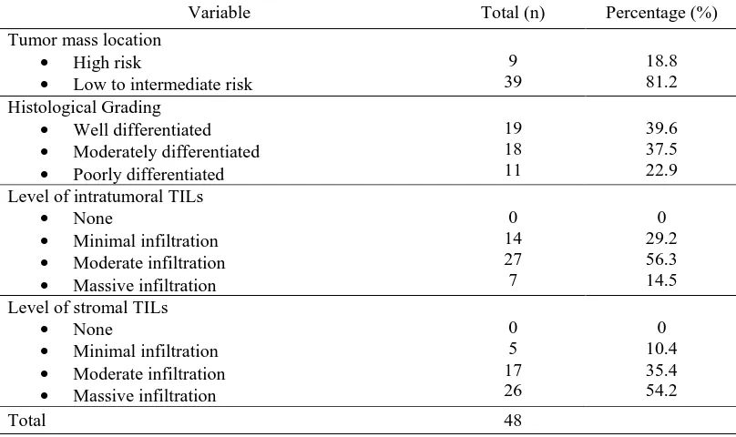
From 48 samples of cSCC which were evaluated ini this study, 9 cases (18.8%) were located at the high risk tumor mass location and 39 cases (81.2%) were located at the low to intermediate risk tumor mass location. Histological evaluation showed that 19 cases (39.6%) were well differentiated tumors, 18 cases (37.5%) were moderately differentiated tumors, and 11 cases (22.9%) were poor differentiated tumors. Evaluation for intratumoral infiltration of TILs showed that 14 cases (29.2%) had minimal infiltration of intratumoral TILs, 27 cases (56.3%) had moderate infiltration of intratumoral TILs, and 7 cases (14.5%) had massive infiltration of intratumoral TILs. Microscopic evaluation for stromal infiltration of TILs showed that only 5 cases (10.4%) had minimal infiltration of stromal TILs, 17 cases (35.4%) had moderate infiltration of stromal TILs, and 26 cases (54.2%) had massive infiltration of stromal TILs. Detailed data are available in table 1 (see below).
Statistical analysis with Kruskal-Wallis method showed that no statistical correlation was found between the tumor mass location with intratumoral TILs infiltration (p-value = 0.824), no statistical correlation was found between the tumor mass location with stromal TILs infiltration (p-value = 0.124), and no statistical correlation between histological grading and intratumoral TILs infiltration (p-value = 0.058). Only stromal TILs infiltration had statistical correlation with histological grading (p-value = 0.027). The data are available in table 2 and table 3 (see below).
IV. DISCUSSION
As mentioned previously, many studies had been done to evaluate the prognostic role of TILs in solid tumors. Only a few studies used the hematoxylin&eosin staining as this staining is the routine staining for histopathological examination and available in every pathological laboratory.
Many authors have suggested that TILs level carries more predictive value than TNM staging for malignant tumors. Studies about TILs as the prognostic value for squamous cell carcinoma had been carried, but the authors only focused for the head and neck squamous cell carcinoma.14
As one of the prognostic factor for cSCC, tumor mass location had been statistically studied in multivariate and statistical data showed that tumors located on the lip and ears have higher risk for local recurrence and metastatis than tumors located on scalp, trunk, perineum, and extremity.13 TILs as
many factors, such as the loss of e-cadherin function for the cell adhesion, the damage of extracellular matrix due to the effect of the proteolytic enzymes, the role of hypoxia-inducible transcription factor (HIF), and the angiogenesis. Tumor metastasis also happens because of the EMT (epithelial to mesenchymal transition) program. Stroma, which is composed of a variety of fibroblast, myofibroblasts, endothelial, myeloid, and lymphoid cells, becomes reactive and releases various signals, including TGF-β, Wnts, and certain interleukins that impinge on nearby carcinoma cells, inducing the latter to activate their previously silent EMT program. Immune cells that play important role for metastasis are neutrophyls and NK cells. Perhaps it can be the reason of no correlation between infiltration of lymphocytes into or surrounding the tumor nests of cSCC with the tumor mass location based on the risk of local recurrence and metastasis.15 We also consider that tumors located on the ears or
lips are close to the lymphatic circulation of head and neck region which supports the metastasis process.
Besides the tumor mass location, UICC and AJCC also regard histological grading of cSCC as an important prognostic factor.11 The prognostic value of TILs itself had been proven in
some studies, especially the infiltration of stromal TILs. Xu, et al, (2017) also studied the role of stromal TILs for squamous cell carcinoma on head and neck. They stated that massive infiltration of stromal TILs were related to the better outcome with lower rate of tumor recurrence, but they did not intend on the correlation of TILs with histological grading in their study.14 In
our study, there is strong correlation between stromal TILs and histological grading or tumor differentiation. We suppose that the lymphocytes which infiltrate surrounding the tumor nest have the antitumorigenic effect for cSCC which inhibit and control tumor growth. Previous studies and supporting literature stated that the subset of lymphocytes which has the antitumorigenic effect is CD8+ T lymphocytes. Further studies using the immunohistochemistry staining to evaluate the predominant subsets of lymphocytes which infiltrate into or around the tumor nests must be held.
This study was retrospective in nature with some limitations. The samples used in this study were obtained from the biopsy and also tumor excision in which the size of tumor and the depth of invasion could not be confirmed. Besides that, the clinical data, including the immunity status, comorbidity, and the previous skin lesion as the predisposing factor were not known. Analysis to the correlation between histopathological variant of cSCC and infiltration of TILs could not be conducted because not all variant were found in our samples. These limitations will be given further consideration in future studies.
V. CONCLUSION
After conducted this study, we conclude some points in the following:
1. No statistical correlation between tumor mass location of cSCC based on the risk of local recurrence and metastasis, level of intratumoral and stromal TILs infiltration.
2. No statistical correlation between histological grading of cSCC and level of intratumoral TILs infiltration
3. There are significant correlation between histological grading and level of stromal TILs infiltration.
We suggest on determining the level of stromal TILs infiltration as one of the prognostic factors which is needed to be reported in the histopathological report for cutaneous squamous cell carcinoma.
REFERENCES
[1] Kraft S, Granter SR, Molecular pathology of skin neoplasms of the head and neck. Arch Pathol Lab Med. 2014; 138: 759-86.
[2] Motaparthi K, Kapil JP, Velazquez EP. Cutaneous squamous cell carcinoma: review of the eighth edition of the american joint committee on cancer staging guideline, prognostic factors, and histopathologic variant. Adv Anat Pathol. 2017; 4(4): 171-94.
[3] Burn T, Breatnach S, Cox N, Griffiths C. Rook’s textbook of dermatology 8th Ed. Singapore: Willey-Blackwell; 2010. p.52.25-27.
[4] Aslam AM. Facial cutaneous squamous cell carcinoma. BMJ. 2016. doi:10.1136/bmj.i1513.
[5] Murphy GF, Beer TW, Cerio R, Kao GF, Nagore E, Pulitzer MP. Squamous cell carcinoma. In Elder DE, Massi D, Scolyer RA, Willemze R. WHO classification of skin tumours. Lyon. 2018. p.35-45.
[6] Bremnes RY, Busund LT, Kilvaer TL, Andersen S, Richardsen E, Paulsen EE, Hald S, et al. The role of tumor-infiltrating lymphocytes in development, progression, and prognosis of non-small cell lung cancer. Journal of Thoracic Oncology. 2015; 11(6): 789-800.
[7] Ahn SG, Jeong J, Hong SW, Jung WH. Current issues and clinical evidence in tumor-infiltrating lymphocytes in breast cancer. Journal of Pathology and Translational Medicine. 2015; 49: 355-63.
[8] Hendry S, Salgado R, Gevaert T, Russel PA, John T, Thapa B, Christie M, et al. Assessing tumor-infiltrating lymphocytes in solid tumor: a practical review for pathologists and proposal for a standarized method from the international immuno-oncology biomarkers working group: part2: tils in melanoma, gastrointestinal tract carcinoma, non-small cell lung carcinoma, and mesothelioma, endometrial and ovarian carcinomas, squamous cell carcinoma of the head and neck, genitourinary carcinoma, and primary brain tumors. Adv Anat Pathol. 2017. Available from:
www.anatomicpathology.com
[9] Patterson JW. Weedon’s skin pathology 4th Ed. China: Churchill
Livingstone; 2016. p. 815-23.
[10] McKee PH, Calonje E, Brenn T, Lazar A. McKee’s pathology of the skin 4th Ed. China: Elsevier Saunders; 2012. p. 1115-34.
[11] Slater D, Walsh M. Standards and datasets for reporting cancers: dataset for the histological reporting of primary invasive cutaneous squamous cell carcinoma and regional lymph node 3rd Ed. London: The Royal College of
Pathologist; 2014. Available from: http://www.rcpath.org›resourceLibrary. [12] Kreike B, Kouwenhove M, Horlings H, Weigelt B, Peterse H, Barterlink H,
van de Vijver MJ. Gene expression profiling and histopathological characterization of triple-negative/basal-like breast carcinoma. Breast Cancer Research. 2007; 9(5). Available from : http://breast-cancer-research.com/content/9/5/R65
[13] Califano JA, Lydiatt WM, Nehal KS, Sullivan BO, Schmults C, Seethal RR, Weber RS, et al. Cutaneous squamous cell carcinoma of the head and neck. In Amin MB, editor. AJCC cancer staging manual 8th Ed. Chicago:
Springer; 2017. p.171-9.
[14] Xu Q, Wang C, Yuan X, Feng Z, Han Z. Prognostic value of tumor-infiltrating lymphocytes for patients with head and neck squamous cell carcinoma. Translational Oncology. 2017; 10(1): 10-6.
[15] Lambert AW, Pattabiraman DR, Weinberg RA. Emerging biological principles of metastasis. Cell. 2017; 168(4): 670-91. Doi:10.1016/j.cell.2016.11.037.
AUTHORS
First Author – dr. Suriany, Resident of Department of Anatomical Pathology, Faculty of Medicine, Universitas
Sumatera Utara, medan, Indonesia, email ID:
ISSN 2250-3153
Second Author – Dr. dr. Delyuzar, M.Ked.(PA), Sp.PA(K), Department of Anatomical Pathology, Faculty of Medicine, Universitas Sumatera Utara, Medan, Indonesia
Third Author – dr. T. Ibnu Alferraly, M.Ked.(PA), Sp.PA, D.Bioeth., Department of Anatomical Pathology, Faculty of Medicine, Universitas Sumatera Utara, Medan, Indonesia
Correspondence Author – dr. Suriany, Resident of Department of Anatomical Pathology, Faculty of Medicine, Universitas Sumatera Utara, Medan, Indonesia, email
ID:suriany.ppds.pa@gmail.com
Table 1. Distribution of cutaneous squamous cell carcinoma based on the tumor mass location, histological grading, and level of tumor-infiltrating lymphocytes.
Variable Total (n) Percentage (%)
Tumor mass location
• High risk
• Low to intermediate risk
9 39
18.8 81.2 Histological Grading
• Well differentiated
• Moderately differentiated
• Poorly differentiated
19 18 11
39.6 37.5 22.9 Level of intratumoral TILs
• None
• Minimal infiltration
• Moderate infiltration
• Massive infiltration
0 14 27 7
0 29.2 56.3 14.5 Level of stromal TILs
• None
• Minimal infiltration
• Moderate infiltration
• Massive infiltration
0 5 17 26
0 10.4 35.4 54.2
Total 48
Table 2. Correlation of level of intratumoral TILs with tumor mass location and histological grading
Variable
Level of Intratumoral TILs
Total p-value Score
0
Score 1
Score 2
Score 3
n % n % n % n %
Tumor mass location
• High risk
• Low to intermediate risk
- -
- -
2 12
22.2 30.8
6 21
67.7 53.9
1 6
11.1 15.3
9 39
0.824
Histological grading
• Well differentiated
• Moderately differentiated
• Poorly differentiated
- - -
- - -
3 6 5
21.4 42.9 35.7
11 10 6
40.8 37.0 22.2
5 2 -
71.4 28.6 -
19 18 11
0.058
Total - - 14 27 7 48
[image:4.612.104.508.176.416.2] [image:4.612.40.583.452.606.2]www.ijsrp.org Table 3. Correlation of level of stromal TILs with tumor mass location and histological grading
Variable
Level of Intratumoral TILs
Total p-value Score
0
Score 1
Score 2
Score 3
n % n % n % n %
Tumor mass location
• High risk
• Low to intermediate risk
- -
- -
2 3
22.2 7.7
4 13
44.5 33.3
3 23
33.3 59.0
9 39
0.124
Histological grading
• Well differentiated
• Moderately differentiated
• Poorly differentiated
- - -
- - -
- 2 3
- 40.0 60.0
5 8 4
29.4 47.1 23.5
14 8 4
53.9 30.8 15.3
19 18 11
0.027
[image:5.612.37.582.82.237.2]