THE MEASUREMENT OF KNEE
JOINT POSITION SENSE
Nicola RELPH
THE MEASUREMENT OF KNEE
JOINT POSITION SENSE
Nicola RELPH
School of Health Sciences
College of Health & Social Care
University of Salford, Salford, UK
Submitted in Partial Fulfilment of the
Requirements of the Degree of Doctor of
Contents
Page
Title page
i
Table of contents
ii
Figures
viii
Tables
x
Acknowledgements
xii
Abstract
xiii
Chapter 1
Introduction
1.1 Anterior Cruciate Ligament (ACL) Injury and Knee Proprioception 2
1.2 Proprioception 2
1.3 Knee Proprioception 3
1.4 Knee Proprioception Measurement Techniques 4
1.5 Age and Knee Proprioception 5
1.6 Gender and Knee Proprioception 6
1.7 Body Mass Index and Knee Proprioception 6
1.8 Physical Activity and Knee Proprioception 6
1.9 Peripheral/ Muscular Fatigue and Knee Proprioception 8
1.10 Osteoarthritis and Knee Proprioception 8
1.11 Thesis Aims and Objectives 9
Chapter 2
2.1.1 The Effect of Anterior Cruciate Ligament Injury on Knee Proprioception 11
2.1.2 The Effect of Anterior Cruciate Ligament Injury on Static and Dynamic Knee Proprioception 12
2.1.3 Summary 23
2.2.1 The Sensorimotor System 28
2.2.2 Peripheral Afferent (Sensory) Pathways 29
2.2.3 Summary 34
2.2.4 Central Efferent Pathways 36
2.2.5 Efferent Information in Muscular Contraction
39
2.2.6 Summary 40
2.3.1 Knee Proprioception 40
2.3.2 The Anterior Cruciate Ligament (ACL) 41
2.3.3 The Posterior Cruciate Ligament (PCL) 42
2.3.4 Additional Knee Joint Mechanoreceptors 43
2.3.5 Summary 44
2.4.1 Measurement of Knee Proprioception 45
2.4.2 Joint Position Sense 49
2.4.3 Threshold to Detect Passive Motion 55
2.4.4 Summary 56
2.5.1 Normative Proprioception Levels 57
2.5.2 Age and Proprioception 57
2.5.3 Gender and Body Mass Index and Proprioception 62
2.5.4 Regular Physical Activity and Proprioception 64
2.5.5 Elite Athletic Populations and Proprioception 68 2.5.6 Fatigue and Proprioception 72
2.6 Thesis Aims and Hypotheses 83
Chapter 3
Methodology
3.0 Introduction 87
3.1 The Test-Retest Reliability of Clinical Knee Joint Position Sense Measurement 89 3.2 Inter-Rater and Intra-Rater Reliability 103 3.3 Learning Effect Analysis to Determine the Required Number of Trials for Clinical Knee Joint Position Sense Measurement in three Conditions 104
3.4 The Consistency, Sensitivity and hence “Optimum Condition” for Clinical Knee Joint Position Sense Measurement – The Effects of Condition, Leg, Direction and
Target Angle 105
3.5 Flow Chart to Illustrate the Decision Making Process to Ascertain the Optimum
JPS Measurement Technique 109
3.6 The Construct Validity of Clinical Knee Joint Position Sense Measurement 113 3.7 Normative Knee Joint Position Sense Based on a UK Population. 116 3.8 Anterior Cruciate Ligament Deficient Knee Joint Position Sense in a
Non-Athletic Population 120
3.9 Anterior Cruciate Ligament Deficient Knee Joint Position Sense in an Elite Athletic Population 123
3.10 Knee Injuries Other than Ligament Injury and Knee Joint Position Sense 126 3.11 Peripheral/ Muscular Fatigue and Knee Joint Position Sense 129
Chapter 4
Results
4.2.1 Non-Athletic Population 138
4.2.2 Elite Athletic Population 138
4.3 Other Knee Injuries and Knee Joint Position Sense 140 4.4 The Effect of Peripheral/ Muscular Fatigue on Knee Joint Position Sense 141
Chapter 5
Discussion
5.0 Introduction 143
5.1 Optimum Environment for Knee Joint Position Sense Measurement 143 5.1.1 Test-ReTest Reliability of Knee Joint Position Sense Measurement. 143 5.1.2 Construct Validity of Knee Joint Position Sense Measurement 147 5.2 Normative Levels of Knee Joint Position Sense Measurement 148 5.2.1 The Effect of Knee Flexion and Extension on Knee Joint Position Sense
Measurement 150
5.2.2 The Effect of Age on Knee Joint Position Sense Measurement 152 5.2.3 The Effect of Activity Levels on Knee Joint Position Sense Measurement 155 5.2.4 The Effect of Gender, Mass, Height and BMI on Knee Joint Position Sense
Measurement 158
5.2.5 The Effect of Self-Reported Knee Condition on Knee Joint Position Sense
Measurement 160
5.2.6 Summary 161
5.3 The Effect of Injury on Knee Joint Position Sense Measurement 161 5.3.1 A Non-Athletic Anterior Cruciate Ligament Population 161 5.3.2 An Elite Athletic Anterior Cruciate Ligament Population 163 5.3.3 The Effect of Additional Injuries on Knee Joint Position Sense Measurement 164
5.3.4 Summary 169
5.5 Clinical and Functional Relevance 173
5.6 Limitations 177
Chapter 6
Conclusion
6.0 Conclusion 190
Appendices
Appendix 1: Published Studies
Appendix 1a: Relph, N., Herrington, L., and Tyson, S. (2014). The effects of ACL injury on knee proprioception: A meta-analysis. Physiotherapy,
100 (3), 187-195. 194
Appendix 1b: The scoring system used in the meta-analysis. 204 Appendix 1c: The characteristics of studies excluded from the meta-analysis 210 Appendix 1d: Relph, N., & Herrington, L. (2015). Inter-examiner, intra-examiner and test-retest reliability of clinical knee joint position sense measurements using an image capture technique. Journal of Sport Rehabilitation (in press). 227
Appendix 1e: Relph, N., & Herrington, L. (2015). Criterion-related validity of knee joint position sense measurement using image capture and isokinetic dynamometry. Journal of Sport Rehabilitation, Technical Report 10.
(http://dx.doi.org/10.1123/jsr.2013-0119). 243
Appendix 1f: Relph, N., & Herrington, L. (2015). The Effect of Peripheral Fatigue on Knee Joint Position Sense. Paper presented at the International Society of
Biomechanics Conference, Glasgow, UK. 255
Appendix 5: Participant Information Sheet for Peripheral Fatigue Study 263 Appendix 6: Participant Informed Consent Form for Peripheral Fatigue Study 265 Appendix 7: Declaration of Originality Form
267
Figures
Page
Figure 1:A PRISMA flow chart of article reduction. 19
Figure 2. The Sensorimotor System 28
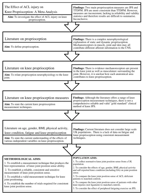
Figure 3. A summary of findings from the literature review and the aims of the 84 thesis.
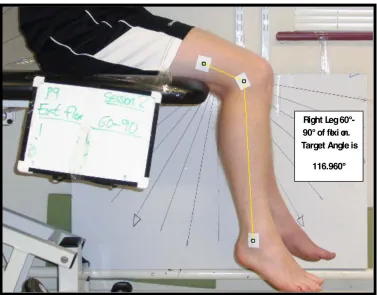
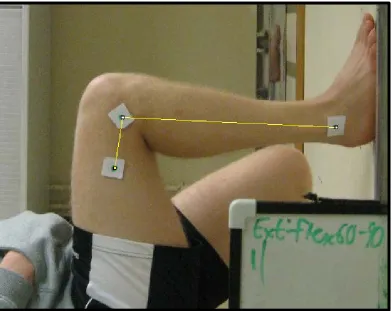
Figure 4. Typical set up and measurement of knee joint angle for sitting JPS
measurements. 90
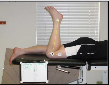
Figure 5. Typical set up and measurement of knee joint angle for Prone
Condition JPS measurement. 91
Figure 6. The Total Trainer Pilates Equipment, model TT2500P and an example of a participant on the equipment during collection of knee JPS. 93
Figure 7. Typical set up and measurement of knee joint angle for active
condition JPS measurement. 93
Figure 8. Mean and Standard Error JPS Flexion and Extension Scores for a
normative population. 134
Figure 9. A significant interaction between Gender and GPPAQ scores from JPS extension data. 134
Figure 10. Correlation between JPS extension absolute error scores and age. 135 Figure 11. Correlation between JPS extension absolute error scores and height. 135 Figure 12. Correlation between JPS extension absolute error scores and BMI. 136 Figure 13. Correlation between JPS extension absolute error scores and KOOS. 136 Figure 14. Correlation between JPS extension absolute error scores and
Lysholm Score. 137
Figure 15. Correlation between JPS extension absolute error scores and
Figure 16. Mean and Standard Error JPS Absolute Error Scores for a non-athletic ACL deficient and normative population. 138
Figure 17. Mean and Standard Error JPS into flexion Absolute Error Scores for an elite-athletic ACL deficient and normative population. 139
Figure 18. Mean and Standard Error JPS into extension Absolute Error Scores for an elite-athletic ACL deficient and normative population. 140
Figure 19. Mean and Standard Error JPS Absolute Error Scores pre and
post fatiguing protocol. 141
Figure 20. Average absolute errors in the elbow ipsilateral matching of 30° targets for different cross sections of the human life span (taken from Goble
et al., 2010). 149
Tables
Page
Table 1. Characteristics of the articles included in the meta-analysis
(Relph et al., 2014). 13
Table 2. Methodological quality score for each of the articles included in 16 the meta-analysis
Table 3. Receptor name, classification, axon type, location and adequate stimuli (adapted from Richards & Selfe (2012) and Lundy Ekman (2013)). 30
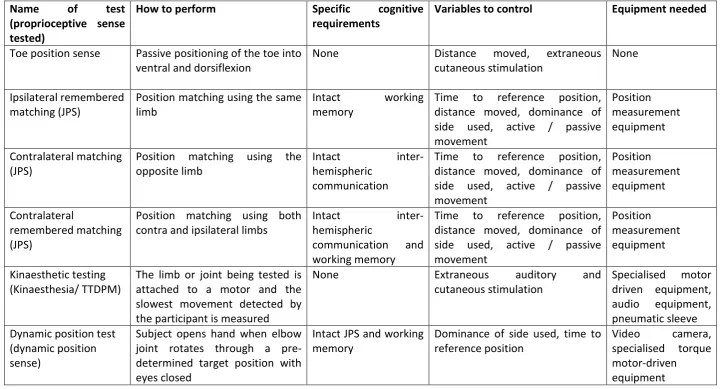
Table 4. Types of clinical proprioceptive testing (adapted from Suetterlin
& Sayer, 2014, p.314). 47
Table 5. Age-related anatomical, physiological, central nervous system and clinical changes in proprioception (adapted from Shaffer and Harrison,
2007, p.197). 61
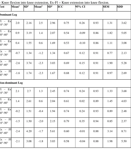
Table 6. Mean (°), standard deviation (SD), 95% confidence intervals (CI), standard error of measurement (SEM), smallest detectable difference (SDD) and intraclass correlation coefficient (ICC) values for the Real Error Score
(RES). 97
Table 7. Mean, standard deviation (SD), 95% confidence intervals (CI), standard error of measurement (SEM), smallest detectable difference (SDD) and intraclass correlation coefficient (ICC) values for the Absolute Error Score
(AES). 98
Table 8. Mean (°), standard deviation (SD), 95% confidence intervals (CI), standard error of measurement (SEM), smallest detectable difference (SDD) and intraclass correlation coefficient (ICC) values for the Real Error
Score (RES). 99
Table 9. Mean (°), standard deviation (SD), 95% confidence intervals (CI), standard error of measurement (SEM), smallest detectable difference (SDD) and intraclass correlation coefficient (ICC) values for the Real Error Score
Table 10. Mean (°), standard deviation (SD), 95% confidence intervals (CI),
standard error of measurement (SEM), smallest detectable difference (SDD) and intraclass correlation coefficient (ICC) values for the Real Error Score
(RES). 101
Table 11. Mean, standard deviation (SD), 95% confidence intervals (CI),
standard error of measurement (SEM), smallest detectable difference (SDD) and intraclass correlation coefficient (ICC) values for the Absolute Error Score
(AES). 102
Table 12. Statistical analyses on the effects of absolute or error scores, leg, range of motion, direction and condition on knee JPS measurements. 107
Table 13. Participant details of study 3.7. 117
Table 14. Normative knee joint position sense values of an adult UK population. 133 Table 15. Mean and standard error JPS absolute error scores for knee injured
Acknowledgements
Foremost, I would like to express my sincere gratitude to my supervisor Dr. Lee Herrington for his motivation, enthusiasm and immense knowledge that provided continuous support to me. Your guidance helped me have the confidence to complete my research and thesis. Thank you. I also would like to give a massive thank you to Prof. Sarah Tyson whose guidance at the beginning of this journey was invaluable.
Thank you to those colleagues past and present who have provided constant advice and kindly read through numerous versions of thesis chapters. To all my participants, including some of those aforementioned colleagues, I thank you for your patience and time.
Abstract
1.1 Anterior Cruciate Ligament (ACL) Injury and Knee Proprioception
It is estimated 250,000 people injure their anterior cruciate ligament each year in the United States alone (Hewett et al., 2007a). One potential mechanism of this injury is reduced knee proprioceptive ability (Hewett et al., 2007a). Further, once the patient has completed rehabilitation, perhaps after reconstructive surgery, evidence suggests a proprioceptive deficiency in the knee is still present (Bonfim et al., 2003, Roberts et al., 2000, Rehm et al., 1998, Barrett 1991a, Carter et al., 1997). Therefore clinical practitioners use ‘proprioceptive exercises’ to attempt to regain pre-injury levels of knee proprioception (Swanik et al., 1997, Ingersoll et al., 2008). However, the success of this treatment is unclear in the literature.
There is strong evidence to support the presence of mechanoreceptors in the anterior cruciate ligament tissue (Barrack et al., 1994). Hence it is intuitive to assume following an injury to this ligament, knee proprioception may be reduced due to the loss of activated mechanoreceptors in the knee joint during motor tasks. Indeed, there is a plethora of literature to support this viewpoint (for example Fischer-Rasmussen and Jensen, 2000, Fremerey et al., 2000, Ozenci et al., 2007, Mir et al., 2008 and Angoules et al., 2011). However, there is also research to counter this theory (for example Remedios et al., 1998, Good et al, 1990, Harter et al., 1992, Dvir et al., 1988, Fischer-Rasmussen et al., 2001, Jensen et al., 2002, Co et al., 1993, Friden et al., 1996), the belief being that a proprioceptive deficit is not present following an ACL injury as other mechanoreceptors in and around the knee joint, particularly the surrounding musculature, may compensate for the loss of ACL afferent information (Beard and Refshauge 2000). The reason for the contradictions in research findings may be due to the vast range of knee proprioception measurement techniques used in ACL deficient population studies. However, without a validated, reliable measure of knee proprioception it is impossible to make a satisfactory conclusion on ACL injury and knee joint proprioception.
1.2 Proprioception
2000). Sherrington (1906a) first published the word proprioception describing it as “a deep field of receptors in which stimuli are traceable to actions of the organism” (p.472). It is believed Sherrington constructed the word from the Latin “proprius” (one’s own) and “reception” (receives). Clinicians have stated global proprioception as “a specialized type of the sense of touch” (Barrack et al., 1994, p.19) and “the sense of position and movements of the limb” (Grigg, 1994, p. 2) and it is thought to be “...used to reference the afferent
information arising from proprioceptors” (Riemann and Lephart, 2002a p.72). However, if
considered in more detail proprioception can be divided into two key aspects of joint homeostasis; joint kinaesthesia (the dynamic sense of movement including joint acceleration, force and velocity) and joint position sense (the static sense of movement) (Ogard, 2011).
Important spatial and temporal afferent information is provided by specialised ‘proprioceptors’ or mechanoreceptors located in and around joints (Hogervorst and Brand 1998). These receptors include muscle spindles, Golgi tendon organs, ruffini nerve endings, pacinian corpuscles, Meissen’s corpuscles and Merkel’s discs (Richards and Selfe, 2012). Receptor afferent information is transmitted by transforming the mechanical energy caused by physical deformation of the joint and muscles to electrical energy of nerve action potential (Stillman, 2002). This information is transmitted to the central nervous system (CNS) and in turn organised and managed in various higher order areas (Biedert, 2000). For example balance and posture are organised at the brain stem but some proprioceptive information is organised at higher levels such as the cerebral cortex and the cerebellum (Biedert, 2000). Motor control commands are sent to relevant muscles around the joint to ensure co-ordinated, effective movement (Riemann and Lephart 2002b). It is clear proprioception has an important role in normal efficient movement therefore clinicians require valid and reliable measurement tools to monitor joint proprioception in patients.
1.3 Knee Proprioception
posterior cruciate ligament also contains these types of mechanoreceptors (Katonis et al., 1991). In addition, other areas of the knee joint including the medial and lateral collateral ligaments and menisci all contain types of mechanoreceptors and hence may play a role in joint proprioception (Solomonow and Krogsgaard, 2001, Pitman et al., 1992). The presence of mechanoreceptors throughout the knee joint suggests proprioceptive afferent information is not only provided by the supporting musculature as once thought (Scott and Loeb, 1994). It is most likely knee joint homeostasis is achieved by accumulation of all mechanoreceptor information, defined as the “final common output theory” (da Fonseca et al., 2004). The majority of tissues in the knee joint and its surrounding muscles provide important afferent information on knee position and movement, therefore it is critical clinicians can measure knee joint proprioception in order to accurately evaluate proprioceptive rehabilitation and pre-screening programmes aimed at preventing knee injury.
1.4 Knee Proprioception Measurement Techniques
There have been a variety of approaches to knee proprioception measurement including investigation of sensory evoked potentials (Courtney et al., 2005), gait analysis adaptations following injury (Devita et al., 1998), electromyography of lower extremity muscles (Houck
consensus on the most appropriate method has not been agreed. There is also a shortage of reliability and validity analysis of knee proprioception measurement techniques. Any, or all of these variables may impact on the measurement of knee proprioceptive ability. As previously stated, it is imperative clinicians can evaluate knee proprioceptive ability effectively and therefore a standardised method of knee proprioception must be established.
1.5 Age and Knee Proprioception
An increase in age is perhaps inevitably correlated to a decrease in certain musculoskeletal and neurological systems (Gilsing et al., 1995). Therefore it is perhaps no surprise research has identified a proprioceptive decline with an increase in age. The results of cross-sectional research evidence shows reductions in both static (JPS) and dynamic (TTDPM) proprioceptive ability with older populations (Kokmen et al., 1978, Pai et al., 1997, Barrett
et al., 1991b, Kaplan et al., 1985, Petrella et al., 1997, Hurley et al., 1998). This has been explained using theory on both peripheral and central adaptations. At the mechanoreceptor level, joint stiffness increases with age (Miwa et al., 1995). This is because of age adaptations in the muscle receptors; the receptors diameters reduce (Herter et al., 2014) and the capsular thickness increases (Swash and Fox, 1972, Mynark and Koceja, 2001). This can create a reduction in sensitivity of muscle spindles and hence proprioception (Herter et al., 2014). Furthermore, the composition of muscle spindles can change which again contributes to desensitisation of the muscle spindles (Suetterlin and Sayer, 2014) and also the total number of effective mechanoreceptors reduces (Shaffer and Harrison, 2007, Aydoğ
et al., 2006, Iwasaki et al., 2003).
Dendrites receive and relay stimuli between neurones and thus are critical to efficient motor control (Lundy-Ekman, 2013, McBean and van Wijck, 2013). At the central level, evidence has suggested the dendrite system is less effective in older patients (Ribeiro and Oliveira, 2010). Furthermore, the nerve conduction velocity decreases, along with a reduction in the number of motor units in adults over 60 years old (Campbell et al., 1973). All potential age related declines may reduce proprioceptive ability (Barrack et al., 1993, Yan and Hui-Chan, 2000). However, there is no normative data available that considers a range of adult ages across a healthy population. This is needed to inform clinicians and their treatment of proprioceptive deficits.
The majority of clinical practitioners will be aware of the difference in ACL injury rates between men and women; women appear to have a higher risk of ACL injury than men (Arendt et al., 1999). However, there is limited research that considers the effect of gender on proprioception and hence if this indeed may be a contributing factor to the increased rate of ACL injury in women. Both Rozzi et al., (1999a) and Nagai et al., (2012) reported some initial reports of a female reduction in knee proprioception. Furthermore authors have considered the effect of the menstrual cycle on proprioception, the theory being the increase in oestrogen interacts with neurotransmitters in the brain which may improve central processing of afferent information (Daniusevičiūtė et al., 2012). However, researcher findings are inconsistent (Fridén et al., 2006, Hertel et al., 2006). Therefore, there is a need for a large scale normative study on knee proprioception that considers any potential effects of gender.
1.7 Body Mass Index and Knee Proprioception
Body mass index (BMI) is a standard measure of mass of a patient with concurrent consideration of height (World Health Organisation, 2000). The effect of BMI on knee proprioception has rarely been considered in the literature. Paschalis et al., (2013) reported proprioceptive deficiencies in overweight and underweight participants compared to a lean control group. Also, Kaya et al., (2014) compared overweight patients with pathology to uninjured overweight controls and reported pathology reduced knee proprioceptive ability. However, to the author’s knowledge this is the full extent of the literature on BMI and proprioception. There are many detrimental consequences of becoming overweight; therefore it may be a reduction of proprioception is also one of these negatives. Clinicians should be aware of the effects of BMI on proprioception to inform their practice. However, more evidence is required in this area.
1.8 Physical Activity and Knee Proprioception
implemented in this research ranges from Tai Chi, golf, swimming, running and strength training. Results are of the same consensus; regular physical activity appears to heighten knee proprioception. In particular with the elderly groups, regular exercise may indeed attenuate the age related decline in proprioception. This is explained by exercise induced adaptations at both peripheral and central areas.
It is thought the latency of the stretch reflex is reduced and the amplitude of the stretch reflex is increases as a result of regular exercise (Hutton and Atwater, 1992). The repetitive nature of exercise may also improve the effectiveness of the gamma motor neuron route (Ribeiro and Oliveira, 2010). This also improves central processing of afferent information (Tsang and Hui-Chan, 2003). Furthermore exercise increases body temperature which has been shown to improve the effectiveness of cutaneous receptors up to temperatures of 37°C (Green, 1977, Gescheider et al., 1997). Therefore regular exercise is thought to improve knee proprioception.
Regular physical activity or training is critical to elite athletic populations. It follows that elite athletes may have enhanced knee proprioception. Indeed, much research provides support for this hypothesis, for example early work by Lephart et al., (1996) and Barrack et al., (1984a, 1984b) reported increased proprioceptive ability in ballet dancers and gymnasts. Other research has replicated this finding with American footballers and archers (Euzet and Gahery 1995) soccer players (Muaidi et al., 2009) swimmers and badminton players (Han
et al., 2013a and Waddington et al., 2013). The reasons for this may be divided into two areas: innate characteristics (Euzet and Gahery, 1995) and the effects of long term training (Ashton-Miller et al., 2001). Elite athletes may be born with superior physiological and neural systems that may enhance proprioception. This may further be enhanced by improvements in central processing of afferent information that may occur during training (Meeuwsen et al., 1993). However, the elevated performance of regular exercisers and elite athletes needs to be confirmed using reliable and valid knee proprioception testing.
1.9 Peripheral/ Muscular Fatigue and Knee Proprioception
fatigue is typically a decrease in the capacity to produce muscular force (Enoka and Duchateau, 2008). The effect of peripheral or muscular fatigue on proprioception has been well considered in the literature (Allen and Proske, 2006, Rozzi et al., 1999b, Torres et al., 2010, Skinner et al., 1986a, Gear, 2011, Ju et al., 2010, Miura et al., 2004, Allen et al., 2010, Ribeiro et al., 2007, Stillman et al., 1999, Ribeiro et al., 2011, Paschalis et al., 2007, 2008, 2013, Marks and Quinney 1993, Dieling et al., 2014). This research provides information on a proprioceptive decline following a bout of maximum exercise to fatigue levels (Torres
et al., 2010, Allen et al., 2010, Ribeiro et al., 2011, Gear, 2011). This may be attributed to impaired excitation of the motor units (Rozzi et al., 2000), an increase in knee laxity (Changela et al., 2012) and an increase in pain (Fortier and Basset, 2012).
However, conversely authors have published evidence that peripheral or muscular fatigue fails to reduce knee proprioceptive ability (Miura et al., 2004, Stillman et al., 1999, Dieling
et al., 2014, Marks and Quinney, 1993). The contrariety of research may be due to
inconsistencies in the fatiguing protocol and knee proprioception measurement. Therefore it is still unclear how peripheral or muscular fatigue impacts proprioceptive ability.
1.10 Osteoarthritis and Knee Proprioception
Osteoarthritis in the most common type of arthritis (Pai et al., 1997) and the knee joint is the most common location for the disease (Sharma et al., 1997). Unfortunately, the pathology typically causes a reduction in knee joint stability and therefore potentially a loss of knee proprioceptive ability (Collier et al., 2004). This result has been demonstrated using both knee kinaesthesia (Lund et al., 2008) and knee joint position sense (Segal et al., 2010). It is thought this can be attributed to impaired articular mechanoreceptors and hence modulated afferent discharge, reduced gamma motor neurone activity and inflammation of the joint (Knoop et al., 2011). However, as stated in previous sections, the measurement of knee proprioception is far from consistent. Therefore clinical practitioners should generalise current research with caution.
1.11 Thesis Aims and Objectives
second aim involves implementation of this tool to report the effects of various independent variables on knee joint position sense ability. The different components of each aim are provided below:
Methodological Aims
To establish a measurement technique that provides the best representation of knee joint position sense ability.
To establish a reliable, consistent and sensitive measurement of knee joint position sense.
To establish a valid measurement technique of knee joint positioning.
To establish the number of trials required for consistent knee joint position sense.
Population Group Aims
To collect normative knee joint position sense from a representative sample of the UK population.
To consider the effects of age, gender, BMI, physical activity and self-reported knee condition on knee joint position sense.
To compare the knee joint position sense of anterior cruciate ligament deficient patients (both non-athletic and elite athletic) to an uninjured matched control group.
To compare the knee joint position sense of patients with any other knee injury (not including ligament damage e.g. OA) to an uninjured matched control group.
2.1.1 The effect of Anterior Cruciate Ligament Injury on Knee Proprioception
The ACL is the most commonly injured knee ligament (Miyasaka et al., 1991). Hewett (2007a) states potentially 250,000 individuals will suffer an ACL injury, with approximately 50,000 needing knee surgeries (Miyasaka et al., 1991) each year in the United States alone. Injuries to the ACL are career threatening for sports professionals and even when rehabilitation is completed, secondary injury problems are common place (Lephart et al., 2000). There is a significantly greater risk of suffering osteoarthritis in the damaged limb, occurring at 10 times a greater rate in ACL-injured athletes, as well as higher risk of injury to the uninjured knee (Bahr and Krosshaug, 2005, Hewett et al., 2006, Johansson et al., 2000). Therefore, it is important to develop effective treatments and preventative strategies for ACL injury.
Evidence suggests an ACL injury significantly reduces the number of effective mechanoreceptors in the ligament (Barrack et al., 1994). Typical surgical practice is to reconstruct and replace the damaged ligament tissue with tendon tissue, which again reduces the number of working mechanoreceptors (Hewett, 2007a).
Mechanoreceptors provide important afferent information regarding position (static) and movement (dynamic) to the central nervous system for processing, this is known as proprioception (Lephart et al., 2000). Therefore it follows that such injuries can be detrimental to proprioception of the knee. The subject of proprioception is steeped in history. For at least 400 years researchers have been investigating how people are able to perceive and accurately control limb movements without visual input (Proske, 2006). Proprioception plays a critical role in normal human performance (Riemann and Lephart, 2002a, 2002b, Stillman, 2002, Barrack and Munn, 2000). Deficits may lead to abnormal movement patterns which may lead to knee misalignment then to other problems such as osteoarthritis (Lephart
et al., 2000). Physiotherapists and other clinical practitioners have therefore used
measurements of proprioception in training and rehabilitation strategies to inform their practice (Hewett et al., 2007b, Ogard, 2011, Stillman, 2000). It is common place for physiotherapists to include proprioceptive exercises in rehabilitation following an ACL injury (Perrin and Irrgang, 2000).
injury with proprioceptive exercises without significant evidence this proprioceptive deficit exists. Therefore a meta-analysis was completed to assess the effects of ACL injury on knee proprioception (see appendix 1a, 1b, 1c). This meta-analysis will be described in the next section.
2.1.2 The effects of Anterior Cruciate Ligament Injury on Static and Dynamic
Knee Proprioception
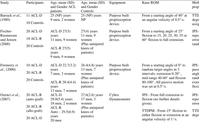
Relph et al., (2014) revealed six studies of sufficient quality and low risk of bias to consider the effect of ACL injury on joint position sense (see table 1 for details). These studies were selected using a meta-analysis protocol. No review protocol exists for meta-analysis of descriptive data, thus the PRISMA guidelines on meta-analysis were followed as far as was practicable for the type of data concerned ( http://www.prisma-statement.org/statement.htm). The following electronic databases were accessed from their inception to September 2013: AMED, CINAHL, PubMed, Medline, PeDro, Sports Discus and the Cochrane Library. Primary journals in the field; The Knee, American Journal of Sports Medicine and the British Journal of Sports Medicine were also manually searched, as were the reference lists of all selected studies to ensure the search was comprehensive. Key terms were: anterior cruciate ligament, proprioception, postural sway, joint position sense, balance, equilibrium or posture using the Boolean operator “OR”. Limits of the search were: English language studies (none of the researchers spoke foreign languages); human studies, adult participants and peer reviewed published full access articles. Unpublished literature and trial registries of current studies were not included in the search. Studies were eligible for inclusion if they 1) investigated proprioception of the knee following ACL injury (conservatively managed or reconstructed) 2) recruited adults (over 16 years) with an ACL injury, including participants with ACL injuries combined with meniscus and/ or collateral ligament damage and 3) included a primary outcome measure of knee proprioception measured by mean angle of error in degrees.
Table 1: Characteristics of the articles included in the meta-analysis (Relph et al., 2014).
Study Participants Age, mean (SD) and Gender ACL patients
Age, mean (SD) and Gender Controls
Equipment Knee ROM Method of measuring
proprioception
Barrack et al., (1989)
11 ACL-D 10 Controls.
25 (NP) years 9 men, 2 women
25 (NP) years NP
Purpose built proprioception device.
From a starting angle of 40° at an angular velocity of 0.5°/s.
TTDPM - Mean angle of error in degrees from 10 trials randomly assigned to flexion or extension Fischer-Rasmussen and Jensen (2000) 20 ACL-D 18 ACL-R 20 Controls ACL-D 27(5) years
11 men, 9 women
ACL-R 27(5) years
9 men, 9 women
27(4) years 11 men, 9 women (Plus uninjured knees of patients) Purpose built proprioception device.
From a starting angle of 25° flexion to 15, 20, 25, 30, 35 or 60° flexion to full extension.
JPS (passive positioning then active repositioning task) – Mean angle of error in degrees from 20 trials randomly assigned to target angles.
Fremerey et al., (2000)
10 ACL-D 20 ACL-R 20 Controls
ACL-D 22.7(3.2) years
7 men, 3 women
ACL-R 28.4(4.4) years
13 men, 7 women
26.4(4.8) years 13 men, 7 women (Plus uninjured knees of patients) Purpose built proprioception device.
From a starting angle of 0° to random target angles in 3 intervals; extension 0-20° , mid-range 40-60° and flexion 80-100°. All passive motion was set at 0.5°/s.
JPS (passive positioning then passive repositioning task) – Mean angle of error in degrees from trials randomly assigned from the
extension range, mid-range and flexion range.
Ozenci et al., (2007) 20 ACL-R (auto-graft) 20 ACL-R (allo-graft) 20 ACL-D ACL-D 29.0(5.4) years 18 men, 2 women ACL-R
Auto – 29.5(6.9) years
20 men
27.6(2.6) years 17 men, 3 women (Plus uninjured knees of patients) Cybex Dynamometer
JPS - From full extension to flexion (no further details given).
TTDPM - From 15° flexion to either flexion or extension at an angular velocity of 1°/s.
20 Controls Allo – 30.2(4.6) years
16 men, 4 women
assigned to either flexion or extension.
Angoules et al. (2011) 20 ACL-R (hamstring) 20 ACL-R (patella tendon)
16 men, 4 women
18 men, 2 women
N/A Con-Trex
Dynamometer
JPS – From full extension (0°) to flexion angles of 15, 45 & 75°.
JPS (passive positioning then active repositioning task) – Mean angle of error in degrees from three trials.
Mir et al. (2008) 12 ACL-R 12 Controls 23(4.75)years 12 men 22(4.35) years 12 men (Plus uninjured knees of patients)
Digital camera, markers.
From a starting angle of 60° flexion to 30° flexion and from a starting angle of 0° flexion to 30° flexion. All motion was at an angular velocity of 10°/s.
JPS (active positioning then active repositioning task) - Mean error angle in degrees over 3 trials.
The full text of the remaining studies was then checked against the selection criteria. Studies with outcome data that did not meet our criteria were excluded at this stage. The selection of appropriate articles was agreed through discussion between two of the researchers and a third party was available to arbitrate if necessary.
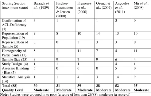
Table 2: Methodological quality score for each of the articles included in the meta-analysis
Scoring Section (maximum score)
Barrack et al., (1989)
Fischer-Rasmussen & Jensen (2000)
Fremerey
et al., (2000)
Ozenci et al., (2007)
Angoules
et al., (2011)
Mir et al., (2008)
Confirmation of ACL Deficiency (3)
3 1 3 1 3 0
Representation of Population (19)
9 8 10 14 13 10
Representation of Sample (5)
3 0 3 3 3 0
Homogeneity of Participants (13)
5 11 11 7 4 11
Sample Size (25) 3 9 7 9 6 4
Study Design (4) 1 1 1 1 4 1
Assessor Blinding / Bias (5)
5 0 0 0 5 0
Statistical Analysis (14)
1 1 4 3 14 9
Total (88) 30 31 39 38 52 35
Quality Level Moderate Moderate Moderate Moderate Moderate Moderate Note: Studies were grouped in to poor (a score of less than 29/88), moderate (a score of
30-58/88) or good (a score of 59+/88) studies based on their final methodological quality score.
Studies that met the eligibility criteria and were of sufficient quality (see table two) were included in the meta-analysis. The following data were extracted: the number of participants, mean angle of error measured using TTDPM and/ or JPS methods and accompanying standard deviation values to include in the meta-analysis and the following comparisons were made:
For joint position sense data:
· ACL injured leg versus contralateral leg control · ACL injured leg versus external control leg
· Patients with a reconstructed ACL versus patients with a deficient ACL For data on the threshold to detect passive motion:
· ACL injured leg versus external control leg
Firstly, comparisons were made using a fixed effect model with an inverse variance method as the outcome measures for both JPS (error matching score) and TTDPM (angular displacement before perception of movement) were consistent between the included studies. However, after consideration of the variability and specifically the heterogeneity values within the JPS and TTDPM protocols, a random effect model with an inverse variance method was also used for comparisons. All data was analysed and presented as forest plots using Review Manager Software (version 5.1). Standard mean difference between groups measured the effect size. Heterogeneity between comparable trials was tested using the chi squared test (level of significance = p< 0.10). Heterogeneity was further tested using I2 percentages to consider the impact potential heterogeneity would have on the meta-analysis.
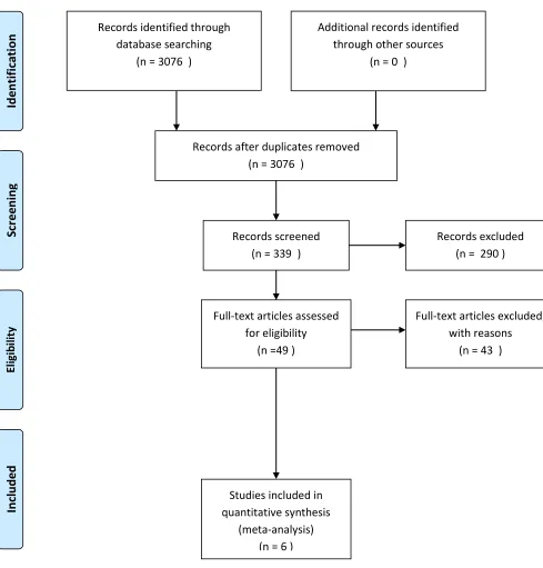
The initial search strategy yielded 3076 articles, 2737 of which did not relate to the research question. Screening of the titles and abstracts of the remaining 339 articles revealed that 290 did not fully meet the inclusion criteria; the main exclusion factor was the use of techniques to measure proprioception other than TTDPM and/or JPS. A further 43 articles were excluded as they provided ‘poor’ quality data with a high risk of bias and/or had missing or inadequate outcome data. The main reasons for missing data were that median data were presented instead of mean data or measures of the variability of the data (standard deviation) were missing. This left six studies which were selected for inclusion in the meta-analysis. The PRISMA flow chart detailing the selection process is shown in figure one.
difference of the mean angle of error was 0.35° (95% CI [0.14 to 0.55]; P= 0.001; I2 = 78%) indicating that the control group had better joint position sense than ACL patients. Three studies (Fischer-Rasmussen and Jensen, 2000, Ozenci et al., 2007 and Angoules et al., 2011) compared ACL reconstructed (n=116) and ACL deficient (not reconstructed) legs (n=100). The pooled standard mean difference of the mean angle error was -0.62° (95% CI [-0.76 to -0.48]; P<0.001; I2 = 42%) indicating that ACL reconstructed patients had better joint position sense.
Records identified through database searching
(n = 3076 )
Sc
reen
in
g
Inc
lude
d
Eli
gib
ilit
y
Ide
nti
fic
at
io
n Additional records identified through other sources
(n = 0 )
Records after duplicates removed (n = 3076 )
Records screened (n = 339 )
Records excluded (n = 290 )
Full-text articles assessed for eligibility
(n =49 )
Full-text articles excluded, with reasons
(n = 43 )
Studies included in quantitative synthesis
[image:34.595.89.578.108.619.2](meta-analysis) (n = 6 )
Figure 1: A PRISMA flow chart of article reduction.
Carter et al., 1997). Poor JPS ability has also been evidenced using passive reproduction methods, in which participants deactivate knee movement using a switch on the apparatus when they feel they have reached the target position (Lee et al., 2009 and Zhou et al., 2008, Friden et al., 1997). Further evidence, using a passive followed by an active reproduction protocol in which participants use their own muscle force to replicate the target angle, provides more evidence of reduced JPS acuity following an ACL injury (Corrigan et al., 1992, Ochi et al., 1999, Katayama et al., 2004, Baumeister et al., 2008). Iwasa et al., (2000) measured JPS using a longitudinal research design and concluded it may take up to 18 months for complete restoration of JPS abilities. However, it should be noted that no pre-injury or normative data was available to make comparisons to post-operative levels. Also, Reider et al., (2003) reported significantly better JPS scores compared to external controls six months post-operative, suggesting rehabilitation may in fact improve JPS to levels above an uninjured population. Knee JPS was measured using passive-active methods discussed previously. Muaidi et al., (2009) considered knee JPS in the transverse place and presented similar results to previous sagittal plane studies, JPS was significantly reduced in ACL injured participants.
Ochi et al., (1999) explained JPS deficits using sensory evoked potentials, their results suggested reconstruction of the ACL preserved mechanoreceptors in the ACL and hence improved JPS compared to ACLs not reconstructed. Baumeister et al., (2008) measured electroencephalography (EEG) signals to consider varied cortical activity during JPS tasks in ACL injured participants. Results indicated ACL participants increased the cortical activity during tasks, this suggests they have higher attention to the task and hence may perceive the task as more complex than uninjured controls. Therefore ACL injured patients may have altered cortical activity and may find motor tasks more complex than pre-injury.
compensate for the loss of ACL afferent signals. However, it may also be possible the methods used to measure proprioception did not involve ACL afferent input and hence deficits were not found.
Furthermore, potential sources of bias must be noted in all studies considering ACL injury and proprioception, regardless of their conclusions. Studies had to be excluded from the meta-analysis (Relph et al., 2014) data due to potential threats to reliability and validity. The majority of studies on this topic do not complete reliability and validity statistics of the measurement tool and therefore it is unclear if the data were viable. There are also issues with missing data in some studies; for example Friden et al., (1997), Roberts et al., (1999), Reider et al., (2003) and Friden et al., (1996) report median data instead of mean data therefore making comparisons to other findings difficult.
Therefore, the suggestion that ACL injuries negatively impact JPS appears intuitive, however the evidence is not as compelling as one might expect. For example within a single study, JPS ability is reduced in some measures, and not in others (Jensen et al., 2005 and Reider et al., 2003). The inconsistency in procedures between studies makes it very difficult to strongly conclude ACL injury does in fact reduce JPS ability. There are two theories to explain the discrepancy in results. Firstly, that the ACL does not play an important role in knee JPS, rather it is the knee musculature around the joint that is the dominant source of afferent information (Beard and Refshauge 2000). Secondly, that the measurement techniques employed are too variable, unreliable and inconsistent. As there is no standardised, reliable measure of JPS, authors have used a vast range of techniques, the majority of which are not tested for reliability and validity. Indeed, lack of reliability and validity measures in many studies led them to be excluded from the meta-analysis (Relph et al., 2014). For full details of articles not included in the meta-analysis (Relph et al., 2014) and evidence of the range of protocols used, please see appendix 1c.
injured legs (n=71) to external control legs (n=30). Results of the fixed effect model indicated a difference in mean angle error of 0.38° (95% CI 0.04 to 0.72; P= 0.03; I2 = 73%) indicating that the external control group had a better TTDPM than the injured leg group. The random effect model results provided opposite findings to the fixed effects model; ACL injured legs were significantly different to ACL uninjured leg (mean angle of error 0.39°; 95% CI [0.24 to 0.54]; P<0.00001; I2 = 50%) but not significantly different to the external control group (angle of error 0.45°; 95% CI [-0.21 to 1.11]; P=0.19; I2 = 73%). Therefore, as with the JPS meta-analysis findings, results are appear to be inconclusive.
There are additional studies that support the findings of the meta-analysis by Relph et al., (2014); however, potential risks of bias may be present. Studies excluded from the meta-analysis had missing data and did not appropriately present reliability and validity statistics on the chosen TTDPM protocols. Therefore these studies are considered here, but with caution. Many studies conclude TTDPM is significantly increased following ACL injury (Lephart et al., 1992, MacDonald et al., 1996, Courtney and Rine, 2006, Friden et al., 1999, Beynnon et al., 1999, Borsa et al., 1997, Corrigan et al., 1992, Reider et al., 2003, Lee et al., 2009, Roberts et al., 1999). In contrast, Valeriani et al., (1996) suggest ACL reconstruction does not restore TTDPM ability, measured using sensory evoked potentials. However, research by Pap et al., (1999), Foonseca et al., (2005), Nishiwaki et al., (2007), Jensen et al., (2002), Fischer-Rasmussen et al., (2001), Risberg et al., (1997) and Wright et al., (1995) conclude TTDPM ability does not reduce following ACL injury. It is difficult therefore to make clear conclusions regarding the effect of ACL injury on TTDPM. It may be TTDPM protocols are less sensitive than JPS to changes in knee proprioception following an ACL injury. Indeed the nature of the protocol, in which the knee is moved dynamically and then the patient responds, may not be sensitive enough to measure ligament deficiencies contributing to proprioception.
Furthermore the potential risk of bias in TTDPM studies is evident from the range of protocols used. As with JPS studies, there has been no standardised TTDPM measurement technique established, hence authors use which ever protocol they feel is most appropriate and often do not provide reliability and validity statistics related to their chosen protocol. This may explain why a number of authors found significant reductions in some TTDPM measures but not others (Friden et al., 1996, Friden et al., 1997 and Roberts et al., 2000, Co
2.1.3 Summary
Results of the fixed effect meta-analysis (Relph et al., 2014) indicated there are statistically significant differences in the proprioception, in terms of JPS acuity and threshold to detection of movement, of patients with ACL injury in that they have poorer proprioception than people without such injuries and poorer proprioception in the injured than uninjured leg. The proprioception of people whose ACL was reconstructed was statistically significantly better than those whose ligament is left unreconstructed (ACL deficient). These differences are seen whether the comparator group is a patient’s uninjured leg, or a control group of people with no injuries; suggesting that either can be used as a control group in future research.
However, results of an additional random effect analysis revealed ACL patients may have worse JPS than their contra-lateral leg, but not compared to an external control group. Therefore, results are inconsistent and no clear conclusions can be made regarding ACL injury and proprioception. This is probably due to the large variation in approaches to JPS measurement, indicated in the analysis by the high I2 scores (these ranged from 42% to 78%). Therefore, a consistent knee proprioception measurement protocol must be developed in order to distinguish whether ACL injury does indeed decline proprioceptive ability.
However, the significant differences that were reported in the meta-analysis were seen most clearly when joint position sense was measured but were less apparent when threshold to detect passive motion measurement techniques were used; the meta-analysis revealed greater differences in joint position sense (JPS) than studies using TTDPM. Furthermore, comparison of the fixed and random effects model results produced opposite findings, again suggesting the result of an ACL injury on dynamic proprioception is not clear. Techniques may be insufficiently sensitive to detect the responses of rapid receptors such as the pacinian corpuscles in the ACL (Barrack and Munn, 2000) as measurements incorporate the participants’ reaction time, which is unrelated to their injury. JPS methods may be more sensitive as these measurements also incorporate the slower responses of the ruffini nerve endings and Golgi tendon organs (Schultz et al., 1984) and allow the conscious perception of joint motion and position. Therefore, joint position sense should be used to measure knee proprioception.
afferent information on the relative position and movement of the knee joint (Riemann and Lephart 2002a, Johansson et al., 2000, Schultz et al., 1984). Therefore, ACL injury may well impair proprioception through disruption to the transmission of this sensory information (Barrack and Munn, 2000). Marks et al., (2007) suggest other articular structures in the knee joint may attempt to compensate for the loss of ACL afferent signals. However, these compensatory signals may be ‘nonphysiologically disorganised’ (Marks et al., 2007 p.42), and hence the central nervous system and consequently joint position are disturbed and the knee become more unstable. The differences in directional (i.e. flexion or extension) proprioception may be due to the location of the injury. The anteromedial bundles of the ACL are most taut in flexion, the posterolateral bundle tautest in extension. Therefore the area of deficiency may determine which direction the deficits in proprioception lie.
ACL injury are due to measurement error and/or the measurement techniques were insufficiently sensitive to detect clinically significant differences (Relph et al., 2014).
Another explanation is that the comparisons included in the meta-analysis could be under-powered because the sample was too small, (again, very few of the studies discussed calculated sample size using power estimations). However the pooled data from the meta-analysis (Relph et al., 2014) involved nearly 200 patients and the 95% confidence intervals of the comparisons made were small, indicating that a lack of power was not an issue. Further research is needed to evaluate the sensitivity and reliability of techniques to measure proprioception at the knee, before they can meaningfully be used as an evaluation tool.
A limitation of research into ACL injury and proprioception is that all data collection is retrospective, which inevitably means pre-injury proprioception is unknown. It is possible that patients who suffered injuries had poorer proprioception which predisposed them to injury. Large scale normative studies are needed to give insight into the distribution of proprioception abilities across the population and whether this predisposes people to ACL injury. Such studies should consider a measurement technique that explores the full range of knee motion and direction using large sample sizes that represent the complete ACL patient population and normative data on proprioception ability.
It must also be noted that heterogeneity of variance in the referenced meta-analysis (Relph
et al., 2104) and the fixed effect model was greater than the recommended level of 50%
(Deeks et al., 2008) in all but one comparison; this may be due to variability in the recruitment strategies across studies. The time since injury when proprioception was measured and the use of rehabilitation programmes was not consistent. Highly varied measurement techniques were also evident, which is a limitation that hampers further analysis. Different pieces of measuring equipment and varied knee movements, in terms of direction and speed of motion, were employed (see appendix 1c). Proprioception increases towards the extremes of range of movement in order to protect the joint from injury (Barrack and Munn, 2000, Borsa et al., 1997), thus studies that do not include measurements across the whole range of movement may either be under- or over- estimating knee proprioception. These inconsistent methods of measuring proprioception could have contributed to the high levels of heterogeneity in the current analysis. However as there is no gold standard method of measuring knee proprioception, this variation was unavoidable.
This section of the thesis examined the effect of an ACL injury on proprioception, in terms of joint position sense and threshold to detect passive motion. The results indicate that patients with ACL injury may have poorer proprioception than people without such
injuries and poorer proprioception in the injured than uninjured leg. The proprioception of people whose ACL is reconstructed may be better than those whose ligament is left
2.2.1 The Sensorimotor System
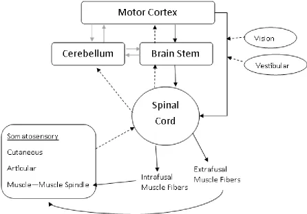
[image:43.595.71.517.384.695.2]The sensorimotor system encompasses the complex relationship between the neurosensory and neuromuscular systems (Lephart et al., 2000). The system incorporates all sensory information from the visual, vestibular and peripheral mechanoreceptors to facilitate joint homeostasis (Riemann and Lephart, 2002a) (see figure 2). Although vision and vestibular inputs are important to joint stability, the peripheral nervous system is of most interest to orthopaedic and musculoskeletal practitioners and hence will be the focus of this thesis. The peripheral nervous system involves the communication and management of peripheral afferent information provided by mechanoreceptors or “proprio-ceptors” (Sherrington, 1906a) located in muscle, tendons, articulations and cutaneous tissue (Lephart et al, 2000). This information is processed by the central nervous system to control muscle activation and joint stabilisation (Riemann and Lephart, 2002a). It is believed that proprioception is an important aspect of this process and provides information on muscle length, tendon tension, joint position, joint movement and deep vibration (Lundy-Ekman, 2013).
In 1833 Sir Charles Bell referred to the ability to detect positions and actions of the hand as the “sixth sense” (McCloskey, 1978). Later the term “kinaesthesia” was coined by Bastian in 1888 and refers to the sense of position and movement of the joints (Proske and Gandevia, 2009). Following this the ground-breaking neurophysiologist Sherrington first published the term “proprioception” describing it as “a deep field of receptors in which stimuli are traceable to actions of the organism” (1906a, p. 472). The word itself is derived from the Latin for one’s own (proprius) and receives (reception). It may be seen as a mysterious sense since we are largely unaware of it in our daily movements but yet it plays a crucial role in motor control (Proske and Gandevia, 2009). Sherrington’s work forms the bases of current physiological understanding of the musculoskeletal senses (Stillman, 2002). He classified all senses and sense organs based on their source of stimulation, suggesting that each receptor type is activated by one accompanying type of stimulus (Sherrington, 1906a). There have been several interpretations of Sherrington’s work and of the term “proprioception”. Some authors regard proprioception as only the acquisition of senses from mechanoreceptors (Grigg, 1994), whilst others regard proprioception as the acquisition and processing of sensory information (Lephart
et al., 2000). The term proprioception will be used in this thesis to describe the process of acquiring and processing sensory information from mechanoreceptors (specifically in the muscle, tendons, articulations and cutaneous tissue) to maintain joint stability and control muscle activation. Evidence has indicated both afferent and efferent information can determine position and movement of the limbs (Ogard, 2011, Gandevia et al., 2006). To note; the more global term of somatosensory sensations would encompass postural equilibrium, tactile, temperature and pain senses (Riemann and Lephart, 2002a) in addition to proprioception and as such will not be considered in detail in this thesis.
2.2.2 Peripheral Afferent (Sensory) Pathways
nerve ending and changes the potential of this nerve ending (Lundy-Ekman, 2013). If this is of sufficient magnitude, a neural signal is propagated towards the central nervous system along ascending pathways. These pathways include the dorsal root ganglion to the spinal cord, to the brain stem and/or the cerebellum and basal ganglia, then from the brain stem to the motor/cerebral cortex (Lephart et al., 1998). A description of each type of peripheral mechanoreceptor is detailed below (also see table three). It is important to note that mechanoreceptors do not work independently of each other, indeed Sherrington himself referred to them as “allies” (Sherrington, 1906b). This concept will be explored later in this section of the thesis.
Table 3. Receptor name, classification, axon type, location and adequate stimuli(adapted from Richards and Selfe (2012)and Lundy Ekman (2013)).
Receptor name Classification Axon Type Location Stimulus Muscle spindle Ia Large myelinated Throughout muscle Muscle stretch Muscle spindle II Medium myelinated Throughout muscle Muscle stretch Golgi tendon
organ
Ib Large myelinated Musculotendinous junction
Strain/ Tension
Pacinian corpuscle
II Medium myelinated Capsule, ligament, menisci, fat pads, skin
Compression
Ruffini ending II Medium myelinated Capsule, ligament, menisci, skin
Stretch, strain
Free nerve ending
Aδ Small myelinated Capsule, ligament, menisci, skin
Nociceptive
Free nerve ending
C Small unmyelinated Capsule, ligament, menisci, skin Nociceptive Meissner’s corpuscles Merkel’s discs Aβ Aβ Medium myelinated Medium myelinated Skin Skin Deformation caused by light touch.
Muscle-Tendon Unit Mechanoreceptors
Muscle Spindles
The number of muscle spindles serving each limb joint significantly declines from proximal to distal positioning, for example there is an estimated 1821 muscle spindles serving the knee joint compared to the 6659 approximate muscle spindles assisting the cervical spine (Scott and Loeb, 1994). Muscle spindles provide important sensory information regarding muscle tension or length of muscle fibres and the velocity of change of muscle displacement (Collins et al., 1998, Edin and Johansson, 1995, Gandevia et al., 1992b, Matthews and Stein, 1969). The muscle bellies contain intrafusal fibres, which are made up of nuclear bag and nuclear chain fibres (Lundy-Ekman, 2013).
Nuclear bag fibres respond directly to quick and sustained spindle stretch (Type Ia), and the frequency of firing increases as stretch increases (Proske et al., 2000). A second smaller group of sensory fibres (annulospiral endings, Type II) attach mainly to the nuclear chain fibres and respond with lower frequencies to sustained stretch (Proske et al., 2000, Matthews, 1987). The currently accepted view is that primary endings (Type Ia) of spindles contribute to sense of position and movement, whereas secondary endings (Type II) respond to position sense alone (Proske, 2006). For most types of receptors in the body an increase in discharge rate corresponds to an increase in stimulus intensity; however an increase in muscle spindle discharge rate represents a longer muscle not an increase in stimulus (Proske, 2006). This is because muscle spindle discharge rates increase in approximate proportion to the size of the lengthening (Proske, 2005). Furthermore, all muscle spindles are recruited at just 25% of maximum contraction, making them very sensitive to the stimulus (Proske, 2006) which is again different to other types of receptors.
(Ribot-Ciscar et al., 2003).Therefore it follows that proprioception would be at an optimum when all involved musculature of a movement contribute to the afferent signal.
There is also strong evidence that suggests muscle contraction during active movement increases muscle spindle activity in contrast to passive movement (Gandevia et al., 1992b). The type of muscle contraction will also influence spindle afferent discharge, for example when muscle spindles fire there is an increased afferent signal during lengthening of a muscle compared to shortening of that muscle (Ribot-Ciscar and Roll, 1998, Matthews, 1987). This suggests that as joints move through a range of motion, the predominant source of muscle spindle afferent information changes, the signals increase towards the lengthened muscle. However, this could also imply that “net” muscle spindle activity across all involved muscles stays constant simply switching between agonist and antagonist depending on the direction of movement. This theory has yet to be confirmed by experimental data.
Golgi tendon Organs in muscle
Golgi tendon organs are situated within muscle bellies near musculotendinous junctions or within tendons and provide additional extrafusal fibres (Type Ib) (Jami, 1992, Stillman, 2000). These mechanoreceptors average length and diameter is 1600µm and 122µm respectively (Jami, 1992). Golgi tendon organs detect differences in tension and force (Proske et al., 2000) but not length (Riemann and Lephart, 2002a), dynamically responding to rapid increases in these two stimuli only. Therefore, due to the high threshold and brief response, it is thought Golgi tendon organs have a protective mechanism near a joint’s extreme range of motion when tension rapidly increases (Johansson et al., 2000). However, previous literature has illustrated Golgi tendon organs may not fire during passive movement (Riemann and Lephart, 2002a) and hence are thought of as purely active mechanoreceptors.
and muscle length afferent information is conflicting, it appears large forces are not accompanied by displacement of the joint. Therefore this may confuse the CNS and reduce proprioceptive ability (Rymer and D’Almeida, 1980, Gandevia et al., 2006). This provides evidence for an important role for both muscle and tendon mechanoreceptors in proprioception during movement.
Articular Mechanoreceptors
Golgi-like Tendon Organs
In contrast to Golgi-tendon organs situated near musculotendinous junction, Golgi tendon organs located in joints provide afferent information on joint angle or position and not force or tension (Solomonow and Krogsgaard, 2001). It is suggested these types of Golgi tendon organs that are found in the knee joint provide constant levels of afferent information throughout the voluntary range of motion (Rymer and D’Almeida, 1980).
Pacinian Corpuscles
Pacinian corpuscles are small ellipsoidal nerve fibres situated close to Golgi-like tendon organs with axon diameters between 8 and 12µm (Lundy-Ekman 2013). They have a low threshold and rapidly adapt to phasic movements earning them the exclusive classification of “dynamic receptors” (Riemann and Lephart, 2002a). Previous literature has shown pacinian corpuscles rapidly sense acceleration and deceleration and hence changes in movement, but not static or constant joint rotations (Johansson et al., 2000). Therefore they detect the onset or termination of movement, but not constant joint displacement.
Ruffini Endings
are most sensitive at maximum flexion and extension positions (McCloskey, 1978, Burgess et al., 1982).
Free Nerve Endings
The majority of free nerve endings are unresponsive during normal joint movement, however, are active when damage or injury occurs in the articular tissue (Johansson et al., 2000). Therefore, research suggests this receptor provides afferent information only once the joint is damaged via nociceptive sensory input (Solomonow and Krogsgaard, 2001, Hogervorst and Brand, 1998).
Cutaneous Mechanoreceptors
Meissner’s corpuscles and Merkel’s discs
Meissner’s corpuscles are responsive to light touch and vibrations. Merkel’s discs are stimulated by skin pressure and hence contribute to proprioception when the skin is stretched (Burgess et al., 1982). Although neither of these receptors is thought of as a true “proprioceptor” the afferents they provide have a minor role in joint position sense and kinaesthesia (Edin and Johansson, 1995, Grigg, 1994, Matthews, 1987, McCloskey, 1978). It is intuitive that the sense of skin movement through stretch or pressure may contribute to our overall proprioception ability, although the presence of Ruffini corpuscles in the skin may also contribute specifically to joint position sense (Edin and Johansson, 1995). However, there is little evidence to fully support this theory. Grigg (1994) suggests this is due to the difficulties in isolating cutaneous receptors and hence measuring specific cutaneous responses to joint movement. Although this can also be disputed using evidence that the afferent signals provided by cutaneous receptors are time and speed dependent; after 1-2 minutes and at speeds under 0.1mm/s cutaneous sensation ceases (Horch et al., 1975, McCloskey, 1978). Cutaneous receptors may therefore be thought of as secondary or facilitating contributors to proprioception (Burgess et al., 1982).
2.2.3 Summary
spindle) afferent information was ascended to the cerebral cortex (Riemann and Lephart 2002a, Matthews, 1987, Proske, 2005). Also, authors at this time believed it to be more logical to look for dominant joint movement receptors within the joints themselves. However, latterly authors have reverted back to the traditional belief of Sherrington’s (1906b) “muscular sense”, that is the most dominant mechanoreceptor in proprioception to be muscle spindles (Scott and Loeb, 1994, Proske and Gandevia, 2009, Proske et al., 2000, Proske et al., 2005, McCloskey, 1978). Indeed feline studies have indicated the knee joint is served by 400 myelinated joint afferents but 4000 myelinated muscle afferents. This principle is supported by evidence suggesting articular receptors may not be as responsive in mid-range movements (Rymer and D’Almeida, 1980, Burke et al., 1988, McCloskey, 1978) and hence cannot be the most important proprioceptor. Furthermore, studies have illustrated joint proprioception is not lost when articular and cutaneous afferent information is blocked (Clark et al., 1979). Vibration and tendon pulling studies have suggested muscle receptors must have the dominant role in afferent signals as an illusion of joint movement can be induced with these techniques (Matthews, 1987, Proske and Gandevia, 2009, Berkinblit et al., 1992). For example Goodwin et al., (1972) published ground-breaking evidence that when a muscle belly is vibrated the participant experiences illusions of movement and position changes, however if this vibration is moved to the joint, no illusion occurs. Thus it is now more popular to believe muscle spindles may be the main afferent provider for proprioceptive processes.