A Simple Approach for Education Using
Virtual Human Dataset
Woo Chaw Seng¹ and Seyed Hadi Mirisaee²
¹ Faculty of Computer Science & Information Technology, University of Malaya, Malaysia ² First Residential College, University of Malaya, Malaysia
Abstract: The virtual tour of human body is a system which is able to generate a three– dimensional image of a human body based on the human dataset. Virtual human body systems prepare an environment for medical practitioners to treat certain medical conditions especially those which require surgery, precision and planning according to individual patients. Current virtual human body systems have some drawbacks and shortages especially as a study aid system, so the proposed system in this research tries to fulfill the shortages as a proper study aid system. The objective of this research is to develop a system that can simulate fly– through of a specific human organ which enables 3D views of the human body in the form of Computed Tomography (CT) scan, Magnetic resonance Imaging (MRI) scan as well as the actual anatomical images. The proposed system also provides cross section view of a particular organ or the whole body which are sectioned either horizontally or vertically and allows selection of organ view or cross section view. Two different methods have been implemented in this research, namely surface rendering and volume rendering to achieve better quality for 3D images. As a conclusion, advantages of proposed system like manipulating the organs, simplicity and low cost makes it suitable to use by medical students for in-depth study of the human anatomy or being used by biology teachers in high schools as part of the teaching guide.
Keywords: Study aid, Cross section view, organ view, surface rendering, volume rendering
Received November 15, 2009; Accepted March 10, 2010
1. Introduction
Virtual reality (VR) can be plainly described as a computer simulated environment which enables user to immerse, navigate, and interact with the virtual environment. VR can be categorized into two major categories which are 'Immersive Virtual Reality' and 'Non - Immersive Virtual Reality[1].
Virtual tour of the human body project is an incredibly interesting project whereby its mission is to develop a system that is able to generate a three – dimensional (3D) image of a human based on the human dataset. In the medical field, it is important for the medical practitioners to have a complete and thorough understanding of the human internal organs as well as their positioning. Virtual human body systems prepare an environment for medical practitioners to treat certain medical conditions especially those which require surgery, precision and planning according to individual patients. Because of the practitioner desire to know even the slightest detail of the human body, a total of three projects have been carried out in the world based on three slightly different human datasets. These projects are: Visible Human Project (1986), Visible Korean Human (1996) and Chinese Visible Human (2001)[2-4].
As the medical field grows continuously, the challenges of medical practitioners increase significantly[5-9]. Current virtual human body systems have some drawbacks and shortages to be used as a suitable study aid such as high cost, requiring high end computers, Shortages in lightening or difficult to use by end users so the proposed system in this research tries to fulfill these shortages. The proposed system can simulate fly– through of a specific human organ including more specifications. It provides cross sectional view of a particular organ or the whole body which are sectioned either horizontally or vertically and allows selection of organ view. The developed system should enable 3D views of the human body in the form of Computed Tomography (CT) scan, Magnetic resonance Imaging (MRI) scan as well as the actual anatomical images.
2. Background
2.1. Virtual Reality in Medicine
The medical field has greatly benefited from the advancement in VR techniques where it can be used extensively to manipulate medical data into the 3D forms[10]. The various VR applications in medical sciences have clearly shown that the expectation towards the VR application differs according to the goals of each medical field. VR in medicine can be categorized in four distinctive fields which are medical education, Surgical Simulation and Planning, virtual endoscopy and Neuron - Psychological Assessment and Rehabilitation.
Illustrations are important in teaching the anatomy, and VR medical applications are great helps to illustrate. With the 3D visualization, clinicians and students are able to understand better the basic anatomy and important physiological principles. Virtual reality applications can be used to train and assess students’ surgical skills. With the pressure to reduce the use of animals and cadaver in medical training, this application is a great substitute since the 3D generated models are according to the human dataset and the application can be used anytime. In the surgery planning area, VR facilitates surgery planning with rendering the 3D model of the patient and simulating the surgery situations.
Endoscopy is an invasive procedure and carries certain risk such as perforation and bleeding. In order to overcome the problem, virtual endoscopy can be performed by using the standard MRI scan or CT scan. By using a VR application, the produced images from the scan are able to be generated as a 3D model and a virtual endoscopy can be conducted through the 3D model.
In rehabilitation, VR is implemented to assist the patients in their physical therapy whereby the patients are able to conduct their physical therapy in the virtual environment with the help of immersion equipments. In the neuron– psychological assessment, the therapist are able to assess the patient based on their reaction in the virtual environment.
2.2. Digital Media Imaging
There are various procedures that are able to produce digital medical images such as: Fluoroscopy, Magnetic Resonance Imaging (MRI), Nuclear Medicine, CT, Ultrasound and X-ray. 2D medical imaging is generally a medical imaging technique that produces a static image output on films. Conventional CT and MRI machines generate an output of 2D images for the physician. Computed Tomography (CT)[11] scans are particularly useful because it is able to show the lung, bone, soft tissue and blood vessels with clarity.
Magnetic Resonance Imaging (MRI)[12] works by directing radio waves at protons in a magnetic field. The nature of the MRI machine makes it best suited for soft (non - calcified) tissue exams. This is because the human body consists of about 70% water which in turn consists of hydrogen atoms that contributes to the image.
2.3. Three Dimensional digital Image Rendering
Three dimensional digital image rendering consist of two steps, surface rendering and volume rendering. Surface rendering is an indirect method of converting volume data into 3D models. It visualizes objects as a hard set of certain basic elements such as voxels or polygons. Isosurface is one of the surface rendering methods. In this rendering method, isovalue is defined as a boundary between the ‘outside’ and the ‘inside’ and all the values greater than the isovalue, are classified as being ‘inside’[13].
Volume rendering is a technique to display 2D images from 3D discretely sampled dataset. Volume rendering works by displaying the gray scale data directly and without first fitting geometric primitives to the sample[14]. Texture Mapping, Volume Ray Casting, and Shear Warp are the different methods of volume rendering.
Texture mapping is implemented by aligning the textures with the volume and it is rendered at an angle to the viewer. First, the volume object is projected to a plenoptic surface using direct surface rendering or direct volume rendering methods[15]. The rendered surface is then converted into a 2D rectangle image. Volume ray casting utilizes a ray of sight which is shot through to the volume data. The volume data is assumed to be enclosed within a bounding primitive. The data is then interpolated at each point which is along the ray of sight [19]. After interpolation, the gradient is computed to acquire the orientation and local surfaces of the volume. Finally, the model is shaded and composited to produce the color and shading for the model.
For shear warp methodology, the viewing transformation is transformed such that the nearest face of the volume is aligned with an off screen image which is buffered with a fixed scale of voxels to pixels. The volume is then rendered and the buffer is warped to the desired orientation and scale with reference to the rendered model.
3. Related Works
purpose in mind which is to provide aid and improve the medical field by incorporating VR applications in order to generate the human anatomy as most accurate as possible and allow users the freedom to explore the system in a way they were never able to explore the actual human anatomy. Many private and government institutions have acquired the published data and developed various applications such as 3D view of the human dataset, anatomical image viewer, virtual dissection as well as virtual endoscopy.
3.1. The Visible Human Project
With the human dataset published by the National Library of Medicine, many applications were developed to utilize the human dataset and one of those applications was developed by Touch of Life Technologies, Inc (ToLTech). Their applications include[16]:
• VH Dissector
• Virtual Palpitation Guide
• Virtual Edge(Dissection guide)
3.2. The Chinese Visible Human
The Chinese Visible Human was conducted by the Third Military Medical University in China. By utilizing the human dataset acquired, many standard applications were constructed[2] such as:
• Virtual endoscopy
• Anatomical image viewer
• 3D view of the human dataset
Nevertheless, one of the more outstanding ones is the virtual acupuncture human which shows the meridian positioning, acupoint positioning, arbitrary cutting-plane visualization, multi-layer dissection, needle puncturing simulation, as well as the common diseases-therapy information.
3.3. The Visible Korean Human
The Visible Korean Human (VKH) is a project conceived with the main idea to produce an enhanced version of the serially sectioned images of the entire cadaver which has been carried out in two other experiments: the Visible Human Project (VHP) and the Chinese Visible Human (CVH). They hope to improve the existing 3D models and software[17]. The human dataset are acquired through a series of steps which are perfected through the knowledge gained from the two other experiments: VHP and CVH[17]. The main reasons which prompted the starting of VKH are[17, 18]:
• The dataset provided by the Visible Human Project is difficult to be adapted to the oriental race because the organs differ in size and shape.
• The VHP has four missing anatomical images between the four blocks where the cadaver was first divided before sectioning.
• The data did not include anatomical structures smaller than 0.2 mm since both the interval and pixel size of the images are 0.33mm or greater.
• The VHP does not include MRI images of the trunk and limbs because only MRI images of the head was scanned. The CT images of the cadaver are also not complete as the lateral part of the upper limb’s CT images was cut off.
• Both the previous projects did not release segmented images which are essential for the production of 3D models.
In the proposed system, the internal organs which will be used in the 3D rendering are obtained from the anatomical structures which are segmented from the Visible Korean Human since VKH is more complete and compatible with oriental race.
4. Methodology
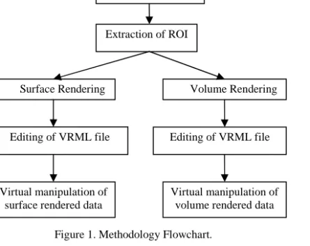
[image:3.594.331.554.414.591.2]In the proposed system, two types of image rendering techniques are applied: surface rendering and volume rendering. The surface rendering method is applied to the images for rendering of the full body view whereas the volume rendering method is applied in rendering the cross - section data which is the Oblique plane and Coronal plane view.
Figure 1. Methodology Flowchart.
Figure 1 outlines the methodology used in developing the system. As observed from the flow chart, the first step is to acquire the data of the human anatomy images as well as the segmented images. The data was provided by the VKH which conducted the experiment and released the full human anatomy dataset. After obtaining the data, the Region of Interest (ROI) extraction begins. Extraction of ROI is important as memory constraint is a factor so by extracting the ROI into another smaller array, it
Original image
Extraction of ROI
Surface Rendering
Virtual manipulation of surface rendered data
Volume Rendering
reduces the amount of used memory and needed time to generate the 3D models in the subsequent steps.
The isosurface technique is implemented at the very last step of the surface rendering pipeline after all the data has been processed to render the 3D model of the full organ. Isosurface rendering generates the surface of the whole organ without any cross section. In volume rendering, the data will go through the pipeline volume rendering. The 3D models will be generated before mapping the 2D texture on the surface which corresponds to the cross section. Texture mapping using Virtual Reality Modeling Language (VRML) was utilized while the other methods require the color data to be stored directly in the VRML file.
After the rendering is completed, the VRML file is edited to combine the various parts of the generated organ separately for the oblique and coronal plane. For generating the full organ, the artery, bones, brain stem, cerebellum, gastrointestinal tract, liver, lungs and skin have to be combined. Besides combining organs, the VRML file is also edited to include viewpoint, orientation and lighting so that the users are able to view the 3D model better. After all the manipulation, the 3D models are ready for user assessment.
4.1. Surface Rendering
Equipped with the provided images by VKH project which consist CT, MRI, anatomical and segmented images, the first task is to gather all the required data and store all the data into a 3D array. After that, the extraction of ROI begins by utilizing the segmented data to extract out the ROI from the CT, MRI and the anatomical image. The segmented images clearly define the boundary of all the organs using different RGB values for different organs. It is used to extract
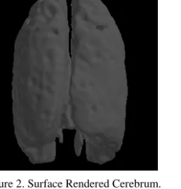
[image:4.594.374.503.342.477.2]the ROI by extracting the pixel values from the CT, MRI and anatomical images with reference to the RGB value in the segmented images. Then data interpolation is implemented on the array. This is important that data without interpolation produces a stair– like shape for the surface. After interpolation, the surface mesh adjustment is conducted; whereby the data is smoothen to produce an even better surface model. Smoothing improves the appearance of the surface mesh as reduces the appearance of sharp ends. In volume mesh generation, it utilizes the isosurface algorithm to generate the 3D model. Different isovalues are provided for different organs to generate the isosurface. The provided isovalues determines the shape of the generated 3D model. After the mesh is generated and saved in VRML format, the VRML file is edited and the 3D models are combined, and the lighting and viewpoints are added. All these are added for the users to have better view of the 3D model. Figure 2 and 3 show the sample of rendered surfaces.
Figure 2. Surface Rendered Cerebrum.
(a)Lower part of 3D model (b)Upper part of 3d model (c)Combined Surface Rendered model Figure 3. Combined Surface Rendered Model of Liver
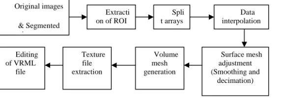
[image:4.594.170.444.513.600.2]Figure 4. Volume Rendering Pipeline
4.2. Volume Rendering
Figure 4 highlights the methodology of volume rendering implemented in constructing the system. Similar to the surface rendering technique, the gathered data of all the images for CT Image, MRI Image, Anatomical Image (Indexed Image and RGB Image), Colormap (for the indexed image of anatomical image) are needed. The data are stored in 3D arrays supported by Matlab. For volume rendering the extraction for the CT, MRI and anatomical images follows the surface rendering methodology. The next step is to split the arrays according to which plane of the organ is being generated. If the oblique plane is being generated, the array will be split equally to two arrays. The interpolated data are subjected to acquire a smooth surface model without the stair–like shape as mentioned in the volume rendering pipeline.
Surface mesh adjustment is conducted to smooth the data in order to reduce the appearance of sharp ends. Decimation is carried out to reduce the number of polygons in order to represent the surface model. Both processes contribute to reduce memory consumption. The volume mesh generation is performed to generate the 3D model of the organs which are split in two. While the part of the organ is exposed, the isosurface only generates the outline of the organ. It will leave the side of the organ which corresponds to the cross section with a gap. End caps are generated to close the gap. The end cap is a flat surface generated to cover a particular gap in surface rendering. After the 3D model is generated, the texture files are created by extracting from the corresponding cross section in order to be displayed from the array containing the ROI. It is useful to avoid the extraction of ROI again. The file is then saved in JPG format which creates a 2D texture file. For the anatomical images the colormap array is utilized to get an accurate color for the indexed image. Finally, the 2D texture is mapped to the 3D model using the end caps which corresponds to the cross section of the organ as the VRML file is edited. Besides mapping the textures, viewpoints and lightings are added in order to display the 3D model clearer.
4.3. Rendering algorithms & Tools
Matlab is the main tool used in developing the system. It provides the means to manipulate matrices, plotting functions and data as well as other capabilities by utilizing the toolbox provided. The commands will be explained in more details for better understanding of the program. The followingalgorithmshows the steps:
• Extract the ROI from the original array which contains the whole set of data
• Crops the new array containing the ROI to produce a smaller array
• Utilized for the surface mesh adjustment where the array is interpolated and smoothen.
• Rotate the array for the purpose of generating the oblique and coronal plane view
• Generate the surface model of the organs.
• Set the colors
• Generate the end caps for the volume organ
VRML is utilized as a method to store the 3D model generated by Matlab. Besides, it is also utilized to create views and orientation to display the 3D models. The views are important to the user in order to have a better inspection of the 3D models. The following algorithm shows the storing process.
• Set the background color for the display
• Determine the lighting for the virtual environment
• Set the viewpoint
The textures are mapped by utilizing the VRML texture mapping function. By adding the VRML nodes, the texture are rotated and mapped to fit the end caps of the 3D model to produce a volume rendered image.
5. Result and Discussion
Extraction of ROI is implemented in both methodologies: surface rendering and volume rendering. Because the starting slice and ending slice of a particular organ is provided, the array is also cropped from the top and bottom to create a smaller array for manipulation using simple processors. All the thirteen organs are able to be displayed based on the selection from the control panel of the system. The controls are easy to use as all the manipulation of the Original images
& Segmented images
Extracti on of ROI
Data interpolation
Surface mesh adjustment (Smoothing and
decimation) Volume
mesh generation Editing
of VRML file
Texture file extraction
3D model is done directly on the display window. The controls are only used to select the organ to view. This control makes the system easy to use for the end users. In the process of rendering the data, slight differences are noticed for the different type of images which are: CT, MRI and Anatomical. As the pixel values for the CT and MRI images are close, the result of the rendering does not differ much. For anatomical images, certain rendered organs had rougher surface. The examples can be observed in figure 5.The CT and MRI images exist in grayscale so the generated 3D models are smoother and the pixel value does not differ much for a particular ROI. However, for the anatomical images, a particular ROI contains many different colors so it contains many different pixel values. Thus the system renders it slightly different. In rendering the skin organ, it is also noticed that there are holes on both side of the rendered organ. For the anatomical and CT skin models, the holes appear near the elbow and the head of the rendered organ. In MRI, it only appears at the head area. The holes are covered by rendering the end caps. In addition, it can be seen that the lung organ, when generated, produces a significant big holes for the anatomical images and those are so obvious. Consequently, the MRI data gives the best rendering results followed by CT images while the anatomical images produces rough surfaces and with holes which are significant in the lungs and skin organs.
With the results obtained, we are on the way to test the system in a medical education environment for feedbacks and further improvements. One of the identified organizations is the Medical Faculty in the University of Malaya.
[image:6.594.66.298.488.694.2](a)CT (b)MRI (c)Anatomical
Figure 5. Comparisons of CT, MRI and Anatomical images for skin
6. Conclusion
This research proposes a study aid system for human body which can simulate fly–through of a specific human organ including more specifications. Some of the features and specification of the proposed system are: suitable lightening and clear view, user friendly controls and compatibility with normal computers which makes it low cost for studying. This system can be served as a study aid which enables the medical students to see a more realistic 3D model of the human dataset instead of the conventional 2D images as it is hard to relate it to the actual 3D organ of the patient. It provides a better understanding for the practitioner if they are able to view not only the surface but the cross section of the organ and understand the interrelationship within the organ.
Two different methods have been implemented in this research, namely surface rendering and volume rendering to achieve the better quality. Surface rendering gave an output of a fairly good 3D model. For volume rendering, it was found texture mapping is the best complement methodology with Matlab. As the provided dataset is comprehensive and meticulously segmented, it contributed to the satisfactory rendering results. By using different rendering tool, various 3D models were generated and the results were able to be compared.
Some of the future enhancements that can be conducted on the system are adding more functionality to the system, enhance the 3D model of the artery to make it smoother and continuous, overcome the memory constraints and put the skin and bones oblique and coronal slice together and generate the organs in interpolated colors instead of setting a single color mesh.
Acknowledgment
We would like thanks Yong Yoke Leng for implementing the application. We also thank Mangalam Sankupellay for the initial idea. We appreciate the special grant RO932/2009A given by the university.
References
[1] Chung, M.S., Another trial for making serially sectioned images, 23 January 2008]; Available from:
http://vkh.ajou.ac.kr/vkh.ajou.ac.kr/presentation/ purpose.files/frame.htm, 2008.
[3] Herfarth, C., et al., "The effect of virtual reality and training on liver operation planning," Swiss surgery vol. 8, no. 2, pp. 67-73, 2002.
[4] KISTI, Visible Korean Human, 23 March 2009]; Available from: http://vkh3.kisti.re.kr/new/, 2009.
[5] Krapichler, C., Haubner, M., Engelbrecht, R., and Englmeier, K.H., "VR interaction techniques for medical imaging applications," Computer Methods and Programs in Biomedicine, vol. 56, no. 1, pp. 65-74, 1998.
[6] Levoy, M., "Display of Surfaces from Volume Data," IEEE Computer Graphics and Applications, vol. 8, no. 3, pp. 29-37, 1988. [7] Mueller, K., IsoSurface Rendering, 12 May
2009]; Available from:
http://www.cs.sunysb.edu/~mueller/teaching/cse3 32/isoSurface.pdf, 2003.
[8] NLM, The Visible Human Project, 15 March
2008]; Available from:
http://www.nlm.nih.gov/research/visible/visible_ human.html, 2009.
[9] Park, S., Chung, M.S., Hwang, S.B., Lee, Y.S., Har, D.H., and Park, H.S., "Visible Korean Human: Improved serially sectioned images of the entire body," IEEE transactions on medical imaging, vol. 24, no. 3, pp. 352-360, 2005. [10] Piantanida, T. and Greenleaf, W., Medical
Applications of Virtual Reality Technology, in The biomedical engineering, CRC Press, Florida, pp. 69.1-69.27, 2004.
[11] Radiology Info., MRI of the Body, 15 May
2008]; Available from:
http://www.radiologyinfo.org/en/info.cfm?pg=bo dymr, 2008.
[12] Riva, G., "Application of virtual environment in medicine," Methods Inf Med, vol. 42, pp. 524-534, 2003.
[13] Shen, P. and Willis, P., "Texture Mapping Volume Objects," in International Conference on Vision, Video and Graphics, Edinburgh, 2005. [14] The free dictionary, Computed tomography
scans, 23 March 2009]; Available from:
http://medical-dictionary.thefreedictionary.com/Computed+tom ography+scans, 2009.
[15] Touch of Life Technologies, VH Dissector, 23
March 2009]; Available from:
http://www.toltech.net/products/vh_dissector/ind ex.htm, 2009.
[16] Visualization and Imaging Research group, Visualization of Chinese Visible Human, 23
March 2008]; Available from:
http://appsrv.cse.cuhk.edu.hk/~crc/cvh.html, 2008.
[17] Waterworth, J., Virtual Reality in Medicine: A Survey of the State of the Art, 8 February 2007;
Available from:
http://www.informatik.umu.se/~jwworth/medpag e.html, 1999.
[18] Xia, J., Ip, H.H., Samman, N., Wong, H.T., Gateno, J., and Wang, D., "Three - dimensional virtual - reality surgical planning and soft - tissue prediction for orthognathic surgery," IEEE Trans Inf Technol Biomed vol. 5, no. 2, pp. 97-100, 2001.
Woo Chaw Seng is a senior lecturer at the Faculty of Computer Science and Information Technology, University of Malaya. His research interests include image processing and mobile applications