Introduction
Imaginal discs in Drosophila and other dipterans are the larval precursors forming most of the adult cuticular body. The discs are formed when the embryonic ectoderm invaginates and fashions into a sac-like structure consisting of two opposing layers: a peripodial epithelium (PE) and disc proper (DP) (Fig. 1A,C) (Auerbach, 1936; Cohen, 1993). During larval development, imaginal cells in both layers proliferate extensively and are patterned. Then, at metamorphosis, cells in the DP differentiate into almost the entire adult cuticle, while PE cells, e.g. in the wing disc, form only ventral and lateral pleura (body wall) and ventral wing hinge (Milner et al., 1984). Because of such nominal contributions towards the adult cuticle, a functional role for the PE during earlier disc patterning and growth went largely unexplored.
Recent studies, however, reveal that PE cells are required for growth and patterning of the DP during larval development. Cho et al. (Cho et al., 2000) have shown that ectopic expression or loss of hedgehog (hh) in the eye PE results in pattern changes in the DP. Similarly, Gibson and Schubiger (Gibson and Schubiger, 2000) have observed that expression of Fringe in the eye PE leads to smaller eyes containing minor pattern abnormalities. In addition, Egfr signaling activity in the PE of the wing disc specifies the notum/hinge of the DP (Pallavi and Shashidhara, 2003).
Despite these studies, little is known about the development of PE cells. The last examination of both PE and DP development was a histological study by Auerbach (Auerbach,
1936) describing the morphology of cells in wing and leg discs. Here, we focus on compartmental organization, proliferation and cell lineage of the PE in wing, eye and leg imaginal discs. We expand upon Auerbach’s study and observe that epithelial morphogenesis (cuboidal-to-squamous shape change in the PE; cuboidal-to-columnar shape change in the DP) occurs in only a subset of cells in the disc epithelia. Furthermore, each disc exhibits a unique temporal and spatial progression of morphogenesis. We find that squamous morphogenesis within the PE of wing and leg discs require both hedgehog(hh) and decapentapelagic (dpp), but not wingless (wg) signaling. Squamous morphogenesis in the PE of these discs causes an anterior shift of the anteroposterior (AP) compartment boundary in the PE relative to the stationary AP boundary in the DP. Finally, through cell lineage tracing, we find that during larval development cells born in the PE move into and reside in the DP.
Materials and methods
Fly stocksEgg collections (1/2 hour) from wild-type stock ‘Sevelen’ were raised at 25°C and staged as hours after egg deposition (AED). For more precise staging during the third instar, we used hours after the second molt based on spiracle morphology. In experiments using temperature-sensitive mutations, the age of larvae was adjusted for development at 25°C (development at 18°C is half as fast and at 29°C is 1.3 times faster).
Imaginal discs of Drosophila provide an excellent system with which to study morphogenesis, pattern formation and cell proliferation in an epithelium. Discs are sac-like in structure and are composed of two epithelial layers: an upper peripodial epithelium and lower disc proper. Although development of the disc proper has been studied extensively in terms of cell proliferation, cell signaling mechanisms and pattern formation, little is known about these same processes in the peripodial epithelium. We address this topic by focusing on morphogenesis, compartmental organization, proliferation and cell lineage of the PE in wing, second thoracic leg (T2) and eye discs. We show that a subset of peripodial cells in different imaginal discs undergo a cuboidal-to-squamous cell shape
change at distinct larval stages. We find that this shape change requires both Hedgehog and Decapentapelagic, but not Wingless, signaling. Additionally, squamous morphogenesis shifts the anteroposterior (AP) compartment boundary in the peripodial epithelium relative to the stationary AP boundary in the disc proper. Finally, by lineage tracing cells in the PE, we surprisingly find that peripodial cells are displaced into the disc proper during larval development and this movement leads to Ubx
repression.
Key words: Peripodial epithelium, Compartments, Epithelial morphogenesis, Clonal analysis, Cell lineage, Drosophila
Summary
Developmental analysis and squamous morphogenesis of the
peripodial epithelium in Drosophila imaginal discs
Kimberly D. McClure and Gerold Schubiger*
University of Washington, Department of Biology, Seattle, WA 98195, USA
*Author for correspondence (e-mail: gerold@u.washington.edu) Accepted 16 September 2005
Development 132, 5033-5042
Published by The Company of Biologists 2005 doi:10.1242/dev.02092
Research article
De
To monitor cell shape changes in the wing disc, both hhts2and wgIL homozygous larvae were grown at the permissive temperature (18°C) until 65 hours AED (6-7 hours prior to the cell shape changes in both epithelial layers), then shifted to the restrictive temperature (29°C) and dissected 12 hours later at early third instar. For leg discs, the temperature shift and dissection occurred 12 hours later. Both hhts2 and wgILare null alleles at the restrictive temperature (Ma, 1993; van den Heuvel, 1993). We used the same temperature shifts and dissection protocols for larvae of en-Gal4/UAS dpp; hhts and en-Gal4/UAS cycD-Cdk4; hhts. Cell morphology was assessed in progeny of Ubx-Gal4 females crossed to UAS-dad males. Ubx>dad larvae were raised at 18°C until second instar, when Ubx-Gal4 becomes limited to the PE (Pallavi and Shashidhara, 2003), then shifted to 25°C until wandering third instar.
We induced GFP-expressing cell clones in imaginal discs using y w hs-flp122; act5C>CD2>Gal4 UAS-GFPNLS(III) to detect dividing cells and determine doubling times of PE and DP cells. To define compartment identity of clones, we used en-lacZ [enXho(Hama et al., 1990)], hh-lacZ [PZ hhp30(U. Heberlein)] and ap-lacZ[aprk560 (Diaz-Benjumea and Cohen, 1993)].
Mitotic recombination was induced using the FLP/FRT method (Xu and Rubin, 1993), by a 1 hour heat shock at 37°C at 38 hours, 48 hours and 72 hours AED, and discs were analyzed at 110 hours AED. Flies of genotype y w hs-flp122; FRT(42D), Arm-lacZ51A, M(2)60E/ P(neoFRT) 42D P{w+mc=Ubi-GFP (S65T)nls}were used to induce large +/+ (M+) clones in a M/+ background. +/+ clones were identified by two copies of GFP and the absence of marker Arm-lacZ. M(2)60E encodes the ribosomal protein L19 (RpL19) with a strong Minute phenotype (Hart, 1993).
To induce clones in the PE of wing discs, we used the PE-specific Ubx-Gal4driver. To study the lineage of PE cells, we treated larvae of UAS-flp-EBD; act5C>stop>nuclacZ/Ubx-Gal4 with estrogen during the second (48-72 hours AED) and third (72-96 hours AED) larval instars (Weigmann and Cohen, 1999). The PE lineage was also examined by temperature-sensitive regulation of Ubx-Gal4 activity using a temperature-sensitive version of the Gal80 protein [tubulin (tub) promoter-Gal80tsfusion (McGuire et al., 2003)]. To induce PE-specific clones, larvae of tub-Gal80ts; UAS flp; Ubx-Gal4/act5C>stop>nuclacZ were raised at 18°C until 48-60 hours AED, then heat pulsed for 6 hours at 29°C. Larvae were then placed back at 18°C until wandering third instar.
Immunocytochemistry
Dissection and fixation of discs has previously been described (Maves and Schubiger, 1998). The following primary antibodies were used in overnight incubations at 4°C in PBNT: rabbit anti-␣-Spectrin (1:1000, D. Branton), mouse BrdU (Becton Dickinson 1:100), rabbit anti-Cyclin A (1:1000, D. Glover), mouse anti-Engrailed 4D9 (1:20, DSHB), mouse anti-Histone (1:2000, Chemicon), mouse or rabbit anti--gal (1:1000, Promega and Cappel, respectively), rabbit anti-p-MAD (1:2000, P. ten Dijke), mouse anti-Ubx (1:100, R. White), rat anti-Ci 2A (1:2000, Motzny and Holmgren) and rabbit anti-Teashirt (1:3000, S. Cohen). BrdU (Sigma, 10 g/ml) incorporation was performed for 30 minutes before fixation (Johnston and Schubiger, 1996). Cell death was identified by Acridine Orange staining (1 g/ml for 5 minutes). Combined confocal images (Bio-Rad MRC 600 system) and composite images of discs were made using Adobe Photoshop 7.0.
Results
Morphogenesis of the PE in different discs
We describe morphogenesis of Drosophila wing and second thoracic leg discs to compare dorsal versus ventral discs, respectively, and the eye disc because of its unique developmental program, where cell division in the eye disc
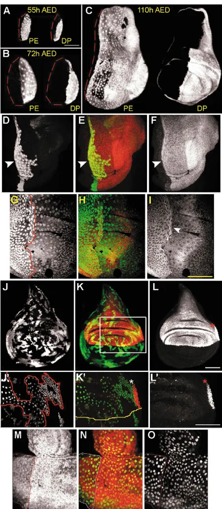
correlates with the movement of the morphogenetic furrow (MF) along the anteroposterior (AP) axis (Wolff and Ready, 1991). We define the PE and DP by location in the disc and morphological differences, using the same criteria and terminology introduced by Auerbach (Auerbach, 1936). In thoracic imaginal discs, cells attain one of three different morphologies by the end of larval development: columnar, cuboidal or squamous (Fig. 1A-D). The ‘lower’ epithelium, which we designate disc proper (DP), has both columnar and cuboidal cells (Fig. 1C). The ‘upper’ peripodial epithelium (PE) has both squamous and cuboidal cells (Fig. 1A). Cuboidal cells of both layers have been termed ‘medial edge’, ‘cubic’ and ‘margin’ cells, we use margin cells because of its original use (Baena-Lopez et al., 2003; Pallavi and Shashidhara, 2003; Zeitlinger and Bohmann, 1999) (Fig. 1B,C). We examined cell morphology in two optical planes: (1) the plane of the PE; and (2) a longitudinal xzsection through the disc center (Fig. 2A-M). Morphology was first examined in wing and eye discs at 48 hours AED and at 72 hours AED in leg discs.
[image:2.612.319.554.74.232.2]In 48 hours AED wing discs, all cells are cuboidal (Fig. 2A, insert) and remain so until 60 hours AED when most DP cells elongate along the apicobasal axis (Fig. 2B, insert). At these early stages, the PE and DP can be distinguished by a pseudostratification of cells in the DP. Later, at 72 hours AED, only cells in the center of the PE, above both the presumptive notum and wing blade, enlarge in diameter to a squamous shape (Fig. 2C). This same sequence of shape changes occur in leg discs, although 12 hours later (Fig. 2G). In both discs, cells at the ventral and lateral margins of the PE remain cuboidal (grey arrowheads in Fig. 2C,D,E wing disc; Fig. 2G,H, leg disc). Meanwhile, most cells in the DP become columnar (Fig. 2C, wing disc; Fig. 2G, leg disc) and folds appear (Fig. 2D,E, wing disc; Fig. 2G,H, leg disc). In wing discs, both the number and the diameter of squamous PE cells Fig. 1.Cell morphologies in wing discs. Pseudo-colorized confocal sections of a wing disc (110 hours AED) stained with ␣-spectrin illustrates cell morphologies in the peripodial epithelium (PE) and disc proper (DP). Disc oriented with anterior leftwards, dorsal upwards. Broken white lines mark the orientations of optical cross-section and longitudinal cross-section. Red, squamous peripodial epithelium (sPE); yellow, cuboidal cells of the margin in the peripodial epithelium (mPE) and disc proper (mDP); blue, columnar cells of the disc proper (cDP). (A) Confocal image of the PE; (B) longitudinal section; (C) confocal section in plane of the DP (5 m below PE); (D) cross-section. Scale bar: 50 m.
De
increase, such that by 96 hours AED, squamous cells cover the entire presumptive wing blade, most of the notum, and dorsal hinge of the DP. In leg discs, squamous PE cells cover the precursors of tarsal segments 2-5, and most of the tibia and femur (Fig. 2H).
In 48 hours AED eye discs, squamous PE cells are found on both sides of the Bolwig’s nerve (red asterisk), while cells of the DP are cuboidal (Fig. 2I). Longitudinal sections of 60 hours AED eye discs reveal the flattened shape of eye PE cells and the close apposition of the two disc layers (Fig. 2J). By this time, cells in the DP are columnar (Fig. 2J). Interestingly, eye discs lack margin cells (Fig. 2I-M), and eye PE cells become cuboidal later in larval development (compare Fig. 2K with 2M). Thus, morphogenesis of the different imaginal discs occurs with disc-specific timing.
AP compartments in wing and leg disc PE
Imaginal discs are subdivided into AP compartments, the expression of selector genes cubitus interruptus (ci) and engrailed (en), maintain A and P compartment identities, respectively (Blair, 2003). After mid-second instar, the dorsal selector gene apterous (ap) further subdivides the DP of wing and haltere discs into dorsal (D) and ventral (V) compartments (Garcia-Bellido et al., 1973).
We examined the expression of compartment-specific genes (en, ciand ap) and performed clonal analysis to define the compartmental organization in the PE. In wing discs at 55 and 72 hours AED, the boundary of en in the PE is slightly anterior to the boundary of enin the DP (Fig. 3A,B). Between
72-96 hours AED, this boundary shifts to the anterior side of the disc (Fig. 3C). ci-expressing margin cells in the PE abut the en-expression domain and are at the anterior side of the PE by 96 hours AED (Fig. 3I). Thus, all squamous PE cells are in the P compartment and overlie cells of both A and P compartments in the DP (Fig. 3C). As in wing discs, we observed that posterior PE cells in the leg disc undergo a similar anterior shift, but this occurs 12 hours later in development (84 hours AED) (data not shown). We speculate that PE cells located in the P compartment increase their surface area during the conversion to a squamous morphology. This may account for the anterior shift of the AP compartment boundary in the PE of both wing and leg discs.
Clonal restrictions define compartmental boundaries (Garcia-Bellido et al., 1973). To test whether clones in the PE, as in the DP, are restricted by compartments, we induced random GFP-marked clones during the first instar and analyzed wing and leg discs at 110 hours AED. In 115 wing discs examined, 22 large PE cell clones were found to straddle the en-expression domain in the PE (Fig. 3D-F). We found a similar clonal restriction within the en-expression domain in the leg PE (n=14 large clones in 68 discs). Furthermore, we observed this clonal restriction even when large PE cell clones were generated using the Minute system (Morata and Ripoll, 1975) (Fig. 3G-I, leg disc; data not shown).
[image:3.612.86.552.71.354.2]Beginning mid-second instar, ap-lacZ is expressed in the dorsal compartment of the wing DP (Blair, 1993; Diaz-Benjumea and Cohen, 1993). ap-lacZ is not expressed in the Fig. 2.Cell shape changes in the PE of different imaginal discs. Temporal sequence of epithelial morphogenesis in wing (A-E), leg T2 (F-H) and eye (I-M) discs at different larval stages. ␣-Spectrin outlines cell morphology (A-M). Each panel displays an image of the PE (left) and longitudinal section (right) taken through the center of the disc (indicated by arrows only in A,F,I). In longitudinal sections, the PE is oriented left of the disc lumen. White arrowheads indicate squamous PE cells, grey arrowheads mark margin cells. Bolwig’s nerve is indicated by red asterisks. Broken yellow lines mark the AP boundary, anterior is towards the left. MF, morphogenetic furrow.
De
PE, except for a small region of posterior margin cells (40+ cells) that border the wing blade primordia and notum of the DP (Fig. 3L,L⬘, red asterisks). Although ap-lacZ-expressing PE cells do not co-express Ubx (data not shown), we argue that these cells are part of the PE based on their apposition to the DP. We induced random GFP-expressing clones at mid-second instar in the background of ap-lacZ. These clones were found within either the ap-lacZ domains of the PE and DP or within the non-ap-lacZdomain of the PE (n=21 discs, Fig. 3J-L,J⬘ -L⬘). Therefore, the PE is nearly ventral in compartment identity except for a small dorsal compartment positioned at the far posterior side of the PE.
DV lineage restriction in the eye PE and the Bolwig’s nerve
In eye discs, clones induced after 48 hours AED define a restriction that divides the eye into dorsal and ventral compartments (Baker, 1978; Campos-Ortega et al., 1978). This disc is further subdivided into a large A and small P compartment during the third instar. Anterior cells differentiate into most of the head capsule, the eye and parts of the antenna, while posterior cells form the remainder of antenna and head (Morata and Lawrence, 1978).
En expression in the eye disc, unlike wing and leg discs, is ubiquitous throughout the DP with higher levels of expression in the posterior region of the antenna (Strutt and Mlodzik, 1996). In the PE, En is observed in a small cluster of cells over the dorsoanterior region of the eye DP, in about half of the discs analyzed (17/34 discs, data not shown). Given the inconsistent and possible transient expression of En in the eye PE, we question its control in posterior identity. However, En is consistently observed in the posterior region of the antennal PE, and random GFP-expressing clones induced at 72 hours AED in the background of lacZ, were restricted by the en-expression domain (n=20 clones, data not shown).
[image:4.612.56.281.222.738.2]To determine whether a DV compartment boundary is present in the eye PE, we first generated clones during the first larval instar with analysis at 120 hours AED. Interestingly, we observed preferential growth of clones in long, two- or three-cell wide strips that frequently aligned with the Bolwig’s nerve and extended over both the eye and antennal domains of the disc (data not shown). Using the Minute system, we found that large +/+ PE cell clones were restricted into D or V compartments. The boundaries of these clones co-localized with the Bolwig’s nerve (Fig. 3M-O). The Bolwig’s nerve is
Fig. 3.Compartment boundaries in the peripodial epithelium. (A-C) Expression of en-lacZin the wing PE and DP during larval development. (A) 55 hours; (B) 72 hours; (C) 110 hours AED. An anterior shift of en-expression in the PE relative to the en-expression in the DP coincides with the formation of squamous PE cells. Scale bar in A: 50 m (A-F,M-O). (D-F) The en-expression domain in the wing PE defines a clonal restriction. (D) A GFP-expressing PE cell clone straddles (arrowhead) but does not cross the en-expression domain (F) in the PE. (E) Overlay of D and F. (G-I) Large M+ PE cell clones respect the AP compartment boundary in the wing PE. (G) A +/+ PE cell clone, generated in a Minute mutant background, is distinguished by two copies of GFP (outlined in red) and grows along but does not cross the anterior Ci expression domain in the PE (arrowhead) in I. (H) Overlay of G and I. (J-L) PE cells in the wing disc belong to the V compartment. (J) Random GFP-expressing PE clones grow adjacent to without crossing the ap-lacZexpression domains (L) in the wing PE or DP. (L) ap-lacZexpression (red asterisk) is limited to 40+ margin cells in the P compartment of the PE. (K) Overlay of J and L. (J⬘-L⬘) Enlargements of the boxed region in K. (J⬘) A PE cell clone (outlined in red) that grows next to but does not cross the ap-lacZ(asterisk in L⬘) expression region in the PE (L⬘). (K⬘) Overlay of J⬘and L⬘. A yellow line marks the DV compartment boundary in the DP (lower layer), asterisk indicates ap-lacZin PE. .The PE cell clone in J-L⬘traverses the DV compartment in the DP. Scale bars: 50 m. (M-O) M+cell clone in the eye PE reveals that the Bolwig’s nerve defines a DV lineage restriction. (M) Heterozygous M/+cells are marked by Arm-lacZ. A +/+ PE cell clone is distinguished by the absence of Arm-lacZ (M) and two copies of GFP (O) and defines a straight line left of the Bolwig’s nerve (arrowheads and outlined in red). (N) Overlay of M and O.
De
the larval optic nerve (Schmucker et al., 1997), that inserts into the PE (F. Cesares and J. Bessa, unpublished), where it acts as a physical barrier separating PE cells into D and V compartments.
Hedgehog signaling is necessary for squamous morphogenesis in the PE of wing and leg discs
Growth and pattern formation in imaginal discs relies upon several secreted signaling molecules, such as hedgehog (hh), wingless (wg) and decapentapelagic (dpp). We therefore examined whether these signals also control the previously described morphological changes in the disc epithelia. For this, we used temperature-sensitive alleles of hh (hhts2) and wg (wgIL). Both hhts2/hhts2 and wgIL/wgIL larvae, grown at the restrictive temperature prior to and during cell shape changes in the disc epithelia, develop severely reduced wing and leg discs compared with discs from their sibling controls (wgIL/CyO y+) (hhts2/TM6B) (Fig. 4A-D; data not shown for leg discs). Despite their small size, wing and leg discs from wgIL/wgIL larvae display normal epithelial morphology with squamous and columnar cells in the PE and DP, respectively (Fig. 4B,B⬘; data not shown for leg disc). Thus, wgsignaling is dispensable for morphogenesis in the disc epithelia. By contrast, wing and leg discs from hhts2/hhts2 larvae fail to form squamous cells (Fig. 4D,D⬘; data not shown for leg disc). Interestingly, loss of hh function does not affect morphogenesis in the wing and leg DP, as columnar cells are still observed
(Fig. 4D⬘; data not shown for leg disc). Thus, Hh function is necessary for specific shape changes that occur in the PE.
As wing and leg discs from hhts2/hhts2 larvae are small in size, squamous cells might not form because of fewer divisions in the discs. To stimulate proliferation in hh-depleted larvae, we overexpressed cyclin-D-Cdk4 (Datar et al., 2000) in posterior cells, which includes all squamous PE cells, using the engrailed-Gal4 (en-Gal4) driver. Experimental larvae shifted to the hh-restrictive temperature develop larger discs than those from hhts2/hhts2 larvae (compare Fig. 4D with 4E) yet no squamous cells were observed (Fig. 4E⬘), indicating that Hh signaling is necessary for the formation of squamous PE cells in wing and leg discs.
Dpp is required for squamous morphogenesis in the PE of wing and leg discs
[image:5.612.196.561.389.693.2]Because shape changes in the PE of wing and leg discs requires hh signaling and that dpp expression is hhdependent, we tested whether dpp function can rescue squamous morphogenesis in hh-depleted animals. For this we use hhts2 larvae, while also overexpressing dpp(UAS dpp) in the posterior compartment using the en-Gal4 driver. We found that activation of dpp in posterior PE cells rescues the hhts2 phenotype as squamous cells are observed in both wing and leg discs (Fig. 4F,F⬘; data not shown for leg disc). This indicates that hh-dependent dpp activity is sufficient for squamous morphogenesis in wing and leg discs. To determine whether dpp signaling is necessary for
Fig. 4. hh and dpp activity are required for squamous morphogenesis in the wing PE. In each panel, confocal sections from top to bottom are of the PE (A-F) and disc cross-section (A⬘-F⬘). Red arrows in A-F indicate cross-section levels in A⬘-F⬘. Red lines indicate region of squamous cells in A⬘-C⬘,F⬘. AP compartment boundary (yellow lines) in the PE (A-F). (A,A⬘) Control disc from larvae heterozygous for wgIL (wgIL/CyO y+). (B,B⬘) Discs that lack wgactivity (wgIL/wgIL) are small yet form squamous PE cells and columnar DP cells (B⬘) (n=22 discs). (C,C⬘) Control disc from larvae heterozygous for hhts2
(hhts2/TM6B). (D,D⬘) In the absence of hh function (hhts2/hhts2), discs lack squamous PE cells, while DP cells become columnar (D⬘) (n=46 discs). (E,E⬘) Discs from hhts2larvae that overexpress cyclin-D-Cdk4are larger than discs from hhts2/hhts2larvae yet squamous cells do not form (E⬘) (n=26 discs). (F,F⬘) Discs from hhts2larvae that overexpress dpprestores squamous morphogenesis in the PE (F⬘) (n=34 discs). (G,G⬘,H,H⬘,I-K) Ubx>dad wing discs have aberrant squamous
morphogenesis. Panels from top to bottom are confocal sections in plane of
DP and disc cross-section. Red arrows in G,H indicate cross-section levels in G⬘,H⬘. Red lines indicate region of squamous cells in H⬘. (G⬘) A PE that consists entirely of cuboidal-shaped cells and (H⬘) a PE with limited squamous cells apposed to clefted folds in the DP. (I-K) Cross-sections of Ubx<dadwing discs, shows that the AP compartment boundary in the PE (red and yellow arrowheads) fails to shift in the absence of squamous morphogenesis (compare with Fig. 3C). (I) ␣-Spectrin staining. (K) En expression. (J) Overlap of I,K. Scale bar: 50 m.
De
squamous morphogenesis, we inhibited PE-specific dpp signal transduction by overexpression of UAS-dad (Tsuneizumi et al., 1997) using the wing disc PE-specific driver Ubx-Gal4. Ubx>dad wing discs are slightly smaller than control discs. Additionally, these discs display aberrant folding patterns in the DP, and, more importantly, a PE made of only cuboidal-shaped cells, often stacked into two or more rows giving the PE a stratified appearance (Fig. 4G⬘,H⬘,I). Four out of 20 wing discs from Ubx>dad larvae displayed a small region of squamous PE cells consistently apposed to clefted folds in the DP (Fig. 4H,H⬘). Interestingly, Ubx>dad wing discs that lack squamous PE cells display only a minor shift of En-expressing PE cells towards the anterior side of the disc (Fig. 4I-K). Therefore, we conclude that hh-dependent dpp signal
transduction is necessary and sufficient for squamous morphogenesis of the PE and also the anterior shift of the AP compartment boundary in wing and leg discs.
To determine whether Dpp signaling is activated in the wing disc as PE cells undergo squamous morphogenesis, we used the antibody that recognizes the phosphorylated form of Mothers against decapentapelagic (p-MAD) (Tanimoto et al., 2000). Prior to squamous morphogenesis, p-MAD is confined to a ventral wedge in both the AP compartments of the PE (Fig. 5A,A⬘). As PE cells become squamous (72 hours AED), p-MAD is still ventral; however, a stripe of expression appears along the length of the PE (Fig. 5B,B⬘). The location of the p-MAD stripe is coincident with the AP compartment boundary in the PE (compare Fig. 5B with Fig. 3B). As more cells acquire a squamous morphology during the third instar, p-MAD expression broadens and is present in the majority of squamous cells in the P compartment of the PE (Fig. 5C,C⬘ ,D-D⬘⬘). Therefore, we conclude that the Dpp signaling pathway is activated during squamous morphogenesis of PE cells in the wing disc.
Interestingly, the PE and DP in the wing disc show different distributions of Dpp activation. In the P compartment of the DP, highest p-MAD expression is reported in cells adjacent to the AP compartment boundary, whereas in the A compartment there is broad activation of Dpp signaling (Tanimoto et al., 2000). By contrast, we observed p-MAD expression limited to squamous PE cells within the P compartment (Fig. 5C,C⬘). Hh expression (hh-lacZ), however, is identical in the two cell layers, with expression observed in all P compartment cells (Fig. 5E).
Differences in cell number between the PE and DP
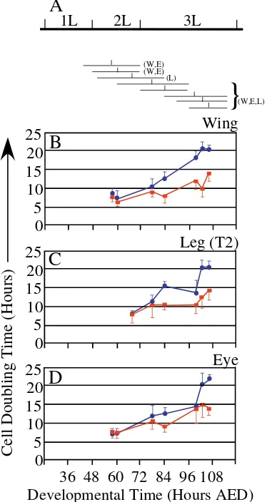
In different imaginal discs, the number of cells that compose the PE and DP differs dramatically by the end of the third larval instar; our analyses, however, indicate that this is not always the case during larval development. We counted the number of cells within the wing PE and DP at 72 hours AED and 110 hours AED. At 72 hours AED, as squamous cells first appear, we counted 291±34 cells in the PE and 996±71 cells in the DP (n=5 discs). These counts are consistent with Steiner’s (Steiner, 1978) from dissociated early third instar wing discs (1024±32 cells). At 110 hours AED, we found 2099±236 cells in the PE and estimated 39,200±1170 cells in the DP (n=10 discs). Thus, DP cells outnumber PE cells by 3:1 at 72 hours AED and 20:1 by the end of larval development. Several hypotheses were tested to account for the increasing discrepancy in cell number between the disc epithelia, including cell death (Acridine Orange) and endomitosis; however, both were excluded because during the third instar less apoptosis was observed in the PE than DP and PE cells remain diploid based on our DNA measurements (data not shown). We induced random GFP-expressing cell clones (act5C>Gal4,UAS GFP) at specific times in larval development (Fig. 6A), to examine whether different rates of cell division contribute to the discrepancy in cell numbers between the disc epithelia. To calculate cell doubling times of both squamous PE and columnar DP cells, we determined the median number of cells per clone after a defined period of growth (Neufeld et al., 1998).
[image:6.612.64.272.73.414.2]Consistent with previous reports (Garcia-Bellido and Merriam, 1971; Madhaven and Schneiderman, 1977), we found that DP cells in the wing disc double every 6.3-8.0 hours Fig. 5. Dpp signaling pathway is activated during squamous
morphogenesis in the wing disc. Color images show p-MAD (red) and ␣-spectrin (green). Black and white images (A⬘-D⬘) show p-MAD and (D⬘⬘) ␣-spectrin alone. Wing discs are from larvae at 67 hours AED (A,A⬘), 72 hours AED (B,B⬘) and 96 hours AED (C,C⬘). (A,A⬘) Prior to squamous morphogenesis, p-MAD is present in a ventral wedge in the PE. (B,B⬘) Early in squamous morphogenesis, a stripe of p-MAD expression appears in the PE (yellow arrowhead). (C,C⬘) During the third instar, p-MAD expression broadens and is observed in the majority of squamous cells of the posterior compartment in the PE. (D-D⬘⬘) Enlargement of region boxed in C. (D-D⬘⬘) p-MAD is observed only in squamous cells of the posterior compartment and is absent from margin cells (C,D⬘) in both A and P compartments of the PE. (E) hh-lacZ expression in the wing PE from a wandering third instar larva. Scale bar in A: 50 m.
De
during the second instar and 10-14 hours during the third instar (Fig. 6B). Cell division rates between the disc epithelia differ only when clones were induced after shape changes in the PE (72-90 hours AED) (Fig. 6B). At this time, squamous PE cells divide much slower, every 17-20 hours (Fig. 6B). Similar observations were made in leg discs (Fig. 6C). Thus, in wing and leg discs, the appearance of squamous cells correlates with slower cell division.
For the eye-antennal disc, we compared doubling times of DP cells positioned anterior to the morphogenetic furrow with PE cells positioned over the presumptive eye domain. As in
wing discs, eye PE and DP cells exhibit similar doubling times during the second larval instar (Fig. 6D). Unlike wing and leg discs, the doubling times of eye PE cells increase much later in the third instar (Fig. 6D).
These results indicate that proliferation in all three discs is coordinated between the two disc epithelia until 72 hours AED. At this time, at least for the wing disc, there is a 3:1 ratio between DP and PE cells. From our clonal analysis, we conclude that during the third instar, wing DP cells have one, but not more than two, additional cell divisions compared with the squamous PE cells. Margin cells in both disc epithelia have identical doubling times and therefore do not contribute to the ratio changes of the DP and PE. Therefore, the DP to PE cell ratio should differ by 12:1 at the end of larval development; however we observed a 20:1 ratio. Thus, different rates of cell division between the PE and DP cannot, on their own, account for the increasing number of cells in the DP.
Displacement of PE cells into the DP and changes of Ubx expression
A significant number of act5C>Gal4,UAS GFP clones generated in wing discs at 48 and 60 hours AED and analyzed at 110 hours AED, contained cells in both epithelial layers [35.4% (n=143 clones) and 27% (n=155 clones)]. Similar results were observed in the leg disc [31.1% (n=103 clones) and 23.9% (n=142 clones)]. The area of these clones largely resided in the PE with a smaller area in the margin of the DP (data not shown). Clones that have cells in both disc epithelia were not fusions of two independent clones because they did not contain twice as many cells as clones limited to a single epithelium (data not shown). Thus, we propose that these clones reflect a displacement of cells from one epithelial layer into the other. If cells from the PE move to DP, this would explain the increasing number of cells in the DP during the third instar.
To test directly whether cells born in the PE end up in the DP, we specifically induced clones in the PE using two independent methods: (1) Ubx-Gal4 activation by a Flp recombinase regulated by estrogen (Weigmann and Cohen, 1999); (2) temperature-sensitive regulation of Ubx-Gal4 using the tubulin-Gal80ts fusion protein (McGuire et al., 2003). In the embryo and first instar larvae, Ubx-Gal4 expression is found in all cells of the wing disc (Pallavi and Shashidhara, 2003), but becomes limited to the PE by the second instar (Baena-Lopez et al., 2003; Pallavi and Shashidhara, 2003). To induce clones only in the wing PE, we activated Flpase by feeding larvae estrogen during the second (48-72 hours AED) or third instar (72-96 hours AED). We expected that the induction of PE-specific clones would be limited to the PE, and indeed clones of squamous PE cells (sPE) and clones of margin cells from the PE (mPE) were recovered (Fig. 7C, 48-72 hours AED). However, we additionally observed clones in the margin cells of the DP (mDP) and clones encompassing both disc epithelia, containing squamous PE cells and margin cells of both disc layers (sPE+mPE+mDP) (Fig. 7A-C). To rule out the possibility that perduring Ubx-Gal4 activity labels margin DP cells (mDP), we performed a lineage analysis of PE cells by temperature-sensitive regulation of Ubx-Gal4 activity using the tubulin-Gal80ts fusion protein (McGuire et al., 2003). Again, a significant number of clones containing squamous PE cells with margin cells from both disc epithelia (sPE+mPE+mDP)
0
5
10
15
20
25
0
5
10
15
20
25
0
5
10
15
20
25
36 48 60 72 84 96 108 Developmental Time (Hours AED)
Cell Doubling Time (Hours)
Wing
Leg (T2)
Eye
B
C
D
1L 2L 3L
A
(W,E) (W,E)
(L)
(W,E,L)
[image:7.612.78.265.210.562.2]}
Fig. 6.Estimated doubling times of squamous PE and columnar DP cells in wing, eye and leg (T2) imaginal discs during larval
development. (A) Cell clones were induced (act5C<Gal4, UAS GFP) by heat-shocking larvae for 10-20 minutes at 37°C at the following times: first (42 hours AED), second (48, 52, 60 hours AED) and third instar (72, 84, 90, 96 hours AED). Below the timeline are eight horizontal lines, the left end of the horizontal lines indicate the time of clone induction the right end indicate time of fixation for clone analysis of each experimental set. W (wing), E (eye) and L (leg) indicate the discs analyzed for each clone induction. A vertical mark in each horizontal line represents the mid-point between clone induction and fixation (=average clone age). Estimated cell doubling times are plotted at this mid-point. At each time interval more than 100 clones were scored in over 50 discs. Data represent median cell doubling times±s.d. Cell doubling times of squamous PE cells (blue circles) and columnar DP cells (red squares) in the wing (B), leg (C) and eye (D) imaginal discs.
De
were observed (data not shown). Both methods indicate that margin cells of the DP share a lineage relationship with cells in the PE. To address whether there is a continuous displacement of cells from the PE into the DP, we fed larvae estrogen during the third instar (72-96 hours AED). Even at this later induction time, clones spanning both disc epithelia (sPE+mPE+mDP) and margin cell clones in the DP (mDP) were observed, yet less frequently (Fig. 7C). This supports the idea that cells born in the PE divide and produce progeny that are displaced into the DP. We propose that this cell movement further increases the number of DP cells late in larval life. Furthermore, while tracing the lineage of PE cells we were able to study the gene expression of these cells as they are displaced into the DP. In clones encompassing both disc epithelia, we find Ubx expression in all squamous cells, rarely in margin cells of the PE and never in cells of the DP (see Fig. S1 in the supplementary material), indicating that Ubx expression is turned off when PE cells migrate into the DP.
To examine alternatively whether DP cells are displaced into the PE, we generated genetically marked cell clones using the FRT/FLP-mediated recombination in larvae heterozygous for Ubiquitous-GFP (UbiGFP/+). Using this method, known as twinspot analysis, we can identify the daughter cells of a single recombinant event, one homozygous wild type (unmarked) and its sister twin (homozygous for UbiGFP). Twinspot analysis
allows us to define the epithelial origin (PE or DP) of the sister clones and also the movement of either clone into the apposing epithelial layer. Clones were induced at 38 and 48 hours AED with analysis at 110 hours AED. At both clonal induction times, we found that clones from the DP did not encompass cells in the margin or squamous PE (n=23 discs at 38 hours AED, n=15 discs at 48 hours AED), indicating that DP cells reside in the same epithelium throughout larval development. However, clones originating in the PE often contained margin cells from both the PE and DP (n=10/23 clones at 38 hours AED, 4/16 at 48 hours AED). Therefore, we conclude that only PE cells move into and reside in the DP.
Discussion
Disc morphogenesis
Little is known about when and how disc cells acquire their diverse morphologies. Although Hh and DPP are well-known for their roles in cell proliferation and patterning, previous studies, in addition to our study here, indicate that they are also active in epithelial morphogenesis. In eye discs, Hh is both necessary and sufficient to initiate cell shape changes that occur in the morphogenetic furrow (Heberlein et al., 1995; Heberlein et al., 1993; Ma, 1993). In wing discs, columnar cells require DPP signaling for normal cytoskeletal organization, shape and pseudostratified organization (Gibson and Perrimon, 2005; Shen and Dahmann, 2005). Our study describes precisely and for the first time when cuboidal, columnar and squamous cell morphologies arise in the epithelia of different imaginal discs. We examine how the genesis of different morphologies in imaginal discs are affected by loss of a non-autonomous signal (wg,hhand dpp). Our study indicates that Hh-dependent DPP signaling is required for squamous morphogenesis in the PE of wing and leg discs. Additionally, we observe that DPP signaling is activated as PE cells transition to a squamous morphology. In agreement with Gibson and Perrimon (Gibson and Perrimon, 2005), our results indicate that the establishment of columnar morphology in the DP of wing and leg discs is independent of DPP signaling activity. Clearly, one question still remains: what is the mechanism which causes DP cells to become columnar? The information from these three studies provides, at least, an initial framework of how epithelial morphogenesis occurs in imaginal discs.
[image:8.612.46.322.73.266.2]As both hh and dpp are expressed in wing and leg discs prior to the onset of squamous morphogenesis in the PE, it is clear that their ability to instruct these shape changes must be regulated by additional temporal signals. An obvious candidate for such a temporal signal is ecdysone, which initiates the onset of the larval molts and adult differentiation (Riddiford, 1993). The ecdysone signal is mediated by a heterodimer complex consisting of the ecdysone receptor (EcR) and RXR-homolog Ultraspriacle (Usp) (Koelle et al., 1991; Thomas et al., 1993; Yao et al., 1993). To test whether squamous morphogenesis is triggered by ecdysone signaling, we induced usp–/– clones and found that cells of such clones still exhibited normal cuboidal-to-squamous shape changes (data not shown). Therefore, the Fig. 7. Clonal relationship between the wing disc epithelial layers.
PE-specific cell clones induced during the second larval instar (48-72 hours AED) using UAS-flp-EBD, Ubx-Gal4 and act5C>stop>nuclacZ. Analysis of clone distribution and frequency was performed in 110 hours AED larvae. (A) Clones with 1-3 cells are present and also a clone with 30+ cells limited to the sPE (yellow asterisks). Cuboidal cell clones are also present at the disc margins in either the PE or DP (arrows), as is a clone that includes squamous and margin cells of both epithelial layers (yellow arrowhead). Scale bar: 50m. (B-B⬘⬘) A z-series of confocal images of the boxed region in A. Teashirt labels squamous PE cells and margin cells in both PE and DP, -gal labels clones. Images in the PE (B,B⬘) and the DP (B⬘⬘) showing a clone that consists of both squamous PE cells and margin cells present in both the disc epithelia. (C) Distribution and frequency of clones born in the wing disc PE during the second (48-72 hours AED) and third (72-96 hours AED) larval instars. sPE, clones of squamous cells in the PE; mPE, clones of margin cells in the PE; mDP, clones of margin cells in the DP; sPE+mPE+mDP clones of squamous PE cells and margin cells in both the PE and DP.
De
temporal cue(s) that initiate disc morphogenesis is independent of ecdysone signaling and remains unknown.
Previous studies document that shape change of epithelial cells can activate certain signaling pathways (Wang et al., 1993; Zhu and Assoian, 1995). Thus, squamous morphogenesis of the PE may enhance planar and/or vertical epithelial signaling to promote growth and patterning of the disc. We made two observations that support this statement. First, where PE cells fail to undergo squamous morphogenesis, both the disc and adult wing show an obvious reduction in size (Gibson et al., 2002). Second, in discs that lack PE-specific DPP signaling, folded clefts in the presumptive wing blade primordia are consistently apposed to a region of squamous PE cells, suggesting communication between the disc epithelia where shape changes do occur. Alternatively, the aberrant apposition of AP compartment boundaries in the PE and DP, owing to a failure in squamous morphogenesis, may result in epithelial abnormalities such as folded clefts in the DP. Resolving the mechanisms by which cell shape can affect disc growth and pattern will integrate both morphogenetic and signaling processes that are crucial for disc development.
Cells in the DP are included in the peripodial lineage
Pallavi and Shashidhara (Pallavi and Shashidhara, 2003) performed a lineage analysis of cells in the wing disc using Ubx-Gal4, UAS flp and act5C>stop>nuclacZ. As Ubx-Gal4 is initially expressed in both disc epithelia prior to the second larval instar (Baena-Lopez et al., 2003; Pallavi and Shashidhara, 2003), cells of both the PE and DP were marked. Their analysis concluded that cells of the PE and DP share a common origin in the disc primordium but later become separate lineages, although cells that make up the PE and DP lineages were never specified. Our results, based on lineage-tracing cells born in the PE, are in overall agreement with their conclusions; however, there are some differences. Although Pallavi and Shashidhara (Pallavi and Shashidhara, 2003) identified cell clones spanning both disc epithelia, they could not determine when or where these clones were born. Furthermore, clones that encompassed cells from both PE and DP were interpreted as either fusions between two independent clones or as clones that originated early in the embryo before separation of the two lineages (PE and DP).
Using four different methods, we found that cells that originate within the PE produce progeny that are a part of the DP. We used both the MARCM and estrogen-inducible systems to perform a clonal analysis specific to cells within the PE. These two methods indicate that cells born within the PE produce daughter cells that contribute to the DP. Additionally, a twinspot clonal analysis leads to a similar conclusion and has the advantage of marking cells more directly than either the MARCM- or estrogen-inducible systems. Thus, our analysis indicates a lineage relationship between margin cells in both the PE and DP, and squamous cells in the wing disc, and provides evidence that together these cells comprise the peripodial lineage (Fig. 7).
As cells are displaced from the PE and into the DP they lose Ubx expression. The loss of Ubx may cause cells to acquire a more distal fate, forging a possible link between displacement and cell fate changes. Similar dynamic cell movements, along with changes in gene expression, were observed in the chick
during somite segmentation (Kulesa and Fraser, 2002). In addition, cell movements and changes in gene expression, similar to what we describe here, were reported by Weigmann and Cohen (Weigmann and Cohen, 1999), who observed that leg disc cells born in the most proximal regions of the disc contribute to more distal leg segments. Finally, we propose that once PE cells are displaced into the DP they may change their cell fate by altered cell signaling. Displaced cuboidal cells at the margins of the disc receive not only planar signals from both epithelial layers, which they are a part of at different stages in larval development, but also vertical signals from overlying PE cells after displacement into the DP. These new planar and/or vertical signals may lead to Ubx repression.We suggest that the mechanisms that play a role in the development of the imaginal discs may be functionally similar to mechanisms that regulate primary neurogenesis in vertebrates. Neural plate formation and patterning cues arise from two sources: a horizontal source within the plane of an epithelium and a vertical source that arises from the underlying mesodermal cells (Weinstein and Hemmati-Brivanlou, 1999; Wilson and Edlund, 2001). Our study suggests that patterning of the imaginal discs is a much more dynamic process with cells exposed to not only signals within the plane of an epithelium but also vertical signals between disc epithelia.
We thank Stephen Cohen and L. S. Shashidhara for fly stocks and/or antibodies. We also thank Fernando Casares for interesting insights. We are grateful to Margrit Schubiger, Barbara Wakimoto, Savraj Grewal, Merrill Hille, Lynn Riddiford for critical reading and comments. This work was supported by the National Institutes of Health grant (GM58282) to G.S., and K.M. was supported by grant number 5T32 HD07183 from the NIH.
Supplementary material
Supplementary material for this article is available at http://dev.biologists.org/cgi/content/full/132/22/5033/DC1
References
Auerbach, C.(1936). The development of the legs, wings and halteres in wild type and some mutant strains of Drosophila melanogaster. Proc. R. Soc. Edinb. B58,787-815.
Baena-Lopez, L. A., Pastor-Pareja, J. C. and Resino, J.(2003). Wg and
Egfr signalling antagonise the development of the peripodial epithelium in Drosophila wing discs. Development130, 6497-6506.
Baker, W. K. (1978). A clonal analysis reveals early developmental
restrictions in the Drosophila head. Dev. Biol.62, 447-463.
Blair, S. S.(1993). Mechanisms of compartment formation: evidence that non-proliferating cells do not play a critical role in defining the D/V lineage restriction in the developing wing of Drosophila. Development119, 339-351.
Blair, S. S.(2003). Lineage compartments in Drosophila. Curr. Biol.13, R548-R551.
Campos-Ortega, J. A., Jurgens, G. and Hofbauer, A. (1978). Clonal
segregation and positional information in late ommatidial development in Drosophila. Nature274, 584-586.
Cho, K. O., Chern, J., Izaddoost, S. and Choi, K. W.(2000). Novel signaling from the peripodial membrane is essential for eye disc patterning in Drosophila. Cell103, 331-342.
Cohen, S. M.(1993). Imaginal disc development. In The Development of
Drosophila melanogaster, vol. 2 (ed. M. Bates and A. Matrinez-Arias). New York: Cold Spring Harbor Press.
Datar, S. A., Jacobs, H. W., de la Cruz, A. F., Lehner, C. F. and Edgar, B. A. (2000). The Drosophila cyclin D-Cdk4 complex promotes cellular growth. EMBO J.19, 4543-4554.
Diaz-Benjumea, F. J. and Cohen, S. M.(1993). Interaction between dorsal
De
and ventral cells in the imaginal disc directs wing development in Drosophila. Cell75, 741-752.
Garcia-Bellido, A. and Merriam, J. R. (1971). Parameters of the wing
imaginal disc development of Drosophila melanogaster. Dev. Biol.24, 61-87.
Garcia-Bellido, A., Ripoll, P. and Morata, G. (1973). Developmental
compartmentalisation of the wing disk of Drosophila. Nat. New Biol.245, 251-253.
Gibson, M. C. and Perrimon, N.(2005). Extrusion and death of
DPP/BMP-compromised epithelial cells in the developing Drosophila wing. Science
307, 1785-1789.
Gibson, M. C. and Schubiger, G. (2000). Peripodial cells regulate
proliferation and patterning of Drosophila imaginal discs. Cell103, 343-350.
Gibson, M. C., Lehman, D. A. and Schubiger, G. (2002). Lumenal
transmission of decapentaplegic in Drosophila imaginal discs. Dev. Cell3, 451-460.
Hama, C., Ali, Z. and Kornberg, T. B.(1990). Region-specific recombination and expression are directed by portions of the Drosophila engrailed promoter. Genes Dev.4, 1079-1093.
Hart, K., Klein, T. and Wilcox, M.(1993). A Minute encoding a ribosomal
protein enhances wing morphogenesis mutants. Mech. Dev.43, 101-110.
Heberlein, U., Wolff, T. and Rubin, G. M.(1993). The TGF beta homolog
dpp and the segment polarity gene hedgehog are required for propagation of a morphogenetic wave in the Drosophila retina. Cell75, 913-926. Heberlein, U., Singh, C. M., Luk, A. Y. and Donohoe, T. J.(1995). Growth
and differentiation in the Drosophila eye coordinated by hedgehog. Nature
373, 709-711.
Johnston, L. A. and Schubiger, G.(1996). Ectopic expression of wingless in imaginal discs interferes with decapentaplegic expression and alters cell determination. Development122, 3519-3529.
Koelle, M. R., Talbot, W. S., Segraves, W. A., Bender, M. T., Cherbas, P.
and Hogness, D. S.(1991). The Drosophila EcR gene encodes an ecdysone
receptor, a new member of the steroid receptor superfamily. Cell67, 59-77.
Kulesa, P. M. and Fraser, S. E. (2002). Cell dynamics during somite
boundary formation revealed by time-lapse analysis. Science298, 991-995. Ma, C., Zhou, Y., Beachy, P. A. and Moses, K.(1993). The segment polarity gene hedgehogis required for progression of the morphogenetic furrow in the developing Drosophila eye. Cell75, 927-938.
Madhaven, M. and Schneiderman, H.(1977). Histological analysis of the
dynamics of growth of imaginal discs and histoblast nests during the larval development of Drosophila melanogaster. Wilhelm Roux’s Arch. Dev. Biol.
183, 269-305.
Maves, L. and Schubiger, G. (1998). A molecular basis for
transdetermination in Drosophila imaginal discs: interactions between wingless and decapentaplegic signaling. Development125, 115-124. McGuire, S. E., Le, P. T., Osborn, A. J., Matsumoto, K. and Davis, R. L.
(2003). Spatiotemporal rescue of memory dysfunction in Drosophila.
Science302, 1765-1768.
Milner, M., Bleasby, A. and Kelly, S.(1984). The role of the peripodial
membrane of leg and wing imaginal discs of Drosophila melanogaster during evagination and differentiation in vitro. Wilhelm Roux’s Arch. Dev. Biol.193, 180-186.
Morata, G. and Ripoll, P. (1975). Minutes: mutants of drosophila
autonomously affecting cell division rate. Dev. Biol.42, 211-221. Morata, G. and Lawrence, P. A.(1978). Anterior and posterior compartments
in the head of Drosophila. Nature274, 473-474.
Neufeld, T. P., de la Cruz, A. F., Johnston, L. A. and Edgar, B. A.(1998). Coordination of growth and cell division in the Drosophila wing. Cell93, 1183-1193.
Pallavi, S. K. and Shashidhara, L. S.(2003). Egfr/Ras pathway mediates
interactions between peripodial and disc proper cells in Drosophila wing discs. Development130, 4931-4941.
Riddiford, L. M.(1993). Hormone receptors and the regulation of insect
metamorphosis. Receptor3, 203-209.
Schmucker, D., Jackle, H. and Gaul, U.(1997). Genetic analysis of the larval optic nerve projection in Drosophila. Development124, 937-948. Shen, J. and Dahmann, C.(2005). Extrusion of cells with inappropriate Dpp
signaling from Drosophila wing disc epithelia. Science307, 1789-1790.
Steiner, E.(1978). Establishment of compartments in the developing leg
imaginal discs of Drosophila melanogaster. Wilhelm Roux’s Arch.180, 9-30.
Strutt, D. I. and Mlodzik, M.(1996). The regulation of hedgehog and
decapentaplegic during Drosophila eye imaginal disc development. Mech. Dev.58, 39-50.
Tanimoto, H., Itoh, S., ten Dijke, P. and Tabata, T.(2000). Hedgehog creates
a gradient of DPP activity in Drosophila wing imaginal discs. Mol. Cell 5, 59-71.
Thomas, H. E., Stunnenberg, H. G. and Stewart, A. F. (1993).
Heterodimerization of the Drosophila ecdysone receptor with retinoid X receptor and ultraspiracle. Nature362, 471-475.
Tsuneizumi, K., Nakayama, T., Kamoshida, Y., Kornberg, T. B., Christian,
J. L. and Tabata, T. (1997). Daughters against dpp modulates dpp
organizing activity in Drosophila wing development. Nature389, 627-631. van den Heuvel, M. H.-S. C., Klingensmith, J., Perrimon, N. and Nusse, R.(1993). Mutations in the segment polarity genes wingless and porcupine impair secretion of the wingless protein. EMBO J.15, 5293-5302. Wang, N., Butler, J. P. and Ingber, D. E.(1993). Mechanotransduction across
the cell surface and through the cytoskeleton. Science260, 1124-1127.
Weigmann, K. and Cohen, S. M. (1999). Lineage-tracing cells born in
different domains along the PD axis of the developing Drosophila leg.
Development126, 3823-3830.
Weinstein, D. C. and Hemmati-Brivanlou, A. (1999). Neural induction.
Annu. Rev. Cell Dev. Biol.15, 411-433.
Wilson, S. I. and Edlund, T.(2001). Neural induction: toward a unifying
mechanism. Nat. Neurosci.4, 1161-1168.
Wolff, T. and Ready, D. F.(1991). Cell death in normal and rough eye mutants of Drosophila. Development113, 825-839.
Xu, T. and Rubin, G. M.(1993). Analysis of genetic mosaics in developing and adult Drosophila tissues. Development117, 1223-1237.
Yao, T. P., Forman, B. M., Jiang, Z., Cherbas, L., Chen, J. D., McKeown,
M., Cherbas, P. and Evans, R. M.(1993). Functional ecdysone receptor
is the product of EcR and Ultraspiracle genes. Nature366, 476-479.
Zeitlinger, J. and Bohmann, D. (1999). Thorax closure in Drosophila:
involvement of Fos and the JNK pathway. Development126, 3947-3956.
Zhu, X. and Assoian, R. K.(1995). Integrin-dependent activation of MAP
kinase: a link to shape-dependent cell proliferation. Mol. Biol. Cell 6, 273-282.