D
E
V
E
LO
P
M
E
N
T
INTRODUCTION
The formation of many organs involves distinct organizing centers acting in concert to pattern the tissue in three dimensions. In the central nervous system (CNS), local organizers pattern different regions along the anteroposterior (AP) and dorsoventral (DV) axes by defining a series of molecular identities that direct differentiation of diverse neuronal cell types (Rubenstein et al., 1994). The mesencephalon (mes) and rhombomere 1 (r1) of the anterior hindbrain are a powerful model system for studying these events, as the key molecules that direct AP and DV patterning are known, and each region forms distinct structures. Dorsally, the mes gives rise to the tectum consisting of the laminated superior and inferior colliculi, while r1 becomes the foliated and layered cerebellum. Ventrally, the mes and r1 are organized into distinct clusters of neurons. The isthmic organizer, positioned at the mes/r1 boundary, directs AP patterning of the mes and r1 via the secreted factor Fgf8 (Wurst and Bally-Cuif, 2001; Zervas et al., 2005). DV patterning of the neural tube involves morphogens that are secreted from dorsal and ventral sources, with sonic hedgehog (Shh) expressed ventrally in the floor plate and underlying notochord (Ingham and McMahon, 2001). Although the molecular events underlying AP patterning in the mes/r1 have been well defined (Wurst and Bally-Cuif, 2001; Zervas et al., 2005), the extent to which Shh regulates DV patterning in the mes/r1 and the involvement of Shh-downstream effectors is poorly understood.
The few studies to date addressing Shh signaling in the mes/r1 have uncovered a role for Shh in inducing ventral cell types, in addition to a general role in proliferation and cell survival in the mes (Zervas et al., 2005). Miss-expression studies in the chick dorsal mes have demonstrated that Shh can induce in a concentration-dependent manner a series of transcription factors normally found in a nested pattern in the ventral mes (Agarwala et al., 2001). Similarly, in vitro explant studies and ectopic expression of Shh in vivo have shown that Shh can induce ventral mes/r1 cell types, including dopaminergic, serotonergic and motor neurons (Agarwala and Ragsdale, 2002; Fedtsova and Turner, 2001; Ye et al., 1998). Both dopaminergic and serotonergic neurons also require Fgfs for their induction (Ye et al., 1998). Analysis of Shh-null mutant mice has been less informative as Shh is required for survival and proliferation of mes precursors within 1 day (E9.0) of initiation of Shhexpression (Agarwala et al., 2001; Chiang et al., 1996; Echelard et al., 1993; Fedtsova and Turner, 2001; Ishibashi and McMahon, 2002). In addition, the remaining mes is completely dorsalized (Fedtsova and Turner, 2001), precluding analysis of potential requirements for Shh signaling at subsequent stages of mes development. Although the only established role for Shh in early patterning of r1 is the induction of ventral serotonergic neurons (Ye et al., 1998), conditional gene inactivation has demonstrated that Shh signaling plays a later role (after E16.5) in regulating granule cell precursor proliferation in the cerebellum (Corrales et al., 2004; Lewis et al., 2004; Corrales et al., 2006). Given the severity of the early mes defects in Shh-null mutants and the largely unresolved role of Shh signaling in early r1 patterning, the precise in vivo requirement for Shh signaling in embryonic mes/r1 development remains to be determined. In particular, it has not been addressed whether Shh signaling plays a role in mes/r1 development after E9.0, and whether and how Shh might regulate patterning of dorsal mes/r1 structures.
Sonic hedgehog regulates Gli activator and repressor
functions with spatial and temporal precision in the
mid/hindbrain region
Sandra Blaess1,2, JoMichelle D. Corrales1,2and Alexandra L. Joyner1,2,3,*
The midbrain and anterior hindbrain offer an ideal system in which to study the coordination of tissue growth and patterning in three dimensions. Two organizers that control anteroposterior (AP) and dorsoventral (DV) development are known, and the regulation of AP patterning by Fgf8 has been studied in detail. Much less is known about the mechanisms that control
mid/hindbrain development along the DV axis. Using a conditional mutagenesis approach, we have determined how the ventrally expressed morphogen sonic hedgehog (Shh) directs mid/hindbrain development over time and space through positive regulation of the Gli activators (GliA) and inhibition of the Gli3 repressor (Gli3R). We have discovered that Gli2A-mediated Shh signaling
sequentially induces ventral neurons along the medial to lateral axis, and only before midgestation. Unlike in the spinal cord, Shh signaling plays a major role in patterning of dorsal structures (tectum and cerebellum). This function of Shh signaling involves inhibition of Gli3R and continues after midgestation. Gli3R levels also regulate overall growth of the mid/hindbrain region, and this largely involves the suppression of cell death. Furthermore, inhibition of Gli3R by Shh signaling is required to sustain expression of the AP organizer gene Fgf8. Thus, the precise spatial and temporal regulation of Gli2A and Gli3R by Shh is instrumental in coordinating mid/hindbrain development in three dimensions.
KEY WORDS: Mesencephalon/rhombomere1, Sonic hedgehog, Dorsal patterning, Gli3 repressor Development 133, 1799-1809 (2006) doi:10.1242/dev.02339
1Howard Hughes Medical Institute and Developmental Genetics Program, Skirball Institute of Biomolecular Medicine, 540 First Avenue, New York, NY 10016, USA. 2Department of Cell Biology, New York University School of Medicine, 540 First Avenue, New York, NY 10016, USA. 3Department of Physiology and Neuroscience, New York University School of Medicine, 540 First Avenue, New York, NY 10016, USA.
*Author for correspondence (e-mail: [email protected])
D
E
V
E
LO
P
M
E
N
T
The downstream components of Shh signaling have been dissected in detail in the spinal cord and forebrain (Jacob and Briscoe, 2003; Ingham and McMahon, 2001). Shh signaling is transduced through the transmembrane receptors patched (Ptch1) and smoothened (Smo). The inhibition of Smo by Ptch1 is relieved by Shh, thus allowing for transcription of downstream target genes via the Gli zinc-finger transcription factors. In mouse, the three Gli proteins have distinct biochemical functions and in vivo requirements. Whereas Gli1is largely dispensable for normal murine development, Gli2and Gli3mutants die at birth (Bai et al., 2002; Johnson, 1967; Mo et al., 1997; Park et al., 2000). Mouse Gli3 protein is primarily cleaved into an N-terminal repressor form (Gli3R), but Shh signaling counteracts this processing and high levels of Shh signaling can induce a weak Gli3 activator (Bai et al., 2004; Wang et al., 2000). By contrast, Gli2 is efficiently converted into a transcriptional activator (Gli2A) by strong Shh signaling and Gli1 is a constitutive activator. Notably, Gli1 is a transcriptional target of Shh signaling (Bai et al., 2002; Bai et al., 2004), providing a precise readout of Gli2A-mediated Shh signaling. Gli3transcription also appears to be negatively regulated by Shh signaling, but probably by an indirect mechanism (Marigo et al., 1996). Thus, Shh signaling can be divided into two basic signaling functions: Shh signaling that acts primarily via induction of Gli2A (Gli2A-mediated Shh signaling), and Shh signaling that inhibits the processing of Gli3 into a repressor (Gli3R-mediated Shh signaling). In the spinal cord, Gli2A is required to induce the most ventral cell types and Gli3R only partially regulates patterning of intermediate regions (Bai and Joyner, 2001; Bai et al., 2004; Chiang et al., 1996; Ding et al., 1998; Matise et al., 1998; Persson et al., 2002). By contrast, in the telencephalon Shh is necessary dorsally and ventrally to generate a Gli3R gradient and Gli2A is not required (Chiang et al., 1996; Fuccillo et al., 2004; Park et al., 2000; Rallu et al., 2002). Given that Gli2A and Gli3R are used to different extents in the spinal cord and forebrain, it is important to address the contribution of Gli2A- and Gli3R-mediated Shh signaling to mes/r1 development. Furthermore, as the temporal requirement for Shh signaling is distinct for ventral verses dorsal forebrain patterning (Fuccillo et al., 2004), it is necessary to explore the temporal contributions of the two signaling functions in the mes/r1. To determine how and when Gli2A- and Gli3R-mediated Shh signaling regulates mes/r1 development, we analyzed the phenotypes of Shh-, Shh;Gli3 and Gli2-null mutants in detail and compared them with mutants in which Smo or Gli2were ablated at E9.0 or at E11.5. This allowed us to distinguish the sequential requirements for total (induction of Gli2A and inhibition of Gli3R) verses Gli2A-mediated Shh signaling. Unlike the spinal cord or telencephalon, we found that both Gli2 and Gli3 play crucial roles in mes/r1 development. Strikingly, Shh is required to regulate the level of Gli3R for both patterning of dorsal structures and overall growth of the mes/r1. Furthermore, Gli3R continues to regulate dorsal patterning after E11, at least in part by controlling expression of Fgf8in the isthmic organizer. By contrast, Gli2A-mediated Shh signaling is required primarily before E11.5 to sequentially induce distinct ventral neurons from medial to lateral. Therefore, a balance between Gli2A- and Gi3R-mediated Shh signaling is instrumental in controlling the size and intricate morphology of all mes/r1-derived structures.
MATERIALS AND METHODS
Mouse lines
The age of embryos was determined by designating noon of the day a vaginal plug was detected as E0.5. The day of birth was designated as P0.
The generation of the Gli2floxed allele is described elsewhere (Corrales et
al., 2006). The other mutant alleles and genotyping are as described: Smo
floxed (Long et al., 2001), Smorecombined(Zhang et al., 2001), R26R lacZ
(Soriano, 1999), Shh-null (Chiang et al., 1996), Gli2zfd-null (Mo et al., 1997),
Gli3Xt-null (Maynard et al., 2002), En1-Cre(Kimmel et al., 2000; Li et al.,
2002) and Nestin-Creallele (Tronche et al., 1999). Gli3Xtwas maintained on
a C57/BL6 background, other mouse lines on an outbred Swiss Webster background.
-Galactosidase staining
Dissected brains were immersion fixed in 4% paraformaldehyde (PFA) for 30 minutes, cryoprotected in 15% and 30% sucrose and embedded in OCT
(Tissue-Tek). -gal activity was detected in 12 m frozen sections by
incubation in X-gal solution at 37°C overnight. For detailed protocols, see http://saturn.med.nyu.edu/research/dg/joynerlab/protocols.html
Histology, TUNEL staining, immunohistochemistry and RNA in-situ hybridization
Embryos or postnatal brains were fixed in 4% paraformaldehyde or Carnoy,
respectively. Paraffin sections (7 m) were processed for TUNEL assay
[ApopTag, Apoptosis detection Kit (Chemicon)], BrdU pulse assays (Graus-Porta et al., 2001), standard antibody staining or RNA in situ hybridization (http://saturn.med.nyu.edu/research/dg/joynerlab/protocols.html). Primary antibodies were: calbindin (1:4000, Sigma/1:2000, Chemicon), anti-BrdU (1:500, Becton Dickinson), anti-Th (1:500, Chemicon) and anti-5-HT (1:500, ImmunoStar). Secondary antibodies were: biotinylated, FITC- or Cy3-conjugated goat-anti-mouse or anti-rabbit (Jackson ImmunoResearch); or Alexa-488-conjugated donkey anti-rabbit (Molecular Probes).
Western blot
The brain (Shh-null mutants) or mes/r1 (controls) of E12.5 embryos was
extracted in RIPA buffer/0.1 mg/ml PMSF. The extract was run on a 4-15% gradient SDS gel (BioRad) and analyzed by western blot (http://saturn.med.nyu.edu/research/dg/joynerlab/protocols.html). The Gli3 antibody was kindly provided by Baolin Wang (Cornell University, Weill Medical College, NY).
RESULTS
Distinct temporal contributions of Shh signaling to growth and patterning of ventral and dorsal mes/r1
Shhis expressed in the mes/r1 starting at E8.0 (Echelard et al., 1993) and we found that it is maintained at least up to E18.5 (see Fig. S1 in the supplementary material and Fig. 5A,E), suggesting that Shh signaling could regulate mes/r1 development throughout embryogenesis. To assess the temporal requirements for Shh signaling, we analyzed mes/r1 development in Shh-null mutants and in conditional mutants in which the Shh receptor Smo(Long et al., 2001) was inactivated using two distinct Cre-driver lines. En1-Cre was used to remove Smospecifically in the mes/r1 between E8.5 and E9.0 (Kimmel et al., 2000; Li et al., 2002) and Nestin-Cre to remove Smo in the entire neural tube after E11.5 (see Fig. S2 in the supplementary material) (Graus-Porta et al., 2001; Tronche et al., 1999; Corrales et al., 2006).
D
E
V
E
LO
P
M
E
N
T
When Shh signaling was removed in the mes/r1 1 day after the initial onset of Shh expression (Smofl/–;En1-Cre mice; referred to as Smo-En1cko for Smoconditional knock-out driven by En1-Cre), a partial rescue of the Shhnull mutant mes/r1 phenotype was observed (compare Fig. 1B,H,N with Fig. 1C,I,O), although the Smo-En1cko mutants died a few hours after birth. A combination of histological and fate-map analysis of Smo-En1cko brains revealed that dorsal and ventral mes/r1-derived structures were clearly discernible at E18.5/P0, but severely reduced in size. In addition, the cerebellum was not foliated and the tectum did not appear to be divided into superior and inferior colliculi (Fig. 1A,C,G,I; see Fig. S3A,B in the supplementary material). Consistent with the latter, analysis of Dmbx1,a homeodomain transcription factor expressed specifically in the superficial layer of the superior colliculus (Gogoi et al., 2002) (see Fig. S3C in the supplementary material) showed superficial expression throughout the remaining tectum in Smo-En1 cko mutants, indicating that the inferior colliculus was absent (Fig. S3D). By contrast, Dmbx1was not detected in Shh-null mutants (data not shown).
Inactivation of Shh signaling 2 days later in Smofl/–;Nestin-Cre
mice (referred to as Smo-Nes cko) resulted in a much milder phenotype and the mutants lived up to 4 weeks (data not shown) (Machold et al., 2003). The main mes/r1 phenotype of Smo-Nes cko mutants was a truncated inferior colliculus and a small, unfoliated cerebellum (Fig. 1D,J). The lack of foliation in the cerebellum of the two Smocko mutants is probably due to the late role of Shh signaling in granule cell precursor proliferation (Corrales et al., 2004; Lewis et al., 2004).
To determine when the phenotypes first arise in the Smocko embryos, we analyzed sagittal sections of E9.5 (Smo-En1cko) and E12.5 (both Smocko) wild-type and mutant embryos. In Smo-En1 cko embryos the size of the mes vesicle was slightly reduced within 12 hours of inactivation of Shh signaling (Fig. 3; data not shown). By E12.5, the mes/r1 area of Smo-En1 cko embryos was reduced severely along both the DV and AP axes, and ventral and dorsal aspects of the isthmus were closely opposed (Fig. 1M,O). In addition, dorsal r1 (the cerebellar anlage) was thinner than in wild-type embryos, and the dorsal posterior mes was truncated. Consistent with
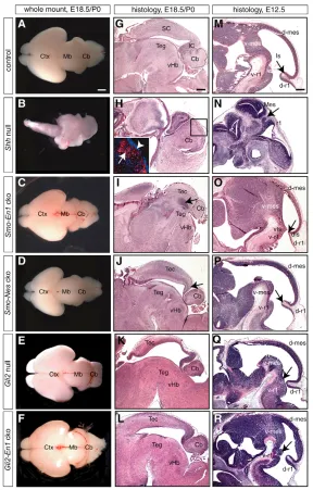
Fig. 1. Mid/hindbrain phenotype in the absence of total or Gli2A-mediated Shh signaling. (A-L) Dorsal view of whole-mount brains (A-F), and Hematoxylin and Eosin staining of midline sagittal sections (G-L) of P0 wild-type (control) and cko embryos or E18.5 null embryos. (B,H) In
Shh-null mutants, cerebral cortex (Ctx), dorsal midbrain (tectum, Tec), ventral Mb (tegmentum, Teg) and ventral anterior hindbrain (vHb) are not discernible, and the cerebellum (Cb) is abnormal. (H, inset) Calbindin-positive Cb Purkinje cells (red, arrow) and Hoechst staining (blue) of the area indicated by the square. A cell dense layer probably corresponds to the Cb external granule cells (arrowhead). (C,I) In Smo-En1cko mutants, the size of the Mb/Cb is reduced, the ventricle is absent (arrow) and the Tec is not divided into superior (SC) and inferior colliculi (IC). (D,J) In
Smo-Nescko mutants, the IC is truncated (arrow) and the Cb is reduced in size. The Ctx is also reduced in size. (E,F,K,L) In Gli2-null and Gli2-En1cko mutants, the Teg, vHb and Cb are reduced in size. The Tec thins in Gli2-null mutants because of hydrocephaly. (M-R) Hematoxylin and Eosin staining of midline sagittal sections of E12.5 wild-type (control) and mutant embryos. (N) In Shh-null mutants, the mes/r1 is severely reduced in size. Dorsal and ventral neural tube is joined at the isthmus (Is) (arrow). (O) A close apposition of ventral and dorsal isthmus (vIs, dIs) is visible in
Smo-En1cko mutants (arrow), and dorsal mes (d-mes) and r1 (d-r1) are truncated. (P) In Smo-Nescko embryos, d-r1 and the posterior d-mes are reduced in size (arrow), but no obvious defect is observed in ventral mes (v-mes) or r1 (v-r1). (Q,R) There is no obvious dorsal phenotype in Gli2-null or
[image:3.612.49.337.57.508.2]D
E
V
E
LO
P
M
E
N
T
the phenotype of Smo-Nescko embryos at P0, the mes/r1 phenotype at E12.5 consisted of a slight reduction of dorsal tissue, primarily in the most medial regions (Fig. 1P). By E16.5, there was a clear truncation of the posterior midbrain and a slight reduction in the size of the cerebellum, but the phenotype was milder than in Smo-En1cko embryos (data not shown). In summary, these data demonstrate that Shh signaling is crucial for development of mes/r1 structures throughout the entire DV axis from E8.0 to after E11.5.
Shh signaling is required sequentially for ventral neuron induction
We next determined the temporal requirement for Shh in generating specific ventral neuronal cell types. In Shh-null mutant embryos, no dopaminergic [tyrosine hydroxylase (Th) positive] or serotonergic [5-hydroxytryptamine (5-HT) positive] neurons were detected (data not shown), consistent with an early loss of all ventral mes/r1 progenitors. Interestingly, in Smo-En1cko embryos in which Shh signaling is removed at E9.0, dopaminergic and serotonergic neurons were generated, but were severely depleted (Fig. 2A-D). Moreover, the removal of Shh signaling 2 days later in Smo-Nescko mutants did not affect the induction of these neurons (Fig. 2E,F). Thus, Shh signaling is required for the specification of dopaminergic and serotonergic neurons before and shortly after E9.0, but not after E11.5.
To determine whether more laterally generated neurons are also differentially affected by sequential inactivation of Shh signaling, we analyzed expression of the transcription factors Islet1 (Isl1) (ventral motoneurons) and Nkx2.2 (ventral-laterally derived neurons). In E18.5 Shh-null mutant embryos (data not shown) and in E10.5 and E12.5 Smo-En1cko embryos, neither transcription
factor was detected in the ventral mes/r1 (Fig. 2I-L and data not shown). Dorsal Isl1-positive cells were, however, maintained in Smo-En1cko mutants (Fig. 2J,L). By comparison, E12.5 Smo-Nes cko mutants had both ventral Isl1- and Nkx2.2-expressing neurons, although they were slightly reduced in number compared with wild-type embryos (Fig. 2M-P). Thus, neuronal subpopulations derived from distinct ventral to lateral domains in the mes/r1 are dependent on Shh signaling in a sequential manner.
Selective inactivation of Gli2A-mediated Shh signaling does not affect the formation of dorsal mes/r1 structures
[image:4.612.48.368.389.739.2]To determine the extent to which Gli2A-mediated Shh signaling is used downstream of Shh for development of ventral and dorsal mes/r1-derived structures, we analyzed the requirement for Gli2A at different time points using Gli2-null mutants and a Gli2 conditional allele (Corrales et al., 2006), in which Gli2 was inactivated at E9.0 (Gli2-En1cko) or E11.5 (Gli2-Nescko). In E12.5 and E18.5 Gli2-null mutant brains, the ventral midbrain and r1 were reduced in size, but not absent (Fig. 1E,K,Q). Consistent with this, a small number of dopaminergic and serotonergic neurons are present in E12.5 Gli2-null mutants (Matise et al., 1998). Furthermore, although the dorsal midbrain was thinner probably because of hydrocephaly at E18.5 (Corrales et al., 2004; Palma and Ruiz i Altaba, 2004), there was no obvious truncation of the inferior colliculus (Fig. 1K,Q). The E18.5 cerebellum was unfoliated and smaller than normal, owing to the requirement for Gli2in granule cell precursor proliferation after E16.5 (Corrales et al., 2004). Comparison of the phenotypes of wild-type, Gli2and Shh-null
Fig. 2. Distinct temporal dependence of medial-to-lateral-derived ventral cell types on Shh signaling.(A-H) Immunohistochemistry for dopaminergic (Th, green) and serotonergic (5-HT, red) neurons on E18.5 sagittal sections. Sections are counterstained with Hoechst (blue), the mid/hindbrain is outlined. (A-H) Both cell types are greatly reduced in Smo-En1cko and Gli2-En1cko but not in Smo-Nescko mutants (arrows). The number of serotonergic neurons in the posterior hindbrain is normal (arrowheads). Arrowhead in C indicates Th-positive neurons in the locus coeruleus. (I-P) In situ hybridization for Nkx2.2
and Isl1on horizontal sections of E10.5 and E12.5 mes. (I-L) The ventral (V) populations of Isl1- and
Nkx2.2-positive cells are not induced in Smo-En1
cko mes (K,L, red arrowheads). (M-P) In Smo-Nes
D
E
V
E
LO
P
M
E
N
T
mutants shows that the contribution of Gli2A signaling to overall embryonic mes/r1 development is restricted to ventral regions (Fig. 1G,H,K,M-O).
Removal of Gli2at E9.0 in Gli2-En1cko mutants resulted in a surprisingly mild phenotype. The size of the ventral mes/r1 appeared grossly normal at E12.5 and E18.5 (Fig. 1L,R). Dopaminergic and serotonergic neurons were present in reduced numbers at E18.5, but were not as severely depleted as in Smo-En1 cko mutants (Fig. 2C,D,G,H). By contrast, the more laterally derived Isl1- and Nkx2.2-expressing neurons were not present in Gli2-En1cko mutants (data not shown). As expected, the cerebellar phenotype at E18.5 was similar to Gli2-null mutants and the tectum appeared normal (Fig. 1K,L) (Corrales et al., 2004). To address whether Gli2-mediated signaling plays a role after E11.5 in mes/r1 development, we analyzed sections of Gli2-Nes cko brains. Histological and marker analysis for Isl1 and Nkx2.2at E12.5 or Th at P0 did not show any obvious reduction in the number of ventral neurons (data not shown). Thus, Gli2A-mediated Shh signaling is only required for induction of ventral mes/r1 structures and primarily before E11.5.
Shh signaling is required for cell survival during the early stages of mes/r1 expansion
To address the cellular mechanism underlying the rapid tissue loss when all Shh signaling is removed after E9.0, we analyzed Smo-cko mutants for cell death and proliferation, as both are affected in Shh null mutants by E9.0 (Ishibashi and McMahon, 2002). Analysis of apoptosis using a TUNEL assay revealed a massive increase in cell death in Smo-En1cko mutants at E9.5, and to a lesser extent at E10.5, throughout the DV axis except in the floor plate. Along the AP axis, cell death was highest near the isthmus (Fig. 3A-D; data not shown). By contrast, no obvious difference in the percentage of proliferating cells in the remaining mes/r1 ventricular zone was
observed with BrdU pulse labeling of E9.5 Smo-En1cko embryos (data not shown). Consistent with the near normal size of Smo-Nes cko brains, no changes in proliferation or apoptosis were detected at E12.5 in these mutants (Fig. 3E,F; data not shown). These results demonstrate that Shh signaling continues to regulate expansion of the mes/r1 between E9.0 and E11.0, but primarily through prevention of cell death.
Increased Gli3R levels are the primary cause for
the growth and dorsal patterning defects in Shh
mutants
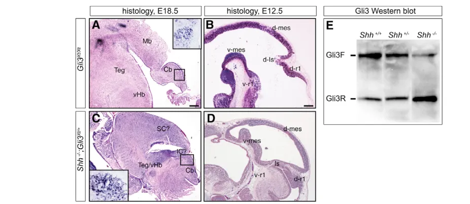
As Gli2A-mediated Shh signaling is not required for mes/r1 growth or dorsal patterning, it is likely that precise regulation of the level of Gli3R is crucial for both processes. If this is the case, then the absence of Gli3R in Gli3Xt-null mutants (Hui and Joyner, 1993; Johnson, 1967) should result in cerebellum and dorsal midbrain defects. Indeed, the dorsal mes area was found to be truncated and the isthmus and r1 enlarged in E12.5 Gli3Xt-null mutants (compare Fig. 4B with
Fig. 1M). At E18.5, the remaining tectum and isthmus were thicker than in wild type and the cerebellum was abnormally shaped in mutants without exencephaly (compare Fig. 4A with Fig. 1G).
We next tested whether the Shh-null phenotype could be partially rescued by removing one copy of Gli3 in Shhnull mutants. Indeed, we found that Shh–/–;Gli3Xt/+mutant embryos had a considerably
[image:5.612.50.545.438.680.2]milder phenotype than Shh-null mutants (compare Fig. 4C,D with Fig. 1H,N). At E12.5, the mes vesicle was almost normal in size and ventral structures were present, but the dorsal and ventral neural tube was fused at the isthmus (Fig. 4D). In contrast to Shh-null mutants, the midbrain was recognizable in E18.5 double mutants and appeared to be divided into inferior and superior colliculi dorsally (Fig. 4C). A more normally structured, but still abnormally shaped cerebellum with calbindin-positive Purkinje cells was also present (Fig. 4C, inset).
D
E
V
E
LO
P
M
E
N
T
To determine whether Gli3R levels are increased relative to full-length Gli3 in the absence of Shh signaling,we performed western blot analysis for Gli3 protein in wild-type and Shhmutant brain extracts (Fig. 4E) (Wang et al., 2000). In E12.5 Shh-null mutant brains, the N-terminal cleavage product was clearly increased compared with wild type and the full-length product was reduced (Fig. 4E). A slight reduction in the Gli3 full-length form was even observed in the mes/r1 of Shh+/–embryos. Hence, the upregulation of Gli3R levels in the absence of Shh signaling is indeed the cause for the lack of overall growth of the mes/r1 and abnormal patterning of dorsal structures.
The requirement for Gli2A- and Gli3R-mediated Shh signaling in patterning gene expression in mes/r1 progenitors changes over time
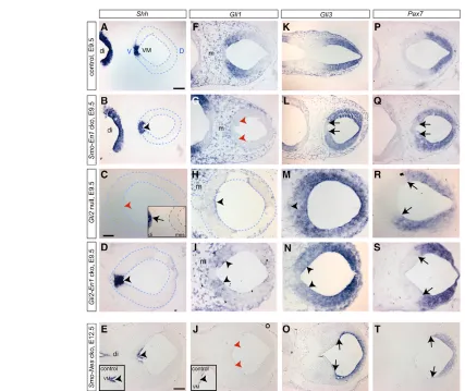
It has previously been shown that markers for dorsal neurons and progenitors (Pax7) are extended ventrally in the mes of Shhnull mutants by E11.5 (Fedtsova and Turner, 2001). To determine the onset of this phenotype and the role of Shh and Gli2 in both setting up and maintaining ventral and dorsal mes/r1 gene expression over time, we analyzed the expression of marker genes that define four DV domains in the mes and r1 from E8.5 to E12.5: Shh expression in the ventral midline, Gli1 expression demarcating the adjacent cells responding to Gli2A-mediated Shh signaling, Gli3 expression comprising the lateral and dorsal mes/r1, and Pax7 expression defining the dorsal plate (Fig. 5A,F,K,P). In E8.5 and E9.5 Shh-null mutants, Shhand Gli1 expression was absent in the mes/r1 (data not shown). The induction of Shhexpression by Shh signaling from the underlying notochord in the mes/r1 must be dependent on Gli2A-mediated signaling, as Shhis not detected in the mes/r1 of Gli2-null mutants (Fig. 5C) (Matise et al., 1998). Interestingly, expression of Shhin the ventral midline was not lost when Shh signaling was ablated at E9.0 (Smo-En1or Gli2-En1cko mutants) or E11.5 (Smo-Nes
and Gli2-Nescko mutants) (Fig. 5B,D,E; data not shown). Thus, Gli2A-mediated Shh signaling is required only to initiate, but not to maintain, Shhexpression.
By contrast,Gli1was initiated but not maintained in Smo-En1and Smo-Nescko mutants once Smowas inactivated (Fig. 5F,G,J; data not shown). Unlike Shh, Gli1was expressed weakly in Gli2-null mutants (Fig. 5H) (Bai et al., 2004) and also in Gli2-En1cko and Gli2-Nes cko mutants (Fig. 5I; data not shown). This residual expression of Gli1reflects a weak activator function of Gli3 in cells close to the source of Shh (Bai et al., 2004). The source of Shh protein in Gli2null mutants is probably the ventral midline of the forebrain (Fig. 5C, inset) (Matise et al., 1998), whereas Shh is provided by the ventral midline of the mes/r1 in Gli2cko mutants (Fig. 5D). In summary, a low level of Gli1is induced in the absence of Gli2in the mes/r1, whereas a high level requires Gli2A-mediated Shh signaling.
Although it has previously been shown that Pax7 expression is expanded into the ventral mes in Shh-null mutants (analysis at E11.5) (Fedtsova and Turner, 2001), it is not known whether Pax7 downregulation depends on Gli2, nor whether Shh signaling is required to sustain this regulation at later stages. Interestingly, whereas in the Shh-null and Smo-En1 cko mutants the Pax7 expression domain was expanded ventrally, the Pax7-positive domain was not extended into ventral areas in Gli2null or cko mutants (Fig. 5P-S; data not shown). This suggests that in Gli2 mutants, the residual GliA-mediated Shh signaling through Gli3A is sufficient to restrict Pax7 expression dorsally (Fig. 5H,I,R,S). Alternatively, only inhibition of Gli3R by Shh is required to repress Pax7 ventrally.
[image:6.612.53.516.57.258.2]In contrast to Pax7, Gli3expression was extended ventrally in the absence of all (Shhnull, Smo-En1cko mutants) and Gli2A-mediated (Gli2null, Gli2-En1 cko mutants) Shh signaling at E9.5 (Fig. 5K-N). However, when Shh signaling was inactivated after E11.5 in the Smo-Nescko or Gli2-Nescko mutants, Gli3and Pax7expression Fig. 4. Regulation of Gli3 repressor (Gli3R) levels is required for dorsal mes/r1 development and growth.(A-D) Hematoxylin and Eosin staining of E18.5 and E12.5 sagittal sections. (A,B) Dorsal mes/r1 development is severely affected in the Gli3Xt/Xt-null mutants. (A) At E18.5, IC and SC are not discernible and the Cb is reduced in size and abnormal. (B) The dorsal isthmus (d-Is) and r1 region are enlarged at E12.5. (C,D) The
D
E
V
E
LO
P
M
E
N
T
continued to be excluded from the ventral mes/r1 similar to wild-type embryos (Fig. 5K,O,P,T; data not shown). Thus, Shh signaling is required only before E11.5 to repress Gli3and Pax7in the ventral mes/r1.
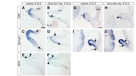
Shh signaling regulates expression of the isthmic
organizer molecule Fgf8
The defects in the inferior colliculus and cerebellum in Smo-En1 cko and Smo-Nescko mutants at P0 (Fig. 1I,J) are similar to defects seen when the isthmic organizer is disrupted (Liu and Joyner, 2001; Zervas et al., 2005). Indeed, the expression domain of Fgf8was shown to be smaller in Shh-null mutants and larger in Gli3-null mutants by E10.5 (Aoto et al., 2002). This could, however, be secondary to loss or expansion of the isthmus in Shh- or Gli3-null mutants, respectively (Fig. 1M,N; Fig. 4B) (Ishibashi and McMahon, 2002). To explore further how and when Shh and Gli3 regulate isthmic gene expression, we analyzed the expression of a
[image:7.612.53.480.54.413.2]D
E
V
E
LO
P
M
E
N
T
of Fgf8,Fgf17 and Spr1 in E12.5 Smo-Nes cko embryos (Fig. 6G-J; data not shown). Importantly, in Gli2-null and Gli2-En1cko mutants no obvious alterations in Fgf8 orSpry1expression were observed at E12.5 (data not shown). Therefore, Shh signaling, through regulation of Gli3R, plays an important role in maintaining normal Fgf8expression in the isthmic organizer from E9.5-E12.5.
DISCUSSION
Regulation of mes/r1 development by Shh signaling is mediated through distinct contributions of Gli2A and Gli3R functions at different stages
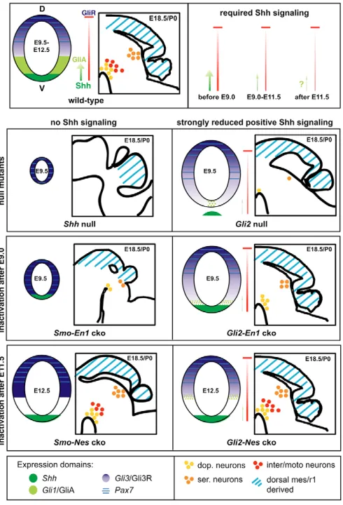
Comparative analysis of the developmental progression of mes/r1 defects in mice with conditional and null mutations in components of the Shh signaling pathway has allowed us to determine the temporal contributions of Gli2A and Gli3R to Shh signaling in mes and r1 development (Fig. 7). In general, high levels of Shh induce Gli2A-mediated Shh signaling and this regulates ventral cell type specification, whereas negative regulation of Gli3R dictates patterning of dorsal structures as well as overall growth in the mes/r1.
The combined results of our analysis and previous studies show that before E9.0, Gli2A-mediated Shh signaling from the notochord induces Shhand Gli1transcription in the ventral midline (Fig. 5C; data not shown) (Bai et al., 2004; Matise et al., 1998). Simultaneously, Shh signaling is required to downregulate ventral Gli3and Pax7expression at the transcriptional level (Fig. 5M; data not shown) (Fedtsova and Turner, 2001). We now demonstrate that Gli3, but not Pax7downregulation requires a high level of Gli2A-mediated Shh signaling. Although, dorsally, Shh signaling does not regulate Gli3 transcription, a gradient of Shh determines the degree
to which full-length Gli3 is processed into Gli3R. Whereas the level of Gli3R is involved in patterning of the tectum and cerebellum, Gli2A-mediated signaling induces a first wave of ventral neurons medially. Finally, Shh signaling regulates cell survival and proliferation before E9.0 (Ishibashi and McMahon, 2002).
By analyzing conditional mutants, we have determined that between E9.0 and E11.5, Shh signaling is no longer required for maintaining Shhexpression in the ventral midline of the mes/r1, but continues to be required to maintain Gli1 expression and aPax7/Gli3 negative domain ventrolaterally. Furthermore, it is Gli2A-mediated Shh signaling that induces a high level of Gli1expression and ventral downregulation of Gli3, but notPax7 transcription. In addition, Gli2A-mediated Shh signaling continues to promote generation of ventral neurons both medially and ventrolaterally. Simultaneously, regulation of Gli3R levels by Shh signaling antagonizes cell death both dorsally and ventrally, and also controls the normal development of dorsal mes/r1 structures, most prominently the inferior colliculus and cerebellum.
After E11.5, we found that Shh signaling is still required to induce Gli1 expression, but not to restrict Pax7 and Gli3 expression dorsally. Similarly, Gli2A-mediated Shh signaling is no longer required to generate the ventral neuronal subpopulations we analyzed. Shh signaling via inhibition of the Gli3R does, however, continue to influence development of the inferior colliculus and cerebellar anlage, but ceases to be necessary for promoting cell survival.
[image:8.612.53.508.56.310.2]Our analysis demonstrates that Gli2A- and Gli3R-mediated Shh signaling is used in a distinct manner in the mes/r1 compared with the forebrain or spinal cord. In the forebrain, although Shh signaling regulates Gli3R production throughout the DV axis, it is only required before E9.0 for normal development of dorsal structures Fig. 6. Reduction in Fgf8 expression and signaling in the absence of Shh. RNA in situ hybridization for Fgf8, Spry1and Wnt1. Analysis was performed on midline sections, as ventral and dorsal isthmus are fused just off the midline in Smo-En1cko mutants (B, inset). (A-F) Dorsal and most posterior mes, isthmus (Is) and r1 are shown (see Fig. 3G) and are outlined where necessary. The thickness of this region is reduced in E10.5 mutants. (A-D) The Fgf8and Spry1domains are severely reduced in Smo-En1cko mutants (B,D arrows). (E,F) Wnt1is expanded posteriorly in Smo
D
E
V
E
LO
P
M
E
N
T
(Fuccillo et al., 2004; Park et al., 2000; Rallu et al., 2002). Furthermore, Gli2A-mediated Shh signaling has only a very minor, if any, role in patterning the telencephalon (Fuccillo et al., 2004; Park et al., 2000; Rallu et al., 2002). In the spinal cord, both Gli2A-and Gli3R-mediated Shh signaling is required, but is restricted to the ventral and intermediate zones, respectively (Bai et al., 2004; Jacob and Briscoe, 2003). By contrast, we have found in the mes/r1, inhibition of Gli3R by Shh plays a sustained role in dorsal mes/r1 structures and Gli2A-mediated Shh signaling induces only ventral cell types.
Gli3R-mediated Shh signaling modulates overall growth and patterning of dorsal mes/r1 structures We identified abnormal patterning of dorsal mes/r1-derived structures in Smo-En1 and Smo-Nes cko mutants at P0, but not in Gli2mutants. In addition, we found that Shh signaling continues to be required for overall cell survival between E9.0 and E11.0, and
that this function does not depend on Gli2. Consistent with a primary role for Gli3R-mediated Shh signaling in mes/r1 growth and dorsal patterning, we showed that the level of Gli3R is increased in Shh-null mutants and that both growth and dorsal patterning are partially rescued in Shh-null mutants when the level of Gli3is reduced. Thus, Gli3R is probably the main downstream effector for these two processes and continues to regulate dorsal patterning after E11.5. However, as Fgf8 signaling is downregulated in the absence of Shh signaling and it has previously been shown that complete removal of Fgf8 in the isthmic organizer results in massive cell death (Chi et al., 2003), we cannot exclude that Gli3R acts at least in part indirectly through regulation of Fgf8. A major effect of decreased Fgf signaling on cell death is unlikely, however, as apoptosis occurred primarily at E9.5 in Smo-En1 cko mutants, a stage when we did not observe a severe reduction in Fgf8expression.
Interestingly, even in the absence of a direct source of Shh within the mes/r1 of Gli2-null mutants, owing to the lack of Shh expression in the ventral midline, Shh secreted from the ventral forebrain is sufficient to promote dorsal development and general growth. It is perhaps surprising that Shh signals can normally reach the dorsal mes/r1, especially after E11.5 when the mes/r1 has undergone substantial growth. Of likely relevance, the dorsal defects in Smocko mutants are most prominent in the region close to the isthmus. It is therefore possible that, as the neural tube is smaller at the isthmic constriction, Shh protein can travel the shorter distance to reach dorsal cells.
Induction of ventral neurons by Gli2A-mediated Shh signaling is coordinated spatially and temporally
Interestingly, we found that the time of dependence of ventral progenitors on Shh signaling correlates with their medial to lateral position in the mes/r1 ventricular zone. The Shh-expressing ventral midline cells, as well as some of the most ventrally induced neurons in the mes/r1 (dopaminergic and serotonergic) are generated in Smo-En1and Gli2-En1cko mutants, demonstrating that they require Shh signaling primarily before E9.0. By contrast, defects in dopaminergic or serotonergic neurons were not observed in Smo-Nesor Gli2-Nescko mutants, suggesting that these neurons are independent of Shh signaling by E11.5. In addition, the more ventrolaterally derived Nkx2.2- and Isl1-positive neurons are not generated in Smo-En1 or Gli2-En1 cko mutants showing they require Shh signaling after E9.0. As these neurons are only slightly reduced in Smo-Nescko embryos, Shh is required primarily between E9.0 and E11.5 for their generation. The temporal requirement for Shh signaling in induction of distinct ventral to lateral-derived neurons is consistent with genetic fate mapping studies in mice that showed that progenitors of dopaminergic and serotonergic neurons respond to GliA-mediated Shh signaling (express Gli1) between E8.0 and E10.0, whereas motoneuron progenitors respond between E9.0 and E10.0 (Zervas et al., 2004) (S.B. and Emilie Dambroise, unpublished). Similarly, in vitro studies demonstrated that the generation of dopaminergic and serotonergic neurons in mes/r1 explants already requires Shh and Fgf8 at E8.0 (Ye et al., 1998).
[image:9.612.54.299.60.420.2]A comparison of the phenotypes of Smoand Gli2cko mutants shows that in addition to Gli2A, Gli3A probably contributes to the induction of some ventral cell types. In particular, the number of dopaminergic and serotonergic neurons is reduced to a greater extent in Smo-En1cko embryos compared with Gli2-En1cko mutants. Consistent with Gli3A contributing to the generation of ventral neurons, Gli3expression is extended ventrally and Gli1is expressed weakly in the ventral mes/r1 of Gli2-En1cko mutants. Alternatively, Fig. 7. Changing temporal requirement for total and Gli2A- and
D
E
V
E
LO
P
M
E
N
T
or in addition, as Shh signaling is maintained in Gli2cko mutants, the level of Gli3R ventrally should be less in Gli2cko than in Smo cko mutants, and this might be sufficient to allow generation of some ventral neurons.
Gli3R-mediated Shh signaling and AP patterning of the mes/r1
The truncation of the inferior colliculus and the smaller cerebellum we observed in Smocko mutants at late gestation are similar to phenotypes seen when Fgf levels are reduced in Fgf17–/–or
Fgf17–/–; Fgf8+/–mutants (Xu et al., 2000). The dorsal phenotype could therefore be an indirect consequence of a decrease in Fgf8 and/or Fgf17. Indeed Fgf8 was previously found to be reduced in Shh-null mutants (Aoto et al., 2002). Furthermore, the finding that Fgf8expression is partially rescued in Shh;Gli3double mutants and increased in Gli3-null mutants suggested that Gli3R-mediated Shh signaling regulates Fgf8 expression. Our studies of Smo-cko mutants argue that Shh regulates isthmic organizer gene expression directly, rather than indirectly through loss or expansion of the isthmus in Shh- or Gli3-null mutants, respectively. We show that Shh signaling continues to be required for Fgf8as well as Fgf17 expression after E9.0, and that the relative reduction in Fgf expression is more severe than the loss of isthmic tissue in Smocko mutants. Furthermore, as we found that Fgf8expression is normal in Gli2mutants, Shh regulates Fgf8 only through antagonizing Gli3R.
Regulation of Fgf8expression by Shh provides a mechanism by which the two key organizers in mes/r1 development can function in unison. Interestingly, interdependence of organizers that express Shh and Fgf8 has been described in the limb and forebrain (Tickle, 2003; Ohkubo et al., 2002). Our studies thus provide further support that cross-regulation of organizers represents a general mechanism to coordinate patterning along different axes during the formation of morphologically complex structures.
Conclusions
Our studies have revealed sequential roles for Shh in DV patterning of the entire mes/r1, and identified the processes requiring induction of Gli2/3A versus downregulation of Gli3R. We demonstrate that GliA-mediated Shh signaling (mainly via Gli2A) is limited to the induction of ventral neurons, and occurs in a medial to lateral sequential manner before E11.5. By contrast, Shh signaling through inhibition of Gli3R is required for overall expansion of the mes/r1 through regulation of cell survival up to E11.0, and for patterning of dorsal structures even after E11.5. Furthermore, the level of Gli3R is crucial in determining the size of the Fgf8expression domain. Sustained Shh signaling thus is necessary not only for DV but also AP patterning of the mes/r1. The formation of all mes/r1-derived structures is therefore intimately linked to the level and extent of Shh signaling.
We thank Andrew McMahon for the Smofloxed mice; Daniel Stephens and Gina Rocco for technical assistance, Emilie Dambroise for fate mapping data and Gunda Schwaninger, Roy Sillitoe and Gordon Fishell for critical reading of the manuscript. We are especially grateful to Mark Zervas for his insightful comments and discussions. S.B. was supported by a post-doctoral fellowship from the DFG. This research was supported by a grant from the NICHD. A.L.J. is an HHMI investigator.
Supplementary material
Supplementary material for this article is available at http://dev.biologists.org/cgi/content/full/133/9/1799/DC1
References
Agarwala, S. and Ragsdale, C. W.(2002). A role for midbrain arcs in nucleogenesis. Development129, 5779-5788.
Agarwala, S., Sanders, T. A. and Ragsdale, C. W.(2001). Sonic hedgehog control of size and shape in midbrain pattern formation. Science291, 2147-2150.
Aoto, K., Nishimura, T., Eto, K. and Motoyama, J.(2002). Mouse GLI3 regulates Fgf8 expression and apoptosis in the developing neural tube, face, and limb bud. Dev. Biol.251, 320-332.
Bai, C. B. and Joyner, A. L.(2001). Gli1 can rescue the in vivo function of Gli2. Development128, 5161-5172.
Bai, C. B., Auerbach, W., Lee, J. S., Stephen, D. and Joyner, A. L.(2002). Gli2, but not Gli1, is required for initial Shh signaling and ectopic activation of the Shh pathway. Development129, 4753-4761.
Bai, C. B., Stephen, D. and Joyner, A. L.(2004). All mouse ventral spinal cord patterning by hedgehog is Gli dependent and involves an activator function of Gli3. Dev. Cell6, 103-115.
Chi, C. L., Martinez, S., Wurst, W. and Martin, G. R.(2003). The isthmic organizer signal FGF8 is required for cell survival in the prospective midbrain and cerebellum. Development130, 2633-2644.
Chiang, C., Litingtung, Y., Lee, E., Young, K. E., Corden, J. L., Westphal, H. and Beachy, P. A.(1996). Cyclopia and defective axial patterning in mice lacking Sonic hedgehog gene function. Nature383, 407-413.
Corrales, J. D., Rocco, G. L., Blaess, S., Guo, Q. and Joyner, A. L.(2004). Spatial pattern of sonic hedgehog signaling through Gli genes during cerebellum development. Development131, 5581-5590.
Corrales, J. D., Blaess, S., Mahoney, E. M. and Joyner, A. L. (2006). The level of sonic hedgehog signaling regulates the complexity of cerebellar foliation. Development133, 1811-1821.
Ding, Q., Motoyama, J., Gasca, S., Mo, R., Sasaki, H., Rossant, J. and Hui, C. C.(1998). Diminished Sonic hedgehog signaling and lack of floor plate differentiation in Gli2 mutant mice. Development125, 2533-2543.
Echelard, Y., Epstein, D. J., St-Jacques, B., Shen, L., Mohler, J., McMahon, J. A. and McMahon, A. P.(1993). Sonic hedgehog, a member of a family of putative signaling molecules, is implicated in the regulation of CNS polarity. Cell
75, 1417-1430.
Fedtsova, N. and Turner, E. E.(2001). Signals from the ventral midline and isthmus regulate the development of Brn3.0-expressing neurons in the midbrain. Mech. Dev.105, 129-144.
Fuccillo, M., Rallu, M., McMahon, A. P. and Fishell, G.(2004). Temporal requirement for hedgehog signaling in ventral telencephalic patterning. Development131, 5031-5040.
Gogoi, R. N., Schubert, F. R., Martinez-Barbera, J. P., Acampora, D., Simeone, A. and Lumsden, A.(2002). The paired-type homeobox gene Dmbx1 marks the midbrain and pretectum. Mech. Dev.114, 213-217.
Graus-Porta, D., Blaess, S., Senften, M., Littlewood-Evans, A., Damsky, C., Huang, Z., Orban, P., Klein, R., Schittny, J. C. and Muller, U.(2001). Beta1-class integrins regulate the development of laminae and folia in the cerebral and cerebellar cortex. Neuron31, 367-379.
Hui, C. C. and Joyner, A. L.(1993). A mouse model of greig
cephalopolysyndactyly syndrome: the extra-toesJ mutation contains an intragenic deletion of the Gli3 gene. Nat. Genet. 3, 241-246.
Ingham, P. W. and McMahon, A. P.(2001). Hedgehog signaling in animal development: paradigms and principles. Genes Dev.15, 3059-3087.
Ishibashi, M. and McMahon, A. P.(2002). A sonic hedgehog-dependent signaling relay regulates growth of diencephalic and mesencephalic primordia in the early mouse embryo. Development129, 4807-4819.
Jacob, J. and Briscoe, J.(2003). Gli proteins and the control of spinal-cord patterning. EMBO Rep.4, 761-765.
Johnson, D. R.(1967). Extra-toes:a new mutant gene causing multiple abnormalities in the mouse. J. Embryol. Exp. Morphol.3, 543-581.
Kimmel, R. A., Turnbull, D. H., Blanquet, V., Wurst, W., Loomis, C. A. and Joyner, A. L.(2000). Two lineage boundaries coordinate vertebrate apical ectodermal ridge formation. Genes Dev.14, 1377-1389.
Lewis, P. M., Gritli-Linde, A., Smeyne, R., Kottmann, A. and McMahon, A. P.
(2004). Sonic hedgehog signaling is required for expansion of granule neuron precursors and patterning of the mouse cerebellum. Dev. Biol.270, 393-410.
Li, J. Y., Lao, Z. and Joyner, A. L.(2002). Changing requirements for Gbx2 in development of the cerebellum and maintenance of the mid/hindbrain organizer. Neuron36, 31-43.
Liu, A. and Joyner, A. L.(2001). Early anterior/posterior patterning of the midbrain and cerebellum. Annu. Rev. Neurosci.24, 869-896.
Long, F., Zhang, X. M., Karp, S., Yang, Y. and McMahon, A. P.(2001). Genetic manipulation of hedgehog signaling in the endochondral skeleton reveals a direct role in the regulation of chondrocyte proliferation. Development128, 5099-5108.
Machold, R., Hayashi, S., Rutlin, M., Muzumdar, M. D., Nery, S., Corbin, J. G., Gritli-Linde, A., Dellovade, T., Porter, J. A., Rubin, L. L. et al.(2003). Sonic hedgehog is required for progenitor cell maintenance in telencephalic stem cell niches. Neuron39, 937-950.
Marigo, V., Johnson, R. L., Vortkamp, A. and Tabin, C. J.(1996). Sonic hedgehog differentially regulates expression of GLI and GLI3 during limb development. Dev. Biol.180, 273-283.
D
E
V
E
LO
P
M
E
N
T
Gli2 is required for induction of floor plate and adjacent cells, but not most ventral neurons in the mouse central nervous system. Development125, 2759-2770.
Maynard, T. M., Jain, M. D., Balmer, C. W. and LaMantia, A. S.(2002). High-resolution mapping of the Gli3 mutation extra-toes reveals a 51.5-kb deletion. Mamm. Genome13, 58-61.
Mo, R., Freer, A. M., Zinyk, D. L., Crackower, M. A., Michaud, J., Heng, H. H., Chik, K. W., Shi, X. M., Tsui, L. C., Cheng, S. H. et al.(1997). Specific and redundant functions of Gli2 and Gli3 zinc finger genes in skeletal patterning and development. Development124, 113-123.
Ohkubo, Y., Chiang, C. and Rubenstein, J. L.(2002). Coordinate regulation and synergistic actions of BMP4, SHH and FGF8 in the rostral prosencephalon regulate morphogenesis of the telencephalic and optic vesicles. Neuroscience
111, 1-17.
Palma, V. and Ruiz i Altaba, A.(2004). Hedgehog-GLI signaling regulates the behavior of cells with stem cell properties in the developing neocortex. Development131, 337-345.
Park, H. L., Bai, C., Platt, K. A., Matise, M. P., Beeghly, A., Hui, C. C., Nakashima, M. and Joyner, A. L.(2000). Mouse Gli1 mutants are viable but have defects in SHH signaling in combination with a Gli2 mutation. Development127, 1593-1605.
Persson, M., Stamataki, D., te Welscher, P., Andersson, E., Bose, J., Ruther, U., Ericson, J. and Briscoe, J.(2002). Dorsal-ventral patterning of the spinal cord requires Gli3 transcriptional repressor activity. Genes Dev.16, 2865-2878.
Rallu, M., Machold, R., Gaiano, N., Corbin, J. G., McMahon, A. P. and Fishell, G.(2002). Dorsoventral patterning is established in the telencephalon of mutants lacking both Gli3 and Hedgehog signaling. Development129, 4963-4974.
Rubenstein, J. L., Martinez, S., Shimamura, K. and Puelles, L.(1994). The embryonic vertebrate forebrain: the prosomeric model. Science266, 578-580.
Soriano, P.(1999). Generalized lacZ expression with the ROSA26 Cre reporter strain. Nat. Genet. 21, 70-71.
Tickle, C.(2003). Patterning systems–from one end of the limb to the other. Dev. Cell4, 449-458.
Tronche, F., Kellendonk, C., Kretz, O., Gass, P., Anlag, K., Orban, P. C., Bock, R., Klein, R. and Schutz, G.(1999). Disruption of the glucocorticoid receptor gene in the nervous system results in reduced anxiety. Nat. Genet. 23, 99-103.
Wang, B., Fallon, J. F. and Beachy, P. A.(2000). Hedgehog-regulated processing of Gli3 produces an anterior/posterior repressor gradient in the developing vertebrate limb. Cell100, 423-434.
Wurst, W. and Bally-Cuif, L.(2001). Neural plate patterning: upstream and downstream of the isthmic organizer. Nat. Rev. Neurosci.2, 99-108.
Xu, J., Liu, Z. and Ornitz, D. M.(2000). Temporal and spatial gradients of Fgf8 and Fgf17 regulate proliferation and differentiation of midline cerebellar structures. Development127, 1833-1843.
Ye, W., Shimamura, K., Rubenstein, J. L., Hynes, M. A. and Rosenthal, A.
(1998). FGF and Shh signals control dopaminergic and serotonergic cell fate in the anterior neural plate. Cell93, 755-766.
Zervas, M., Millet, S., Ahn, S. and Joyner, A. L.(2004). Cell behaviors and genetic lineages of the mesencephalon and rhombomere 1. Neuron43, 345-357.
Zervas, M., Blaess, S. and Joyner, A. L.(2005). Classical embryological studies and modern genetic analysis of midbrain and cerebellum development. Curr. Top. Dev. Biol.69, 101-138.