2875
Introduction
Products of the hedgehog gene family are a group of secreted peptides that have an intimate role in growth, patterning and morphogenesis of many regions in the developing embryo (Ingham and McMahon, 2001). Drosophila hedgehog (hh) was the first member to be identified and characterised (Lee et al., 1992; Mohler and Vani, 1992; Tabata et al., 1992; Tashiro et al., 1993). In the mouse, three homologues have been isolated: sonic hedgehog (Shh) (Echelard et al., 1993; Chang et al., 1994), Indian hedgehog (Ihh) (Echelard et al., 1993) and desert hedgehog (Dhh) (Echelard et al., 1993; Bitgood and McMahon, 1995; Bitgood et al., 1996). Shh function is essential for normal embryonic development; Shh–/–mice have
defects in the neural tube, central nervous system and limbs. These mice have primary failure of division associated with the primordial eye and forebrain structures, producing holoprosencephaly and severe craniofacial anomalies (Chiang et al., 1996).
Patched 1 (Ptc1; Ptch1 – Mouse Genome Informatics) and smoothened (Smo) are transmembrane proteins thought to form a receptor complex for hedgehog ligands (Stone et al., 1996; Marigo et al., 1996). The mechanism of this interaction is poorly understood (Kalderon, 2000; Ingham and McMahon, 2001; Nybakken and Perrimon, 2002); however, genetic
studies indicate that, in the resting state, Ptc1 inhibits the activity of Smo, and binding of Hedgehog proteins to Ptc1 releases this inhibition, allowing signal transduction (Chen and Struhl, 1996; Quirk et al., 1997; Chen and Struhl, 1998). Hedgehog signalling is mediated principally within vertebrate cells by Gli-family zinc-finger transcription factors (Ruiz i Altaba, 1999), of which in the mouse there are three: Gli1, Gli2 and Gli3 (Hui et al., 1994). Gli proteins control cell fate, growth and patterning in vertebrates, having both activating and inhibitory roles (Grindley et al., 1997; Lee et al., 1997; Platt et al., 1997). More recently, further novel components in the hedgehog signalling pathway have been isolated. Hedgehog-interacting protein (Hhip1, also known as Hip1) encodes a membrane glycoprotein capable of binding all mammalian hedgehog proteins and attenuating the signal (Chuang and McMahon, 1999), and Gas1 (growth arrest-specific gene) encodes a glycosylphosphatidylinositol-linked membrane glycoprotein demonstrated to have an antagonistic effect on Shh signalling in the somites (Lee et al., 2001).
Teeth in mammals form on the first branchial arch derivatives, the maxillary and mandibular processes and the frontonasal process. Early tooth development is characterised by reciprocal interactions between the oral epithelium and the underlying neural crest-derived ectomesenchyme of the first
The signalling peptide encoded by the sonic hedgehog gene is restricted to localised thickenings of oral epithelium, which mark the first morphological evidence of tooth development, and is known to play a crucial role during the initiation of odontogenesis. We show that at these stages in the murine mandibular arch in the absence of epithelium, the Shh targets Ptc1 and Gli1 are upregulated in diastema mesenchyme, an edentulous region between the sites of molar and incisor tooth formation. This ectopic expression is not associated with Shh transcription but with Shh protein, undetectable in the presence of epithelium. These findings suggest that, in diastema mesenchyme, restriction of Shh activity is dependent upon the overlying epithelium. This inhibitory activity was demonstrated by the ability of transplanted diastema epithelium to downregulate Ptc1 in tooth explants, and for isolated diastema mesenchyme
to express Ptc1. A candidate inhibitor in diastema mesenchyme is the glycosylphosphatidylinositol-linked membrane glycoprotein Gas1. Gas1 is normally expressed throughout mandibular arch mesenchyme; however, in the absence of epithelium this expression was downregulated specifically in the diastema where ectopic Shh protein was identified. Although Shh signalling has no effect upon Gas1 expression in mandibular arch mesenchyme, overexpression of Gas1 results in downregulation of ectopic Ptc1. Therefore, control of the position of tooth initiation in the mandibular arch involves a combination of Shh signalling at sites where teeth are required and antagonism in regions destined to remain edentulous.
Key words: Shh, Gas1, Tooth development, Diastema, Mandibular arch
Summary
Restriction of sonic hedgehog signalling during early tooth
development
Martyn T. Cobourne1, Isabelle Miletich2and Paul T. Sharpe2,*
1Department of Craniofacial Development and Orthodontics, GKT Dental Institute, King’s College London, Floor 28,
Guy’s Hospital, London SE1 9RT, UK
2Department of Craniofacial Development, GKT Dental Institute, King’s College London, Floor 28, Guy’s Hospital,
London SE1 9RT, UK
*Author for correspondence (e-mail: paul.sharpe@kcl.ac.uk)
Accepted 10 March 2004
Development 131, 2875-2885
Published by The Company of Biologists 2004 doi:10.1242/dev.01163
branchial arch (Peters and Balling, 1999; Tucker and Sharpe, 1999; Jernvall and Thesleff, 2000). In the mouse embryo at around embryonic day (E) 11.5, individual thickenings in the branchial arch epithelium mark the first morphological signs of tooth development. These thickenings undergo localised proliferation to form an epithelial bud that, together with local condensations of ectomesenchyme, form the tooth germ. Tooth germs progress through a well-characterised path of differentiation, in which the ectomesenchyme gives rise to the tooth pulp and dentine-producing odontoblasts, and the epithelium differentiates into enamel-secreting ameloblasts.
During the initiation process, expression of Shh is localised to the epithelial thickenings of future teeth (Bitgood and McMahon, 1995; Hardcastle et al., 1998), and there is in vitro evidence to suggest that Shh acts as a mitogen, inducing proliferation as these thickenings form a tooth bud (Hardcastle et al., 1998; Sarkar et al., 2000; Cobourne et al., 2001). Indeed, inhibition of Shh signalling in mandibular explants from E10.5 results in a failure of bud formation and an arrest of tooth development (Sarkar et al., 2000; Cobourne et al., 2001). Further, conditional knockout of Shh in the developing tooth germ from E12.5 leads to a reduction in overall size of the developing tooth bud (Dassule et al., 2000). Given this localised and crucial role of Shh signalling during early odontogenesis, it is clearly important to restrict the sites of activity along the developing oral axis specifically to the sites of tooth development. This is well illustrated in the developing mouse, as mice have lost their antemolar dentition during evolution, and the incisor regions are separated from molar regions by a diastema or non-tooth-forming edentulous region. We have used the developing mandible as a model to investigate the relationship between downstream mediators of the Shh pathway and non-transcriptional regulation of hedgehog signalling during the initiation of odontogenesis. We find that following removal of the oral epithelium, the endogenous source of Shh, both Ptc1 and Gli1 are upregulated in the diastema of isolated mandibular mesenchyme. This upregulation was associated with Shh protein detected at a distance from the tooth-forming regions. Conversely, Gas1 was specifically downregulated in isolated diastema mesenchyme in regions corresponding to ectopic Ptc1. Electroporation of Gas1 inhibited ectopic Ptc1 expression in the diastema, suggesting that Gas1 plays a role in limiting the activity of Shh signalling along the early tooth-forming axis of the mandibular arch. Interestingly, neither ectopic Ptc1 expression nor Shh protein was detectable in the diastema of mandibular processes in the presence of epithelium. Moreover, the ability of transplanted diastema epithelium to inhibit Ptc1 in developing tooth germs, and for isolated diastema mesenchyme to express Ptc1, suggests that in the developing mandible, although Shh protein is present within non-odontogenic diastema mesenchyme, signalling activity by this protein is inhibited by the epithelium.
Materials and methods
Explant cultures
For explant cultures, CD-1 pregnant mice were sacrificed with cervical dislocation. Timed matings were set up such that noon of the day on which vaginal plugs were detected was considered as E0.5. Mandibles were dissected from embryos under a stereomicroscope
and, if necessary, treated with 2 units/ml Dispase (GibcoBRL) in order to separate the epithelial and mesenchymal components. Mandibular processes or isolated mesenchyme were cultured as previously described (Ferguson et al., 1998). For Shh blocking experiments, culture medium was supplemented with 5E1, a monoclonal antibody against the biologically active amino-terminal signalling fragment of Shh (Shh-N) (Ericson et al., 1996), at 130 µg/ml (Cobourne et al., 2001); for controls, identical concentrations of 2H3, an unrelated antibody directed against neurofilaments, were used (Dodd et al., 1988). Alternatively, agarose beads soaked in either 5E1, 2H3 (both at 130 µg/ml) or Shh protein (1.25 µg/µl rat Shh; Ontogeny) were applied to the tissue. After the required period of culture, explants were prepared for whole-mount or radioactive-section in situ hybridisation (Ferguson et al., 1998). For cartilage analysis in explant cultures, mandibles were harvested from a line of transgenic mice engineered with a Proα1(II) collagen promoter driving a chondrocyte-specific β-galactosidase reporter (Proα1(II)-lacZ, a gift of B. De Crombrugghe) (Zhou et al., 1995). Following culture, cartilage was visualised with X-gal staining (Sanes et al., 1986).
Transplant experiments
For recombination experiments, mandibular diastema or tongue epithelium was harvested from ROSA-26 mice (exhibiting ubiquitous expression of β-galactosidase) (Zambrowicz et al., 1997) and transplanted onto developing incisor tooth germs of intact mandibular explants derived from CD-1 mice. This allows transplant localisation following staining with X-gal (Sanes et al., 1986). For transplantation of isolated diastema mesenchyme, tissue was taken from a mouse line exhibiting ubiquitous expression of green fluorescent protein (GFP) under the control of a β-actin promoter (GFP-mice) (Hadjantonakis et al., 1998) and transplanted into intact tongues derived from CD-1 mice. This allows transplant visualisation under fluorescence.
In situ hybridisation
Whole-mount digoxigenin-labelled and double-labelled (digoxigenin-fluorescein) whole-mount in situ hybridisation was carried out according to Shamim et al. (Shamim et al., 1998). Radioactive section in situ hybridisation using 35S-UTP radiolabelled riboprobes was performed as described by Wilkinson (Wilkinson, 1992). Antisense riboprobes were generated from mouse cDNA clones that were gifts from several laboratories: Shh (Echelard et al., 1993); Ptc1 (Goodrich et al., 1996); Hhip1 (Chuang and McMahon, 1999); Gli1 (Hui et al., 1994); Barx1 (Tissier-Seta et al., 1995); Gas1 (Lee and Fan, 2001);
Ihh (Echelard et al., 1993).
Immunohistochemistry
Immunohistochemistry was carried out according to Gritli-Linde et al. (Gritli-Linde et al., 2001). Primary antibody (Shh rabbit polyclonal IgG, Ihh rabbit polyclonal IgG; Santa Cruz) was detected using a biotinylated monoclonal anti-rabbit IgG (Sigma) and visualised with DAB (Vector Laboratories). Slides were counterstained with Haematoxylin (Vector Laboratories).
Electroporation
experiment was performed using 3 µg/µl of pcAβ-IRES-mGFP to assess any effect of electroporation or GFP expression on Ptc1 expression. All explants were cultured for 24 hours before further processing.
Results
Shh-responsive genes are dependent upon mandibular epithelium
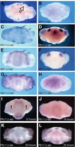
Shh has been demonstrated as the inductive signal for Ptc1, Gli1 and Hhip1 in the mandibular process (Dassule and McMahon, 1998; Hardcastle et al., 1998; Cobourne and Sharpe, 2002). An investigation was carried out to determine the effect upon these downstream genes of removing the oral epithelium, the endogenous source of Shh. E11.5 mandibular processes were cultured for 24 hours in both the presence and absence of epithelium, and assayed using whole-mount in situ hybridisation. At E11.5, normal expression of Shh was restricted to the epithelium of the developing incisor and molar teeth (Fig. 1A, small arrows). The edentulous diastema, between these tooth-bearing regions, showed no expression of Shh (Fig. 1A, large arrow). In the presence of epithelium, the normal expression of Ptc1, Gli1 and Hhip1 was found to be restricted to regions of the developing teeth in a diffuse pattern within odontogenic epithelium and mesenchyme (Fig. 1C,E,G; arrowed). Removal of the epithelium from E11.5 mandibular explants, followed by 24 hours of culture, demonstrated an absence of Shh from the mesenchyme (Fig. 1B). This was expected, as the epithelium is the source of Shh transcription. However, in the case of Ptc1 and Gli1, the expression domains of these genes changed dramatically. Removal of epithelium resulted in localised, bilateral expression in isolated regions of mandibular arch mesenchyme (Fig. 1D,F; arrowed). By contrast, Hhip1 expression was lost in isolated mesenchyme after 24 hours (Fig. 1H).
These dramatic changes in the expression domains of Ptc1 prompted an investigation into the dynamics of this system. Timed culture of E11.5 isolated mandibular arch mesenchyme and analysis by whole-mount in situ hybridisation revealed that Ptc1 upregulation was preceded by downregulation of expression. Downregulation began in the molar regions after approximately 12 hours (Fig. 1I; arrowed; n=7), and was maximal in both incisor and molar regions after around 16 hours (Fig. 1J; n=8). Localised upregulation was identified at around 20 hours of culture (Fig. 1K; n=6), becoming established after approximately 22 hours (Fig. 1L; n=8). By contrast, Shh transcription was never detected in isolated mandibular mesenchyme using either whole-mount or radio-labelled (data not shown) in situ hybridisation at any time period following loss of epithelium.
Ectopic expression of Ptc1 is restricted to diastema mesenchyme
[image:3.612.311.567.71.572.2]In the absence of epithelium, localised upregulation of Ptc1 and Gli1 was restricted to the proximal regions of the mandibular axis, in either the molar or diastema-forming regions of mesenchyme. Following epithelial removal, isolated mesenchymal explants undergo considerable change from their original shape during a 24 hour culture period, and, for this reason, direct comparison with normal explants was
inconclusive in definitively establishing the region of ectopic expression. The exact location of ectopic Ptc1 was therefore identified using molecular markers. The experiments were repeated and expression of Barx1 was analysed in conjunction with Ptc1. Barx1 is a homeobox-containing gene known to be restricted specifically to molar-forming mandibular arch mesenchyme (Tissier-Seta et al., 1995); at E11.5 this expression is fixed, even in the absence of epithelium (Fig. 2A) (Ferguson et al., 2000). Double-labelling of mesenchymal explants with both Ptc1 and Barx1 confirmed that ectopic Ptc1 expression extended more distally than the molar-restricted Barx1 domain (Fig. 2B; arrow). This suggested that ectopic Ptc1 was present in the diastema, but did not exclude the possibility that it extended throughout both regions. However, analysis of adjacent sections using radio-labelled in situ hybridisation demonstrated that the Ptc1 and Barx1-expressing regions were distinct along the proximodistal axis (Fig. 2C,D). Together, these findings suggested that Ptc1 upregulation was occurring in isolated diastema mesenchyme after 24 hours of culture, in a region distinct from the proximal molar-forming mesenchyme.
Ptc1 and Gli1 are induced by ectopic Shh signalling in the diastema
The presence of ectopic Ptc1 and Gli1 transcription in the diastema of isolated mandibular mesenchyme in the absence of Shh was suggestive of a localised source of Shh signalling within this region. However, it is possible that these genes might also be the targets of additional hedgehog family members or alternative signalling pathways within the mandibular arch. Isolated E11.5 mandibular arch mesenchyme was therefore cultured in the presence of the Shh-blocking antibody 5E1, or control 2H3 antibodies. 5E1 is capable of blocking Shh signalling in several regions of the developing embryo, including mandibular explants, whereas 2H3 has no effect (Cobourne et al., 2001). After 24 hours in the presence of 2H3, ectopic Ptc1 and Gli1 was detected in the mesenchyme of the diastema region (Fig. 2E,G; n=7), whereas in cultures exposed to 5E1, no expression was detected (Fig. 2F,H; n=6, n=8, respectively). This provided strong indirect evidence that Shh signalling was responsible for inducing expression of these genes in diastema mesenchyme in the absence of Shh transcription.
[image:4.612.312.560.72.512.2]The deposition of Meckel’s cartilage in the middle of the first branchial arch has been shown to be under control of the epithelium. The epithelium inhibits chondrogenesis; if it is removed, large amorphous masses of cartilage are found instead of a narrow rod (Kollar and Mina, 1991). The possibility therefore existed that ectopic Ihh signalling from such regions might be responsible for the observed Ptc1 and Gli1 induction seen in isolated mandibular mesenchyme. The morphology of Meckel’s cartilage was investigated at E11.5, in both the presence and absence of epithelium, using Proα1(II)-lacZ transgenic mice (Zhou et al., 1995). Although Meckel’s cartilage was found to extend further distally in mandibles cultured for 24 hours in the absence of epithelium, no ectopic masses of cartilage were detected (Fig. 2I,J; n=8). Therefore it was unlikely that the consistent, bilateral and symmetrical upregulation of Ptc1 and Gli1 observed in the diastema region was due to Ihh signalling from ectopic cartilage. This was further confirmed by an absence of Ihh
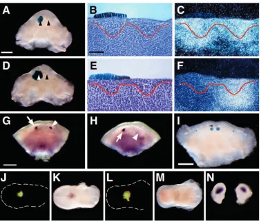
Fig. 2. Ectopic Ptc1 expression is localised to diastema mesenchyme. (A-D) E11.5 mandibular mesenchyme cultured for 24 hours. (A) Barx1 marks the molar regions. (B) Double-labelled (digoxigenin-fluorescein) in situ hybridisation marks Ptc1 (blue staining) diastema regions as distinct from Barx1-expressing (red staining) molar regions. (C) Adjacent sections through Ptc1-expressing domain (upper) demonstrate an absence of Barx1 (lower). (D) Adjacent sections through Barx1-expressing molar region (upper) demonstrate an absence of Ptc1 (lower). (E-H) Ectopic expression of Ptc1 and Gli1 in the diastema is due to Shh signalling. E11.5 mandibular mesenchyme cultured for 24 hours in the presence of 2H3 control antibody (E,G; normal Ptc1, Gli1 expression, respectively) or 5E1 Shh-blocking antibody (F,H; loss of Ptc1, Gli1 expression, respectively). (I,J) E11.5 mandibular explants cultured from Proα1(II)-lacZ transgenic mice demonstrating lacZ expression in Meckel’s cartilage. (I) In the presence of epithelium Meckel’s cartilage extends as two symmetrical rods. (J) In the absence of epithelium Meckel’s cartilage extends further distally (arrowed), but no ectopic cartilages are visible after 24 hours. (K,L) Ihh expression is absent at E11.5 in the mandibular arch in both the presence (K) and absence (L) of epithelium after 24 hours. Scale bars: in A, 600
transcription in Meckel’s cartilage during these early stages of mandibular development (Fig. 2K,L).
Shh protein distribution in the mandibular process The indirect evidence to suggest a source of Shh being responsible for inducing Ptc1 and Gli1 transcription in isolated mandibular diastema mesenchyme led to an attempt at localising the distribution of Shh protein within the mandibular axis during early tooth development. At E11.5 during initiation, Shh was detected throughout the epithelial thickenings of the future teeth but was absent from the underlying mesenchyme. Interestingly, this protein generally had a wider distribution in the epithelium than that of Shh transcripts (Fig. 3A,B). During the early bud stage, Shh was detectable in an expanded region of bud epithelium and also in mesenchymal cells situated around the bud tip; however, Shh transcription was again more localised, detected only in a small group of epithelial cells situated at the tip of the tooth bud (Fig. 3C,D). By E13.5 at the late bud stage, Shh protein distribution had increased, being strongly detected in the outer regions of
bud epithelium and in the surrounding mesenchyme, whereas Shh expression remained localised to the epithelial cells situated at the tip of the tooth bud (Fig. 3E,F). By E14.5 at the early cap stage, Shh protein was present within the enamel knot epithelium, internal enamel epithelium and mesenchyme of the dental papilla (Fig. 3G). At this stage, Shh transcripts remained highly localised to the epithelial cells of the enamel knot (Fig. 3H). Therefore, during the very earliest stages of odontogenesis, Shh protein is able to move beyond Shh-expressing cells within the odontogenic epithelium to cells within the mesenchymal component that express downstream mediators of Shh signalling. Thus, early tooth germs appear to be a viable source of Shh protein, detectable within adjacent mesenchyme.
[image:5.612.118.500.313.643.2]Having established that Shh protein was detectable in the mesenchyme surrounding early tooth buds, an attempt was made to localise a source of Shh responsible for ectopic expression of Ptc1 and Gli1 seen in isolated mandibular mesenchyme. In E11.5 mandibles cultured for 24 hours with the epithelium intact, Shh was present in the epithelium of
Fig. 3. Shh protein is detectable in epithelium and mesenchyme of the developing tooth germ in a wider distribution than epithelial-restricted Shh. (A,C,E,G) Immunohistochemistry; (B,D,F,H) section in situ hybridisation. (A,B) E11.5 epithelial thickening. (C,D) E12.5 early bud. (E,F)
E13.5 late bud. (G,H) E14.5 cap. Shh protein is undetectable in the diastema in the presence of epithelium. (I,J) E11.5 mandibles cultured for 24 hours. (I) Section through the early molar epithelial thickenings (arrows indicate Shh protein in odontogenic epithelium). (J) Section through the diastema demonstrating a lack of detectable Shh protein. (K,L) Immunohistochemistry. Timed-culture of E11.5-isolated mandibular mesenchyme demonstrating the dynamics of Shh detection in the diastema. (K) Shh is first detectable after approximately 20 hours. (L) Shh is strongly detected in the diastema after 24 hours. Shh immunohistochemistry does not cross-react with Ihh. (M,N) E16.5 mandibular condyle. (M) Ihh; (N) adjacent section stained for Shh demonstrating no cross-reaction with Ihh. mc, Meckel’s cartilage; t, tongue. Scale bars: in A, 50
early tooth thickenings, but no protein was detected in the diastema (Fig. 3I,J; n=6). By contrast, in E11.5 mandibular mesenchyme isolated from the epithelium, regions of Shh immunoreactivity were seen after approximately 20 hours of culture, with strong localised expression occurring following 24 hours (Fig. 3K,L; n=3, n=4, respectively). Although transcription of Ihh was not detected at E11.5 in the mandibular process, the possibility existed of cross-reactivity between Ihh and Shh antibodies within this system. In order to eliminate this, the condylar regions of E16.5 murine mandibles were analysed with immunohistochemistry for both proteins. Lack of cross-reactivity was confirmed with the detection of Ihh at the head of the developing condyle using Ihh antibodies, but a failure to detect it using Shh antibodies (Fig. 3M,N; n=4).
Inhibitory properties of diastema epithelium
The observation that Shh protein was only active and detectable in the diastema of isolated mandibular mesenchyme suggested that Shh activity was being inhibited in the presence of epithelium. If this was the case, then isolated diastema epithelium might be expected to downregulate endogenous Shh-induced Ptc1 expression if transplanted onto presumptive tooth germs. Isolating diastema epithelium from E11.5 mandibular processes and transplanting it unilaterally over early incisor tooth buds tested this. Control transplants were performed using tongue epithelium, and, in both cases, donor epithelium was taken from ROSA-26 mice to demonstrate localisation of the transplants (Fig. 4A,B). After 24 hours, Ptc1 expression was examined and found to be normal in the incisor regions exposed to tongue epithelium (Fig. 5A-C; n=5). By contrast, Ptc1 was downregulated in incisor regions exposed unilaterally to diastema epithelium (Fig. 5D-F; n=4). Diastema epithelium thus had the ability to inhibit endogenous Shh protein activity within these tooth germs. At E11.5, expression of Shh remained normal in isolated incisor regions exposed to tongue epithelium (Fig. 5G; arrowhead, compare with normal expression arrowed; n=5). However, this expression was lost following the transplantation of diastema epithelium (Fig. 5H; arrowhead, compare with normal expression arrowed; n=5). This suggested that Shh signalling in the developing tooth germ might be required to maintain Shh transcription during initiation; a finding confirmed by the ability of 5E1-soaked beads to inhibit Shh transcription at E11.5 (Fig. 5I; n=7). This provided evidence of an autoregulatory loop, where Shh
signalling was required to maintain Shh transcription in the dental epithelium during the initiation of tooth development.
Together, these experiments suggest that Shh protein within diastema mesenchyme might be rendered biologically inactive by the overlying epithelium. However, as Shh protein was not detectable by immunohistochemistry in the presence of epithelium it was important to rule out the possibility of local transport or selective accumulation of Shh in the diastema following removal of the epithelium. Therefore, E11.5 diastema mesenchyme was harvested from mandibular processes of GFP-mice, immediately following removal of the epithelium, and plugged into E13.5 tongues taken from wild-type mice. The plugged mesenchyme could therefore be located under fluorescence (Fig. 5J,L). Diastema mesenchyme showed strong endogenous expression of Ptc1 after 24 hours (Fig. 5K; n=9), whereas no expression of Shh was ever seen in these transplants (Fig. 5M; n=7). In addition, diastema explants were also separated from the incisor and molar regions prior to removal of the epithelium, ensuring that the process of epithelial removal in whole explants did not contribute to changes in Shh protein distribution. In these explants, Ptc1 expression remained localised in the diastema (Fig. 5N).
Therefore, diastema epithelium was capable of blocking the activity, either directly or indirectly, of Shh protein normally present, but undetectable, within the underlying mesenchyme; in the absence of epithelium, this protein was both detectable and capable of inducing Ptc1. Furthermore, it was active pre-existing Shh protein that was able to achieve this because no transcription of Shh was ever detected in regions of diastema mesenchyme.
Regulation of Shh activity by Gas1
[image:6.612.48.448.582.731.2]It is clear that the epithelium plays an important role in restricting the activity of Shh along the early mandibular axis. Recently, Gas1 has been demonstrated to have an antagonistic effect on Shh signalling in the somites (Lee et al., 2001). We therefore analysed the expression of Gas1 in the murine first branchial arch. At E11.5, Gas1 was noticeably absent from epithelium and mesenchyme of the developing incisor and molar regions, but strongly expressed in the non-odontogenic regions of mandibular arch mesenchyme, including the diastema (Fig. 6A,B; arrowed). This expression pattern was consistent with Gas1 acting as an additional inhibitor of Shh signalling in non-odontogenic mesenchyme. Interestingly,
further analysis of E11.5 isolated mandibular mesenchyme demonstrated a localised and progressive downregulation of Gas1 in the diastema region between the developing incisor and molar regions between 18-24 hours of culture. However, Gas1 expression was maintained in the peripheral non-odontogenic mesenchyme throughout this time course, indicating differential regulation of Gas1 transcription by the epithelium in odontogenic and non-odontogenic regions of the mandibular arch (Fig. 6C-F). Section in situ hybridisation confirmed that Gas1 downregulation corresponded to the diastema regions of ectopic Ptc1 expression. The progressive Ptc1 upregulation occurred between 18-24 hours, in contrast, Gas1 was progressively downregulated in this region over the same time-course (Fig. 6G-L).
These findings suggested the possibility that in the presence of epithelium Gas1 might inhibit Shh in the diastema, but, following epithelial removal, a loss of Gas1 expression specifically in the diastema would then allow ectopic Shh
signalling in this region. To test this, Gas1 expression was restored in the diastema of isolated mandibular arch mesenchyme by electroporation and the expression of Ptc1 assayed by in situ hybridisation. Consistent with Gas1 being able to inhibit Shh activity, Ptc1 expression was found to be downregulated in these explants (Fig. 6M,N; n=6), whereas in control cultures electroporated with a GFP construct, no downregulation was observed (Fig. 6O,P; n=9). Furthermore, Shh-soaked beads were unable to downregulate Gas1 expression in mandibular mesenchyme, eliminating the possibility that downregulation of Gas1 in Ptc1-expressing regions of isolated mandibular mesenchyme was due to the ectopic Shh signalling activity (data not shown).
Discussion
[image:7.612.121.499.71.393.2]regions, and this expression is important, both during initiation of tooth development and at later stages during morphogenesis (Hardcastle et al., 1998; Dassule et al., 2000; Sarkar et al., 2000; Cobourne et al., 2001). Given this role, it is clearly crucial that regions of Shh expression are closely controlled along the developing oral axis of the mandibular process if odontogenesis is to occur in the correct regions of epithelium. In turn, non-transcriptional antagonism of this signalling pathway will also be an important mechanism in ensuring the correct temporo-spatial control of tooth germ initiation.
In this study, the expression domains of downstream Shh targets were investigated in the mandibular arch following
removal of the source of Shh transcription, namely the oral epithelium. Removal of the epithelium from E11.5 mandibular explants resulted in ectopic expression of Ptc1 and Gli1 in isolated regions of mesenchyme after 24 hours. By contrast, Hhip1 was downregulated over the same time period. Subtle differences in regulation of Shh target gene expression have previously been reported in the first branchial arch derivatives, where Msx1 is required for Shh to induce Ptc1, but not Gli1 in dental mesenchyme (Zhang et al., 1999). Shh also interacts with Prx genes (Ten Berge et al., 2001) and Tbx1 (Garg et al., 2001) in the first arch. Together, these data imply that interactions of genes both upstream and downstream of Shh are controlled and coordinated by several inductive signalling pathways.
Definitive identification of the regions of ectopic Ptc1 and Gli1 expression in isolated mandibular mesenchyme was not straightforward; the mandibular process undergoes a considerable shape change during culture without the integrity of an intact epithelium, isolated mesenchyme lacks histologically reproducible landmarks and there are few molecular markers for specific regions of mesenchyme devoid of epithelium. The use of double-labelled whole-mount and section in situ hybridisation with a specific molar-marker, Barx1, identified these bilaterally symmetrical ectopic regions as distal to the presumptive molar regions, corresponding to the diastema. Barx1 is expressed in the molar and proximal-most regions of mandibular arch mesenchyme, and these expression domains are both established and fixed by E11.5 (Ferguson et al., 2000). The regions of ectopic expression were clearly more distal to the Barx1-expressing zone; however, the possibility of any degree of overlap existing between these two regions could not be definitively excluded.
[image:8.612.45.299.216.742.2]Evidence existed to suggest that Shh signalling was responsible for the ectopic gene expression seen in isolated diastema. Firstly, Ptc1 and Gli1 are only known to be targets of Shh in the first branchial arch (Hardcastle et al., 1998; Dassule and McMahon, 1998). Secondly, upregulation of these genes was blocked in the presence of 5E1 antibody, a known inhibitor of Shh (Ericson et al., 1996). As Shh transcripts were never detected in isolated mandibular arch mesenchyme at any time point following the loss of epithelium, this implied that there must be an active source of Shh within the mesenchyme, capable of inducing both Ptc1 and Gli1. Serial sections of E11.5 mandibular explant cultures, both with and without
Fig. 6. Expression and regulation of Gas1 in the murine first arch. (A,B) Coronal sections through the branchial regions at E11.5 (downregulation of Gas1 in odontogenic mesenchyme arrowed). (C-L) Timed culture of E11.5 mandibular mesenchyme. (C-F) Gas1 progressively downregulates in the diastema (arrowed), whereas expression is maintained in the non-odontogenic mesenchyme underlying the incisor and molar regions. (G-L). Adjacent section in situ hybridisation demonstrates progressive Gas1 downregulation (G,I,K) and Ptc1 upregulation (H,J,L) in isolated diastema mesenchyme between 18 and 24 hours of culture. (M-P) E11.5 mandibular mesenchyme cultured for 24 hours following co-electroporation of Gas1 and GFP constructs (M,N), or electroporation of a GFP construct alone (O,P). Electroporation fluorescence (M,O); Ptc1 expression (N,P). Note downregulation of
Ptc1 in the presence of ectopic Gas1 expression (N, arrowed), as
opposed to controls (P). Scale bars: in A, 200 µm for A,B; in C, 600
epithelium, were investigated using immunohistochemistry to detect the presence of any ectopic Shh protein. No Shh was detected in the diastema of the mandibular process in the presence of epithelium; however, examination of isolated mandibular mesenchyme revealed localised Shh accumulation in the diastema regions after approximately 18 hours of culture. These regions increased in immunoreactivity over the next 6 hours.
What is the source of the Shh detected in diastema mesenchyme? The absence of Shh transcription suggests this protein must originate from other known regions of Shh production at these stages. In the mandibular arch, the most obvious sources would be the developing molar and incisor tooth germs that border the diastema. In support of this, the accumulation of Shh protein in both the epithelium and condensing ectomesenchyme of early tooth buds, at a distance from regions of transcription, suggested that the tooth germs could act as a source of Shh along the mandibular axis. However, both Ptc1 and Hhip1, two members of the pathway that are known to bind Shh, show high-level transcription in the odontogenic mesenchyme surrounding the tooth germs (Hardcastle et al., 1998; Cobourne and Sharpe, 2002). This point not withstanding, Shh has been demonstrated to diffuse further than areas where Ptc1 is expressed at high levels (Gritli-Linde et al., 2001), and it would appear that in the mandibular process Shh is able to move beyond these fields of sequestration into the diastema. Interestingly, rudimentary tooth primordia present in the vole maxillary diastema have been demonstrated to express Shh prior to their apoptotic removal (Keränen et al., 1998), and transient Shh expression has also been shown at E11.5 in mouse diastema epithelium (Dassule and McMahon, 1998). In this study, no specific expression of Shh was detected in the diastema epithelium of the mouse mandible. However, if Shh transcription does occur in the diastema epithelium at earlier stages, albeit transiently and at low levels, theoretically this would provide an additional potential source of the Shh protein demonstrated in the underlying mesenchyme of the diastema.
The failure to detect Shh protein in diastema mesenchyme in the presence of epithelium implies that this protein is either present, but being masked (and therefore non-functional), in whole explants, or that Shh can rapidly accumulate in these regions following epithelial removal. Certainly, loss of epithelium results in downregulation of Ptc1 and Hhip1 in odontogenic mesenchyme surrounding early tooth germs and, as the products of these genes normally sequester Shh, this downregulation might facilitate movement of protein into the diastema. However, diastema mesenchyme transplanted into a tongue host immediately following epithelial removal was able to express Ptc1 but not Shh, suggesting that Shh was normally present, but undetectable in the presence of epithelium. It should be noted that tongue epithelium does contain small sources of Shh within the gustatory papillae from E12.5 (Hall et al., 1999); however, this expression is highly localised and would not be responsible for the high-level Ptc1 expression seen in experimental transplants. Together with the detection of ectopic Ptc1 and Gli1 in the diastema of mandibular arch mesenchyme, and the association of this expression with Shh protein, these data indicate the presence of a Shh-inhibitory mechanism in diastema mesenchyme that is responsible for repressing Shh activity. This inhibition would be reliant upon
the epithelium, confirmed by the ability of diastema epithelium to inhibit the activity of Shh when transplanted over whole incisor tooth germs. However, the transplantation of diastema epithelium onto early incisors at E11.5 was also demonstrated to inhibit Shh transcription, which could explain the downregulation of Ptc1 expression seen in diastema transplants carried out on early bud stage incisor tooth germs. But this capability of diastema epithelium to downregulate Shh transcription might not entirely account for the dramatic downregulation of Ptc1 seen in these transplanted tooth germs. Shh protein was readily detectable in the epithelium and mesenchyme at the tip of these developing teeth, and this protein might be expected to continue inducing Ptc1 expression in the absence of Shh transcription over the period of culture. Ptc1 expression was dramatically downregulated in the diastema-transplanted tooth germs, suggesting that the diastema epithelium is able to mask the activity of pre-existing Shh protein.
A key question is the mechanism of action of any putative Shh inhibitor in the diastema of the mandibular arch, but clearly this process requires an intact epithelium. Several members of the Bmp family of signalling peptides are expressed in the diastema epithelium during these early stages of tooth development (Åberg et al., 1997), and Bmp4 is involved in negatively regulating Shh in the mouse tooth germ (Zhang et al., 2000). However, recombinant Bmp4 protein was unable to repress Ptc1 transcription in isolated diastema mesenchyme (data not shown). More recently Gas1 has been demonstrated to have an antagonistic effect on Shh signalling in the somites: overexpression of Gas1 in pre-somitic mesoderm inhibits the Shh-induced markers Ptc1 and Pax1 (Lee et al., 2001). Gas1 forms a unique and distinct physical complex with the active signalling form of Shh through binding contributions made by the carbohydrate moiety and polypeptide chain (Lee et al., 2001). Gas1 is expressed in a variety of embryonic tissues in a complementary, but partially overlapping, domain to Ptc1 and it has been suggested that it may act as an additional inhibitor of Shh in regions where Ptc1 is not upregulated (Lee and Fan, 2001). However, the exact mechanism whereby Gas1 affects Shh function is not fully understood. It has been postulated that it might act via a direct physical interaction with the receptor complex, through sequestration of the signalling protein itself, or even as a new receptor for Shh (Mullor and Ruiz i Altaba, 2002).
because it is in a complex with Gas1 or another unidentified protein. Clearly, an area of further investigation would be the use of immunoprecipitation experiments to demonstrate a physical relationship between Shh and Gas1 in whole diastema regions. What is also not understood is how loss of the overlying epithelium only results in downregulation of Gas1 in the diastema and not the tooth-forming regions. These observations raise the possibility that epithelial induction of Gas1 is compartmentalised along the oral axis. In the somite, Gas1 expression is known to be induced by several members of the Wnt family of signalling molecules (Lee et al., 2001), and, although several Wnt genes do demonstrate regionally restricted expression in first arch epithelium, none have currently been identified whose expression is restricted to the diastema (Sarkar and Sharpe, 1999).
The data presented in this study demonstrate that Shh
signalling is closely controlled along the oral axis of the first branchial arch during the early stages of tooth development. Not only are mechanisms in place to restrict activity of this peptide around the tooth-forming regions, but also to ensure that signalling is antagonised in those regions where teeth do not develop.
This work was supported by the Wellcome Trust, the Medical Research Council and the Royal College of Surgeons of England. We are grateful to Abigail Tucker for critical reading of the manuscript. 5E1 and 2H3 antibodies were purchased from the Developmental Studies Hybridoma Bank and originally developed by Tom Jessell.
References
Åberg, T., Wozney, J. and Thesleff, I. (1997). Expression patterns of bone morphogenetic proteins (Bmps) in the developing mouse tooth suggest roles in morphogenesis and cell differentiation. Dev. Dyn. 210, 383-396. Bitgood, M. J. and McMahon, A. P. (1995). Hedgehog and Bmp genes are
coexpressed at many diverse sites of cell-cell interaction in the mouse embryo. Dev. Biol. 172, 126-138.
Bitgood, M. J., Shen, L. and McMahon, A. P. (1996). Sertoli cell signalling by Desert hedgehog regulates the male germline. Curr. Biol. 6, 298-304. Chang, D. T., Lopez, A., von Kessler, D. P., Chiang, C., Simandl, B. K.,
Zhao, R., Seldin, M. F., Fallon, J. F. and Beachy, P. A. (1994). Products, genetic linkage and limb patterning activity of a murine hedgehog gene.
Development 120, 3339-3353.
Chen, Y. and Struhl, G. (1996). Dual roles for patched in sequestering and transducing Hedgehog. Cell 87, 553-563.
Chen, Y. and Struhl, G. (1998). In vivo evidence that Patched and Smoothened constitute distinct binding and transducing components of a Hedgehog receptor complex. Development 125, 4943-4948.
Chiang, C., Litingtung, Y., Lee, E., Young, K. E., Corden, J. L., Westphal, H. and Beachy, P. A. (1996). Cyclopia and defective axial patterning in mice lacking Sonic hedgehog gene function. Nature 383, 407-413. Chuang, P. T. and McMahon, A. P. (1999). Vertebrate Hedgehog signalling
modulated by induction of a Hedgehog-binding protein. Nature 397, 617-621.
Cobourne, M. T. and Sharpe, P. T. (2002). Expression and regulation of hedgehog-interacting protein during early tooth development. Conn. Tiss.
Res. 43, 143-147.
Cobourne, M. T., Hardcastle, Z. and Sharpe, P. T. (2001). Sonic hedgehog regulates epithelial proliferation and cell survival in the developing tooth germ. J. Dent. Res. 80, 1974-1979.
Dassule, H. R. and McMahon, A. P. (1998). Analysis of epithelial-mesenchymal interactions in the initial morphogenesis of the mammalian tooth. Dev. Biol. 202, 215-227.
Dassule, H. R., Lewis, P., Bei, M., Maas, R. and McMahon, A. P. (2000). Sonic hedgehog regulates growth and morphogenesis of the tooth.
Development 127, 4775-4785.
Dodd, J., Morton, S. B., Karagogeos, D., Yamamoto, M. and Jessell, T. M. (1988). Spatial regulation of axonal glycoprotein expression on subsets of embryonic spinal neurons. Neuron 1, 105-116.
Echelard, Y., Epstein, D. J., St-Jacques, B., Shen, L., Mohler, J., McMahon, J. A. and McMahon, A. P. (1993). Sonic hedgehog, a member of a family of putative signaling molecules, is implicated in the regulation of CNS polarity. Cell 75, 1417-1430.
Ericson, J., Morton, S., Kawakami, A., Roelink, H. and Jessell, T. M. (1996). Two critical periods of Sonic Hedgehog signaling required for the specification of motor neuron identity. Cell 87, 661-673.
Ferguson, C. A., Tucker, A. S., Christensen, L., Lau, A. L., Matzuk, M. M. and Sharpe, P. T. (1998). Activin is an essential early mesenchymal signal in tooth development that is required for patterning of the murine dentition. Genes Dev. 12, 2636-2649.
Ferguson, C. A., Tucker, A. S. and Sharpe, P. T. (2000). Temporospatial cell interactions regulating mandibular and maxillary arch patterning.
Development 127, 403-412.
Garg, V., Yamagishi, C., Hu, T., Kathiriya, I. S., Yamagishi, H. and Srivastava, D. (2001). Tbx1, a DiGeorge syndrome candidate gene, is regulated by sonic hedgehog during pharyngeal arch development. Dev.
Biol. 235, 62-73.
[image:10.612.57.270.73.384.2]Goodrich, L. V., Johnson, R. L., Milenkovic, L., McMahon, J. A. and Scott, Fig. 7. Shh signalling along the dental axis during initiation of
odontogenesis. (A) In the presence of epithelium, Shh transcription is limited to the incisor and molar-forming epithelium, and is
established by autoregulation (dashed line). Shh protein diffuses from epithelium into mesenchyme, where it induces both Ptc1 and
Hhip1. Ptc1 and Hhip1 protein restrict Shh signalling to the sites of
M. P. (1996). Conservation of the hedgehog/patched signaling pathway from flies to mice: induction of a mouse patched gene by Hedgehog. Genes Dev. 10, 301-312.
Grindley, J. C., Bellusci, S., Perkins, D. and Hogan, B. L. (1997). Evidence for the involvement of the Gli gene family in embryonic mouse lung development. Dev. Biol. 188, 337-348.
Gritli-Linde, A., Lewis, P., McMahon, A. P. and Linde, A. (2001). The whereabouts of a morphogen: direct evidence for short- and graded long-range activity of hedgehog signaling peptides. Dev. Biol. 236, 364-386. Hadjantonakis, A. K., Gertsenstein, M., Ikawa, M., Okabe, M. and Nagy,
A. (1998). Generating green fluorescent mice by germline transmission of green fluorescent ES cells. Mech. Dev. 76, 79-90.
Hall, J. M., Hooper, J. E. and Finger, T. E. (1999). Expression of sonic hedgehog, patched, and Gli1 in developing taste papillae of the mouse. J.
Comp. Neurol. 406, 143-155.
Hardcastle, Z., Mo, R., Hui, C. C. and Sharpe, P. T. (1998). The Shh signalling pathway in tooth development: defects in Gli2 and Gli3 mutants.
Development 125, 2803-2811.
Hui, C. C., Slusarski, D., Platt, K. A., Holmgren, R. and Joyner, A. L. (1994). Expression of three mouse homologs of the Drosophila segment polarity gene cubitus interruptus, Gli, Gli-2, and Gli-3, in ectoderm- and mesoderm-derived tissues suggests multiple roles during postimplantation development. Dev. Biol. 162, 402-413.
Ingham, P. W. and McMahon, A. P. (2001). Hedgehog signaling in animal development: paradigms and principles. Genes Dev. 15, 3059-3087. Jernvall, J. and Thesleff, I. (2000). Reiterative signaling and patterning
during mammalian tooth morphogenesis. Mech. Dev. 92, 19-29. Kalderon, D. (2000). Transducing the hedgehog signal. Cell 103, 371-374. Keränen, S. V., Åberg, T., Kettunen, P., Thesleff, I. and Jernvall, J. (1998).
Association of developmental regulatory genes with the development of different molar tooth shapes in two species of rodents. Dev. Genes Evol. 208, 477-486.
Kollar, E. J. and Mina, M. (1991). Role of the early epithelium in the patterning of the teeth and Meckel’s cartilage. J. Craniofac. Genet. Dev.
Biol. 11, 223-228.
Lee, C. S. and Fan, C. M. (2001). Embryonic expression patterns of the mouse and chick Gas1 genes. Mech. Dev. 101, 293-297.
Lee, C. S., Buttitta, L. and Fan, C. M. (2001). Evidence that the WNT-inducible Growth arrest-specific gene-1 encodes an antagonist of Sonic hedgehog signalling in the somite. Proc. Natl. Acad. Sci. USA 98, 11347-11352.
Lee, J. J., von Kessler, D. P., Parks, S. and Beachy, P. A. (1992). Secretion and localized transcription suggest a role in positional signaling for products of the segmentation gene hedgehog. Cell 71, 33-50.
Lee, J., Platt, K. A., Censullo, P. and Ruiz i Altaba, A. (1997). Gli1 is a target of Sonic hedgehog that induces ventral neural tube development.
Development 124, 2537-2552.
Marigo, V., Davey, R. A., Zuo, Y., Cunningham, J. M. and Tabin, C. J. (1996). Biochemical evidence that patched is the Hedgehog receptor. Nature 384, 176-179.
McLarren, K. W., Litsiou, A. and Streit, A. (2003). DLX5 positions the neural crest and preplacode region at the border of the neural plate. Dev.
Biol. 259, 34-47.
Mohler, J. and Vani, K. (1992). Molecular organization and embryonic expression of the hedgehog gene involved in cell-cell communication in segmental patterning of Drosophila. Development 115, 957-971.
Mullor, J. L. and Ruiz i Altaba, A. (2002). Growth, hedgehog and the price of GAS. BioEssays 24, 22-26.
Nybakken, K. and Perrimon, N. (2002). Hedgehog signal transduction: recent findings. Curr. Opin. Genet. Dev. 12, 503-511.
Peters, H. and Balling, R. (1999). Teeth. Where and how to make them.
Trends Genet. 15, 59-65.
Platt, K. A., Michaud, J. and Joyner, A. L. (1997). Expression of the mouse Gli and Ptc genes is adjacent to embryonic sources of hedgehog signals suggesting a conservation of pathways between flies and mice. Mech. Dev. 62, 121-135.
Quirk, J., van den Heuvel, M., Henrique, D., Marigo, V., Jones, T. A., Tabin, C. and Ingham, P. W. (1997). The smoothened gene and hedgehog signal transduction in Drosophila and vertebrate development. Cold Spring
Harb. Symp. Quant. Biol. 62, 217-226.
Ruiz i Altaba, A. (1999). The works of GLI and the power of hedgehog. Nat.
Cell Biol. 1, E147-E148.
Sanes, J. R., Rubenstein, J. L. and Nicolas, J. F. (1986). Use of a recombinant retrovirus to study post-implantation cell lineage in mouse embryos. EMBO J. 5, 3133-3142.
Sarkar, L. and Sharpe, P. T. (1999). Expression of Wnt signalling pathway genes during tooth development. Mech. Dev. 85, 197-200.
Sarkar, L., Cobourne, M., Naylor, S., Smalley, M., Dale, T. and Sharpe, P. T. (2000). Wnt/Shh interactions regulate ectodermal boundary formation during mammalian tooth development. Proc. Natl. Acad. Sci. USA 97, 4520-4524.
Shamim, H., Mahmood, R. and Mason, I. (1998). In Situ Hybridization to RNA in Whole Embryos. In Molecular Embryology: Methods and Protocols (ed. P. T. Sharpe and I. Mason), pp. 623-633. New Jersey: Humana Press. Stone, D. M., Hynes, M., Armanini, M., Swanson, T. A., Gu, Q., Johnson,
R. L., Scott, M. P., Pennica, D., Goddard, A., Phillips, H. et al. (1996). The tumour-suppressor gene Patched encodes a candidate receptor for Sonic hedgehog. Nature 384, 129-134.
Tabata, T., Eaton, S. and Kornberg, T. B. (1992). The Drosophila hedgehog gene is expressed specifically in posterior compartment cells and is a target of engrailed regulation. Genes Dev. 6, 2635-2645.
Tashiro, S., Michiue, T., Higashijima, S., Zenno, S., Ishimaru, S., Takahashi, F., Orihara, M., Kojima, T. and Saigo, K. (1993). Structure and expression of hedgehog, a Drosophila segment-polarity gene required for cell-cell communication. Gene 124, 183-189.
Ten Berge, D., Brouwer, A., Korving, J., Reijnen, M. J., van Raaij, E. J., Verbeek, F., Gaffield, W. and Meijlink, F. (2001). Prx1 and Prx2 are upstream regulators of sonic hedgehog and control cell proliferation during mandibular arch morphogenesis. Development 128, 2929-2938.
Tissier-Seta, J. P., Mucchielli, M. L., Mark, M., Mattei, M. G., Goridis, C. and Brunet, J. F. (1995). Barx1, a new mouse homeodomain transcription factor expressed in cranio-facial ectomesenchyme and the stomach. Mech.
Dev. 51, 3-15.
Tucker, A. S. and Sharpe, P. T. (1999). Molecular genetics of tooth morphogenesis and patterning: the right shape in the right place. J. Dent.
Res. 78, 826-834.
Wilkinson, D. G. (1992). In Situ Hybridisation: A Practical Approach. Oxford, UK: IRL Press.
Zambrowicz, B. P., Imamoto, A., Fiering, S., Herzenberg, L. A., Kerr, W. G. and Soriano, P. (1997). Disruption of overlapping transcripts in the ROSA beta geo26 gene trap strain leads to widespread expression of beta-galactosidase in mouse embryos and hematopoietic cells. Proc. Natl. Acad.
Sci. USA 94, 3789-3794.
Zhang, Y., Zhao, X., Hu, Y., St Amand, T., Zhang, M., Ramamurthy, R., Qiu, M. and Chen, Y. (1999). Msx1 is required for the induction of Patched by Sonic hedgehog in the mammalian tooth germ. Dev. Dyn. 215, 45-53. Zhang, Y., Zhang, Z., Zhao, X., Yu, X., Hu, Y., Geronimo, B., Fromm, S.
H. and Chen, Y. P. (2000). A new function of BMP4: dual role for BMP4 in regulation of Sonic hedgehog expression in the mouse tooth germ.
Development 127, 1431-1443.