1825
Introduction
During skin development, epidermal and dermal cells continuously receive various environmental cues for differentiation and patterning, and finally form tightly interconnected structures. Communication between cells happens through cell-surface receptors, cell-adhesion molecules, extracellular matrix proteins and chemical messaging via secreted molecules (McElwee and Hoffman, 2000). Hair follicles (HFs) are tiny epidermal appendage structures that develop through a series of epithelium-mesenchyme interactions. Recent findings obtained from analyzing mutant mice revealed various signaling molecules that are involved in HF development (Huelsken et al., 2001; Karlsson et al., 1999); however, the precise function of each of these molecules in the process of HF development is only partially understood.
Tumor necrosis factor receptor; super-family molecules, such as ectodysplasin receptor (EDAR), X-linked ectodysplasin-A2 receptor (XEDAR) and TROY, and their ligand, ectodysplasin (EDA); and other related signal transduction molecules reportedly initiate guard hair follicle and some skin appendage development (Laurikkala et al., 2002; Srivastava et al., 2001; Kojima e al., 2000). Wnt signals and β-catenin were suggested to play important signaling roles in later stages of HF development (Gat et al., 1998; Huelsken et al., 2001; Kishimoto et al., 2000).
Bone morphogenetic protein (BMP) signaling has been found to contribute to various developmental processes, including rapid proliferation and morphogenetic movements during the gastrulation period (reviewed by Mishina, 2003; Furuta et al., 1997). During HF development, BMPs are expressed in the hair placode and surrounding mesenchyme, and may participate in stimulating HF induction. For example, BMP family members such as BMP2, BMP3, BMP4 and BMP7 are expressed in the skin and HFs from the embryonic stage to adult (Lyons et al., 1989; Takahashi and Ikeda, 1996; Stelnicki et al., 1998; Wilson et. al., 1999; Botchkarev et al., 2001). The BMP-neutralizing protein Noggin, which interacts with BMP4, triggers HF induction in the hair placode, suggesting that BMP downregulation may be a cue to start HF development (Botchkarev et al., 1999; Botchkarev et al., 2002) (reviewed by Botchkarev, 2003). In hair placode development, BMPs and sonic hedgehog (SHH) expressions are genetically located downstream of β-catenin signaling (Noramly et al., 1999; Huelsken et al., 2001). SHH also regulates HF growth and morphogenesis (Oro and Higgins, 2003; Callahan and Oro, 2001; Chuong et al., 2000; Dlugosz, 1999). β-Catenin is necessary in determining the fate of stem cells that form follicular keratinocytes, whereas this is not the case for an epidermal fate decision (Jamora et al., 2003).
At the initiation of the anagen phase, follicle progenitor cells of the epidermis induce mesenchymal condensation to form dermal papilla, and then generate proliferating matrix cells.
Interactions between ectodermal and mesenchymal extracellular signaling pathways regulate hair follicle (HF) morphogenesis and hair cycling. Bone morphogenetic proteins (BMPs) are known to be important in hair follicle development by affecting the local cell fate modulation. To study the role of BMP signaling in the HF, we disrupted Bmpr1a, which encodes the BMP receptor type IA (BMPR1A) in an HF cell-specific manner, using the Cre/loxP system. We found that the differentiation of inner root sheath, but not outer root sheath, was severely
impaired in mutant mice. The number of HFs was reduced in the dermis and subcutaneous tissue, and cycling epithelial cells were reduced in mutant mice HFs. Our results strongly suggest that BMPR1A signaling is essential for inner root sheath differentiation and is indispensable for HF renewal in adult skin.
Key words: Bone morphogenetic protein, Type I receptor, Hair follicle cycling, Mouse
Summary
BMPR1A signaling is necessary for hair follicle cycling and hair
shaft differentiation in mice
Munehiro Yuhki1,4, Masahisa Yamada1,2,*, Masako Kawano1, Takuji Iwasato3, Shigeyoshi Itohara3, Hisahiro Yoshida5, Masaharu Ogawa1and Yuji Mishina6
1Laboratory for Cell Culture Development, RIKEN Brain Science Institute, Saitama 351-0198, Japan 2Molecular Neuropathology Group, RIKEN Brain Science Institute, Saitama 351-0198, Japan 3Laboratory for Behavioral Genetics, RIKEN Brain Science Institute, Saitama 351-0198, Japan
4Division of Gene Function in Animals, Nara Institute of Science and Technology, Nara 630-0192, Japan
5Immune System Development Group, RIKEN Research Center for Allergy and Immunology, Kanagawa 230-0045, Japan 6Laboratory of Reproductive and Developmental Toxicology, National Institute of Environmental Health Sciences,
Research Triangle Park, NC 27709, USA
*Author for correspondence (e-mail: masahisa@brain.riken.go.jp)
Accepted 7 January 2004
Development 131, 1825-1833
Published by The Company of Biologists 2004 doi:10.1242/dev.01079
Those epidermal cells further differentiate into hair shaft cells. Additionally, some of these stem cells also form the basal layer of the epidermis. Recent evidence suggests that the initiation of feather placodes in chickens is controlled by positive and negative signals mediated by FGFs and BMPs, respectively (Song et al., 1996; Jung et al., 1998). BMPs signaling is exerted through a complex of type I (BMPRI) and type II receptors (BMPRII). BMPR1A (alternatively known as ALK3) is one of three type I receptors for BMPs. Unfortunately, disrupting
Bmpr1a in mice blocked mesoderm formation and resulted in
intrauterine death before embryonic day 7.5 (E7.5) (Mishina et al., 1995), preventing further investigation into the function of BMP signaling in HF development through BMPR1A, the receptor with the highest affinity for BMP4.
In this study, we examined the role of BMP signaling through BMPR1A during generation and maturation of HFs using mutant mice with Bmpr1a deleted in HFs in skin. We found abnormal HF differentiation and reduced cell growth of interfollicular epidermal cells in the fetal skin of these hair-specific Bmpr1a knockout mice (Hair-Bmpr1a KO mice). In postnatal Hair-Bmpr1a KO mice, a reduction in the number of HFs was apparent in 2-week-old and 10-month-old mutant mice which were hairless in affected regions. Epithelial cells in the hair matrix were located separately covering the luminal surface of open hair canals; and, in the hair shaft, inner root sheath (IRS) development was severely impaired. Furthermore, HF epithelial cells of the older mutant mice also had hardly incorporated BrdU. Taken together, our results strongly suggest that BMPR1A signaling is not only essential for the differentiation of the HF in the developmental stage, but also important for epidermal cell proliferation or differentiation in hair cycle renewal during adult life.
Materials and methods
Generation of mouse lines
Emx1-Cre mice and floxed-Bmpr1a mice had a mixed
129SvJ:C57BL/6 (3:1) background. Mice were genotyped as previously described (Mishina et al., 2002; Iwasato et al., 2000). For evaluation of the Cre activities, Emx1Cre/+mice were bred with CAG-CAT-Z reporter mice (Sakai and Miyazaki, 1997), and double
heterozygous (Emx1-Cre/LacZ) mice were obtained. We crossed the female Emx1Cre/Cremice with male heterozygous Bmpr1a-null mutant
(Bmpr1a+/–) mice (Mishina et al., 1995) to obtain double
heterozygous (Emx1Cre/+Bmpr1a+/–) mice, and further crossed male
Emx1Cre/+Bmpr1a+/–mice with female homozygous floxed-Bmpr1a
(Bmpr1aflox/flox) mice (Mishina et al., 2002) to obtain four types of
mice: Emx1Cre/+ Bmpr1aflox/–; Emx1+/+Bmpr1aflox/–; Emx1Cre/+ Bmpr1aflox/+; and Emx1+/+Bmpr1aflox/+. Emx1Cre/+Bmpr1aflox/–mice
are specific Bmpr1a knockout mice, hereafter referred to as
Hair-Bmpr1a KO mice. The Hair-Hair-Bmpr1a KO mice were viable for 1 year.
This study was carried in accordance with the ‘Guide for the Care and Use of Laboratory Animals’ in the Guidelines of the Society for
Neuroscience and authorized by the Animal Care and Use Committee
of RIKEN.
Immunohistochemistry
Frozen cryosections (15 µm) were treated with 3% H2O2, incubated
with blocking buffer (PBS containing 0.01% Triton X and 1.5% normal goat serum), and incubated overnight at 4°C with monoclonal mouse bromodeoxyuridine (1:100, DAKO), anti-phospho-histone H3 (rabbit polyclonal, 1:300, Upstate), anti Met (goat polyclonal antibody, SANTA CRUZ) and PDGFR-α(rat monoclonal
antibody, Pharmingen). Sections were incubated in biotinylated anti-mouse IgG (1:300, Vector Laboratories), horseradish peroxidase conjugated anti-rabbit IgG (MBL) and horseradish peroxidase conjugated anti-rat IgG (Jackson Immunoresearch) antibody diluted in blocking buffer, and DAB reaction. Primary antibodies used were monoclonal anti β-Catenin (1:500, Sigma, 15B8), polyclonal anti-keratin 5 (1:1000, Covance), polyclonal anti-anti-keratin 6 (K6; kindly provided by Dr Shimomura) (Aoki et al., 2001), anti-TRAF6 (goat polyclonal antibody, Santa Cruz), anti-notch (goat polyclonal antibody, Santa Cruz) and anti-EGFR (goat polyclonal antibody, Santa Cruz). FITC-conjugated goat anti-mouse IgG (1:200, Jackson Immunoresearch), Cy3-conjugated goat anti-rabbit IgG (1:800, Jackson Immunoresearch) and Cy3-conjugated anti-goat IgG (Chemicon) second antibodies were used. Sections were counterstained with Hoechst (Calbiochem).
Analysis of apoptosis
Apoptotic cells were detected by TUNEL assay on 4% paraformaldehyde fixed cryosections of E15-16 limbs by using the ApopTag Peroxidase kit (Intergen).
In situ hybridization
In situ hybridization using digoxigenin-labeled (Roche) cRNA probes was performed on cryosections. Riboprobes for Bmpr1a and Shh were generated as described previously (Mishina et al., 1995; Kato et al., 2001). Skin sections were fixed in 4% formaldehyde, acetylated with 0.5% acetic anhydride in 0.1 M triethanolamine (pH 8.0) for 10 minutes, and rinsed in PBS. The slides were prehybridized in hybridization buffer without cRNA probe at room temperature for 2 hours and hybridized using a hybridization buffer (50% formamide, 5×SSPE, 1 mg/ml yeast tRNA, 0.2% SDS) containing 1 µg/ml cRNA probe at 60°C overnight. Slides were washed in 2×SSC containing 50% formamide at 60°C for 1 hour. Hybridization was detected using an anti-DIG Fab (Roche) coupled to alkaline phosphatase using NBT/BCIP.
X-gal histochemistry staining
Frozen sections (15 µm) were fixed in 4% paraformaldehyde (pH 7.5) for 20 minutes, then washed and incubated with PBS containing 0.01% sodium deoxycholate, 0.02% NP40 and 1 mM MgCl2(lacZ
wash buffer) for 3 hours. For combined X-gal staining with Hematoxylin-Eosin, overnight X-gal staining was processed with PBS containing 1 mg/ml X-gal (5-bromo-4-chloro-3-indoyl-βD-galactopyranoside), 5 mM K-ferrocyanide, 5 mM K-ferricyanide, 0.01% sodium deoxycholate, 0.02% NP40, 1 mM MgCl2 at 37°C
protected from light, then Hematoxylin-Eosin staining was processed.
BrdU-labeling experiments
BrdU (3 mg/100 g body weight, cell proliferation KIT, Amersham Bioscience) was injected to mice subcutaneously, and skin samples were taken 3 hours after injection, fixed in 4% paraformaldehyde and processed for immunohistochemistry.
Electron-micrograph
For electronmicroscopic observation of the skin, a postnatal day 30 Hair-Bmpr1a KO mouse and a control mouse (Emx1Cre/+Bmpr1aflox/–)
Results
Cre-mediated recombination in skin and Hair-Bmpr1a KO mice
To study the function of BMPR1A signaling in the skin, we used a mouse line with a conditional allele of Bmpr1a, in which exon 2 was floxed (Mishina et al., 2002). The Emx1-Cre mouse strain was previously generated by knocking-in Cre recombinase at the Emx1 locus (Iwasato et al., 2000), and this mouse strain expresses Cre in the skin, but not in surrounding tissues, including bone (see below). We bred Emx1-Cre mice with CAG-CAT-Z reporter mice to obtain double heterozygous mice (Emx1-Cre/LacZ mice), and stained histological sections with X-gal. Cre-mediated recombination was expressed over the entire epidermal layer from E13.5 and, 1 day later, guard hair skin follicles around forelimbs and hindlimbs started to
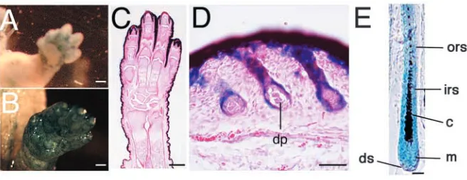
express β-galactosidase activity (Fig. 1A, data not shown). In P0 of Emx1-Cre/LacZ mice, Cre-mediated recombination occurred in skin and hair follicles that were derived from the same stem cells in the skin of the limbs (Fig. 1B-D). In HFs, Cre-mediated recombination occurred in a group of postmitotic cells forming the inner root sheath (IRS), outer root sheath (ORS), hair cortex and hair matrix (Fig. 1E). By contrast, Cre-mediated recombination did not occur in HF mesenchymal components such as the dermal papilla (Fig. 1D) and the dermal sheath (Fig. 1E). Older mice (10 months old) showed the same staining pattern of X-gal (data not shown). Interestingly, Cre activity in these mice was found only in parts of the skin, namely the skin of the limbs, and both the dorsal and ventral side of the basal region of the limbs (Fig. 1A,B, and data not shown).
We then crossed the Emx1-Cre mice with floxed-Bmpr1a mice to delete exon 2 of Bmpr1a to generate a functional null allele in a hair-specific manner (see Materials and methods). Progeny that were trans-heterozygous for floxed-Bmpr1a and Bmpr1a null, and also carried Emx1-Cre (Emx1Cre/+
[image:3.612.239.567.71.198.2]Bmpr1aflox/–; Hair-Bmpr1a KO hereafter) were viable for more than 1 year, but were 10-20% smaller than control mice. Hair loss was observed at Cre-mediated recombination sites of the skin of the limbs (Fig. 2A, arrowheads). The mice showed Cre activity in the same manner as described previously (Fig. 1). Interestingly, these mice also had malformed digits and claws (Fig. 2F,G). Apoptotic cell death was reduced in interdigit areas of E15-16 mutant embryos Fig. 1. Analysis of mice expressing
Cre-recombinase under control of the Emx1 locus. Whole-mount tissue from E13.5 Emx1-Cre/LacZ mice was stained with X-gal (A). Whole-mount tissue from P0 Emx1-Cre/lacZ mice was stained with X-gal (B) and a frozen section from P0
Emx1-Cre/lacZ mice was stained with X-gal,
and counterstained with Hematoxylin-Eosin (C,D). Section from P14 Emx1-Cre/lacZ mice was stained with X-gal (E). c, cortex; dp, dermal papilla; ds, dermal sheath; irs, inner root sheath; m, matrix; ors, outer root sheath. Scale bars: 500 µm in A-C; 50 µm in D; 20 µm in E.
Fig. 2. specific ablation of the Bmpr1a gene in mice. Hair-Bmpr1a KO mice (right) lack external hairs at the limbs
(arrowheads) at P30 (A). (B-D) The right hindlimbs of control mice at P30: (B) whole limb; (C) instep; (D) outstep.
[image:3.612.51.313.413.739.2](Fig. 2J,K) that may explain these abnormalities. In situ hybridization data showed that Bmpr1a was expressed throughout the epidermal layer of HF, ORS, IRS and hair matrix regions in control mice (Fig. 2L,M). Yet, the exon 2-specific probe failed to detect expression of Bmpr1a in the mutant, suggesting that Cre-mediated recombination was completed by P14 (Fig. 2N).
Hair defects in Hair-Bmpr1a KO mice
BMPs are known to be environmental factors for the development of HFs. However, the specific role of BMP signaling through individual receptors in the diversification of epithelial cells and differentiation of HFs function is poorly understood. To explore BMPR1A signaling involvement in the development of HF epithelial cells, we performed morphological analyses during skin development in mutant mice. In mice, initial development of HFs starts from E13 to E18, and extends to postnatal day 14 (P14). In secondary HF development, the postnatal first anagen phase begins around P25 and lasts until P40. The patterning of skin was abnormal in Hair-Bmpr1a KO mice at the end of the initial phase at P14 (Fig. 3A-D). At the end of this first growth phase, around P14,
new hair bulbs start to appear in the subcutaneous space of control mice. The morphology of HFs in the skin of
Hair-Bmpr1a KO mice at P14 showed impaired development in hair
[image:4.612.218.559.71.473.2]formation during this same period. Most of the hair failed to pierce the epidermal surface, remaining stacked and distorted in the subcutaneous region (Fig. 3B). Only 10% of HFs had hair shafts and these were abnormally shaped (Fig. 3C). The distorted hair canals of most of the hair follicles had ectopic keratinized centers (Fig. 3D). In the second anagen phase at P30, newly generated HFs were observed in the deep layer of the subcutaneous region of control mice (Fig. 3E). Although developing HFs were observed in the subcutaneous region of Hair-Bmpr1a KO mice, the amount was significantly less, and retracting epithelial hair shafts had separated from the dermal papilla (Fig. 3E-H,M). Moreover, the hair shaft of KO mice did not extend to the epidermal ceiling. Instead, small cysts formed in the hair canals (Fig. 3H). Any hair that had developed disappeared during this postnatal hair cycle. In addition, unlike control mice, in the skin of Hair-Bmpr1a KO mice, an insignificant number of newly generated HFs could be found in deeper regions of the subcutaneous skin. (Fig. 3I,J,M). Most of the hair canals had cysts including keratinized debris, which Fig. 3. Abnormal hair follicle formation in
Hair-Bmpr1a KO mice. Hematoxylin-Eosin (HE)-stained sections of hindlimbs from Hair-Bmpr1a KO mice are shown in the developmental stage of HF. First anagen phase of P14 control mice (A). Hair follicles in the control skin retract into dermis, maintaining close contact between the dermal papilla and the epithelial hair shaft in P14 control mice (arrow). Abnormally differentiated hair shafts were observed in P14 Hair-Bmpr1a KO mice (B-D).
Retracting epithelial hair shafts that separate from the dermal papilla and form cysts in hair canals were observed in P14
Hair-Bmpr1a KO mice (arrow, B). Section from
P30 control mice shows second anagen phase of HF (E). In P30 Hair-Bmpr1a KO mice (F-H), nearly all of the canals have small cysts (arrowhead, H) in the HF. Section from older (10-month-old) control mice was processed with HE staining (I). The hair canals were wider and large cysts
(arrowhead) were observed in hair canals in older Hair-Bmpr1a KO mice (J-L). The number of HFs per mm2was counted in P14,
P30 and mature mice (n=7 per group) (M). Data are given as mean±s.e.m.; *P<0.05; **P<0.01; Student’s t-test. d, dermis; s, subcutis; pc, panniculus carnosus. Scale bars: 100 µm in A,B,E,F,I,J; 50 µm in
was enlarged and surrounded by the multilayered epithelium (Fig. 3J-L).
Hair follicle epidermal cell cycling in Hair-Bmpr1a KO mice
To determine whether HF developmental defects in
Hair-Bmpr1a KO mice resulted from an impairment of the cell
proliferating ability of HF epidermal cells, we further examined HF development during the anagen phase in KO and control mice. Phosphorylated histone (PH) immunostaining signals showed cells to be in the M phase. Control mice had dividing epithelial cells in HFs; however, in Hair-Bmpr1a KO mice, PH-positive cells were abnormally located in HF hair canals at P30 (Fig. 4A,B). The number of PH-positive cells per HF was significantly reduced in Hair-Bmpr1a KO mice at P30 and at P14 (Fig. 4G). In 10-month-old (older) Hair-Bmpr1a KO mice, both PH immunosignals (Fig. 4C,D,G) and BrdU labeling indices (Fig. 4E,F) were significantly reduced. The number of HFs was also significantly reduced in Hair-Bmpr1a KO mice (Fig. 3M). Taken together, our data suggests that the proliferating ability of hair follicle epidermal cells was severely impaired in Hair-Bmpr1a KO mice.
Although the loss of the cell proliferating ability may explain the absence of new HF development in Hair-Bmpr1a KO mice, the distorted shapes and appearance of abnormal cysts prompted further analysis of HF epidermal cell differentiation. In fact, the IRS structure, which was clearly detected by anti IRS-keratin 6 antibody in control mice, was severely impaired with observable hair loss in Hair-Bmpr1a KO mice at P14 (compare Fig. 5A,B with E,F). By contrast, the expression of keratin 5, which marks the ORS, appeared normal in KO mice hair follicles at P14 (compare Fig. 5C,D
with G,H). The electron density indicated that multiple layers of ORS with trichohyalin granules (th) were located beside a medulla-(me) like structure in Hair-Bmpr1a KO mice at P30 (compare Fig. 5I with J). However, the structure of the IRS, including Henle’s layer (he), Huxley’s layer (hu) and the cuticle of IRS (ci) disappeared, as did the cortex of the hair shaft (c) in Hair-Bmpr1a KO mice (Fig. 5I,J). These results indicated that the hair of
Hair-Bmpr1a KO mice failed to differentiate into the distinct
structures of the IRS. This finding suggests that BMPR1A signaling may be the crucial factor in controlling the cell fate of epithelial cells in the matrix. We also carried out additional immunohistochemical experiments to identify differentiation marker(s) of IRS. We found that the expression of notch, an early differentiation marker of IRS (Kopan and Weintraub, 1993), was relatively normal in the epidermal cells in the Hair-Bmpr1a KO mice (Fig. 6). This implies that BMPR1A signaling is necessary for the differentiation of IRS to form follicular keratinocytes, and that stem cells of the hair matrix only differentiate into a single lineage (outer root sheath) in the absence of BMPR1A.
Bmpr1a deletion affects the expression of β-catenin and Shh
[image:5.612.51.323.72.366.2]To further explore the mechanism by which BMPR1A signaling acts as a regulator in HF induction, we studied the expression profiles of several factors essential for HF development such as Noggin, Shh and β-catenin (Botchkarev et al., 1999; Kulessa et al., 2000; Callahan and Oro, 2001; Huelsken et al., 2001; Jamora et al., 2003). Shh, the regulator of proliferation in HFs, is also know to be a downstream factor of β-catenin signaling (Chiang et al., 1999; Huelsken et al., 2001). The expression of Shh is normally found at the tip of the placode and is restricted to the anterior or posterior hair matrix in mature anagen HF in the control mice (Fig. 7A,C,E) (Gat et al., 1998). Shh was expressed in the distal hair bulb even in 10-month old Hair-Bmpr1a KO mice (Fig. 7B,D,F). Furthermore, the increased expression area of β-catenin in the KO mice was detected by immunohistological analysis (Fig. 7I,J). Interestingly, the expression of β-catenin in the epidermis of Hair-Bmpr1a KO mice was wider than in control mice (Fig. 7I,J). Translocation of β-catenin into the nucleus associates with β-catenin signaling activity. In Hair-Bmpr1a KO mice, the translocation of β-catenin into the nucleus was significantly (P<0.05) reduced (control mice 3.5±0.5%; Hair-Bmpr1a KO mice, 1.9±0.5%; mean±s.e.m.% of translocated β-catenin per Fig. 4. Altered proliferation of hair follicle cells in Hair-Bmpr1a
total β-catenin positive cells; n=15 each group). The skin of Hair-Bmpr1a KO mice appears to have normal skin cycling with no abnormalities observed in the basal layer of skin (Fig. 7H,J). These results suggest that the expression of β-catenin is correlated with wider ORS formation in Hair-Bmpr1a KO mice. Reduced nuclear β-catenin localization may be associated with impaired keratin genesis.
The expression of several other growth regulators (PDGFRα, TRAF6, EGFR, notch and Met), which are important in the morphogenesis of HF development, was also examined. The expression levels of all these genes in HF epidermal cells in Hair-Bmpr1a KO mice seemed to be comparable with control mice HF (Fig. 6). These results are intriguing because both PH-positive cells and BrdU incorporation indices were severely decreased in the hair bulb in 10-month-old KO mice (Fig. 4D,F,G). These data suggested that expression of Shh or other growth regulators are not associated with the cell proliferating ability of Hair-Bmpr1a KO mice.
Discussion
A number of studies indicate that BMPs control hair maturation (reviewed by Botchkarev, 2003; Furuta et al., 1997; Jung et al., 1998; Kulessa et al., 2000), however, the nature of BMP signaling contribution through individual receptors
remained unknown. In this paper, we demonstrate that epithelial cells forming the hair matrix are individually located and cover the luminal surface of opened hair canals, and that, in the hair shaft, inner root sheath (IRS) development was severely impaired in Hair-Bmpr1a KO mice. Furthermore, epithelial cells that remained HFs in older mutant mice did not significantly incorporate BrdU. Taken together, our results strongly suggest that BMPR1A signaling is not only essential for HF differentiation during developmental, but also important for epidermal cell proliferation and, later, the differentiation in hair cycle renewal in adults.
An abnormal pattern of HF structure was observed in
Hair-Bmpr1a KO mice in both the first and second anagen phases
of hair cycle development. Still, part of the HF induction normally occurred in Hair-Bmpr1a KO mice during the first anagen. Because, the induction of HF starts at about E14 in the limbs, where Emx1-Cre is expressed as early as E13.5, it is likely that BMPR1A signaling is not required to initiate hair follicle genesis. EDA, EDAR and TRAF6 could be the important triggers for this induction (Laurikkala et al., 2002; Srivastava et al., 2001; Kojima et al., 2000). Relatively normal expression levels of these genes found in Hair-Bmpr1a KO mice support this idea. However, we cannot completely exclude the possibility that some of the cells in HFs may retain enough BMPR1A to induce normal HF development.
How does BMPR1A signaling contribute to HF development? Kulessa et al. have previously reported that disrupting the BMP signal in HF-specific mice overexpressing
Noggin selectively inhibited the formation of the medulla and
[image:6.612.43.379.71.434.2]the hair shaft. However, Hair-Bmpr1a KO mice showed impaired IRS formation, as indicated by a lack of keratin6 expression, and despite the fact that notch expression, an early marker of IRS (Kopan and Weintraub, 1993), was relatively normal. Thus, although IRS progenitor cells in Hair-Bmpr1a
Fig. 5. Formation of the IRS in Hair-Bmpr1a
KO mice and control mice. Frozen sections of hindlimb skin were prepared from control mice (A-D) and Hair-Bmpr1a KO mice (E-H) at P14. Expression of keratin 6 (A,B,E,F) in the IRS, and expression of keratin 5 (C,D,G,H) in the ORS were detected by
KO mice are likely to be generated, further differentiation involving IRS-keratin 6 expression in the absence of BMPR1A signaling was impaired. These results suggest that BMP signaling, which is essential for early differentiation of IRS, may not be transduced by BMPR1A, and that overexpressed
Noggin interferes with the other receptors mediating signaling
in the early stage of IRS differentiation.
We found malformation of HFs, impaired cell cycling of matrix cells, and complete hair loss in the second anagen. Impaired HF development can be seen in P14 Hair-Bmpr1a KO mice because HF formation in these mice was already impaired in the first anagen. We found notch expression in abnormal HF in Hair-Bmpr1a KO mice, which suggests that the commitment to the IRS cell lineage occurs normally. Although, IRS-keratin 6, a late differentiation marker for both Huxley’s and Henle’s layers, was present in the hair follicle bulb of Hair-Bmpr1a KO mice (Fig. 5E), the structures were missing (Fig. 5J). These differentiation impairments strongly suggest that BMPR1A signaling plays an essential role in the differentiation of IRS. Abnormal HFs in our mice have only multiple layers of ORS and an immature hair shaft that produce cysts composed of keratinized debris. Therefore, our results suggest that the abnormal differentiation of IRS leads to abnormal HF
formation that may eventually impair the cell cycling of epithelial cells.
From these finding, we suspect that bulge formation, originating from HF stem cells, is also affected in the absence of BMPR1A signaling. But, how does the absence of Bmpr1a contribute to the formation of the bulge structure? The skin of
Hair-Bmpr1a KO mice appears to have
[image:7.612.51.405.67.456.2]normal skin cycling with no abnormalities observed in the basal layer of skin except hair loss (Fig. 7H,J). However, bulge structure is therefore likely to unformed in adequate region, which interfere with receiving the key regulator regeneration signals. Interestingly, the expression area of TRAF6, which is genetically down stream of XEDAR or TROY (Laurikkala et al., 2002; Srivastava et al., 2001; Kojima et al., 2000), was increased in the HFs of the Hair-Bmpr1a KO mice compared with control (Fig. 6). The constitutive expression of TRAF6 may represent the inhibition of TRAF6 signaling in the Hair-Bmpr1a KO mice, if feedback regulation is the case. TRAF6 mutant mice are hypohidrotic (anhidrotic), with ectodermal dysplasia (HED) (Naito et al., 2002). Moreover, impaired TRAF6 signaling may contribute to the pathogenesis of a certain type of abnormality in the formation of HF and sebaceous gland that has impaired formation of bulge architecture. Further investigations of Hair-Bmpr1a KO mice and TRAF6 mutant mice might reveal a connection between these two signaling pathways. The development of a perinatal stage-specific disruption system for Bmpr1a in a HF-specific manner would facilitate this comparison.
A similar phenotype, in which the inner root sheath is impaired in a similar fashion as, in Hair-Bmpr1a KO mice, was recently observed in mice harboring a mutation of the β-catenin gene (Huelsken et al., 2001), suggesting that β-β-catenin is necessary for the determination of the cell fate of stem cells to form follicular keratinocytes. Notably, abnormal hair shaft
Fig. 6. The expression of growth
regulators in Hair-Bmpr1a KO mice and control mice. Expression of EGFR, notch, TRAF6, PDGFRαand Met in HFs from P30 Hair-Bmpr1a KO mice and P30 control mice (as indicated) was analyzed by immunohistochemistry (shown in green for antibodies, Hoechst blue for EGFR, notch and TRAF6; brown for antibodies for PDGFRαand Met). The expression levels of all of these regulators in Hair-Bmpr1a KO mice were
differentiations, subsequent loss of hair, and cyst formation in widened canals in the β-catenin mutant mice are the same phenotypes as seen in our mice. Interestingly, the expression of cytekeratin 17, as a marker of the ORS, is upregulated in mutant mice. Based upon the results of β-catenin KO mice, which show the loss of BMP and Shh expression in the mice, the expression of BMP and Shh is controlled by β-catenin in a complex process determining the fate of skin stem cells (reviewed by Oro and Scott, 1998; Barsh, 1999; Huelsken et al., 2001). In Hair-Bmpr1a KO mice, the expression areas of β-catenin and Shh were increased. These results suggest that BMPR1A signaling and β-catenin signaling may be responsible for different functions in HF development and presumably interact with each other. These signals may be using a common downstream pathway; however, it is also possible that they act in parallel to regulate HF differentiation. In situ hybridization demonstrated broad expression of
Bmpr1a in HFs. The reduction of IRS was obvious in Hair-Bmpr1a KO mice; yet, the ORS formed multiple layers in the
mice. In human studies, both BMPR1A and BMPR1B were detected in the HFs in adult and fetal skin (Hwang et al., 2001). Perhaps BMP signaling, through BMPR1B, compensates for the loss of BMPR1A signaling during the generation of the ORS. An unsolved problem of interest is how to define the unique function of BMP signaling through each type I receptor in the HF. Analyzing BMPR1B KO mice may address this question. Taken together, this suggests that BMPR1A signaling has a role in differentiation during maturation of the IRS from hair matrix epithelial cells. Abnormal hair cycling occurs in the absence of BMPR1A signaling, regardless of the relatively normal expression levels of other growth regulation genes for HFs, suggesting that BMPR1A signaling is one of the key components for hair cycling.
Note added in proof
Since the submission of this paper, one paper has appeared demonstrating that importance of BMP signaling for development of IRS in the mouse (Kobielak et al., 2003).
We thank Drs C. Itakura and M. Ray for helpful discussions and careful reading of our manuscript before submission. Thanks to Ms Y. Okumura, S. Yamamoto and B. L. La Madeleine for their expert technical assistance, and to Ms T. Yoshida (Brain Science and Life Technology Research Foundation) for Ultrastructure analysis. The anti-K6 antibody was a very generous gift from Dr Y. Shimomura, and CAG-CAT-lacZ reporter mice were provided by Dr Miyazaki. This work was supported by The Ministry of Education, Culture, Sports and Technology Grant-in-Aid for Young Scientists (B) to M.Y.
References
Aoki, N., Sawada, S., Rogers, M. A., Schweizer, J., Shimomura, Y., Tsujimoto, T., Ito, K. and Ito, M. (2001). A novel type II cytokeratin, mK6irs, is expressed in the Huxley and Henle layers of the mouse inner root sheath. J. Invest. Dermatol. 116, 359-365.
Barsh, G. (1999). Of ancient tales and hair less tails. Nat. Genet. 22, 315-316. Botchkarev, V. A., Botchkareva, N. V., Roth, W., Nakamura, M., Chen, L. H., Herzog, W., Lindner, G., McMahon, J. A., Peters, C., Lauster, R., McMahon, A. P. and Paus, R. (1999). Noggin is a mesenchymally derived stimulator of hair-follicle induction. Nat. Cell. Biol. 1, 158-164.
Botchkarev, V. A., Botchkareva, N. V., Nakamura, M., Huber, O., Funa, K., Lauster, R., Paus, R. and Gilchrest, B. A. (2001). Noggin is required for induction of the hair follicle growth phase in postnatal skin. FASEB J. 15, 2205-2214.
Botchkarev, V. A., Botchkareva, N. V., Sharov, A. A., Funa, K., Huber, O. and Gilchrest, B. A. (2002). Modulation of BMP signaling by noggin is required for induction of the secondary (nontylotrich) hair follicles. J. Invest. Dermatol. 118, 3-10.
Botchkarev, V. A. (2003). Bone morphogenetic proteins and their antagonists in skin and hair follicle biology. J. Invest. Dermatol. 120, 36-47. Callahan, C. A. and Oro, A. E. (2001). Monstrous attempts at adnexogenesis:
regulating hair follicle progenitors through Sonic hedgehog signaling. Curr. Opin. Genet. Dev. 11, 541-546.
Chiang, C., Swan, R. Z., Grachtchouk, M., Bolinger, M., Litingtung, Y., Robertson, E. K., Cooper, M. K., Gaffield,W., Westphal, H., Beachy, P. A. and Dlugosz, A. A. (1999). Essential role for Sonic hedgehog during hair follicle morphogenesis. Dev. Biol. 205, 1-9.
Chuong, C. M., Patel, N., Lin, J., Jung, H. S. and Widelitz, R. B. (2000). Sonic hedgehog signaling pathway in vertebrate epithelial appendage morphogenesis: perspectives in development an evolution. Cell Mol. Life Sci. 57, 1672-1681.
Dlugosz, A. (1999). The Hedgehog and the hair follicle: growing relationship. J. Clin. Invest. 104, 851-853.
[image:8.612.66.271.69.461.2]Furuta, Y., Piston, D. W. and Hogan, B. L. M. (1997). Bone morphogenetic Fig. 7. Expression of Shh and β-catenin in Hair-Bmpr1a KO mice
proteins (BMPs) as regulators of dorsal forebrain development. Development 124, 2203-2212.
Gat, U., DasGupta, R., Degenstein, L. and Fuchs, E. (1998). De novo hair follicle morphogenesis and hair tumors in mice expressing a truncated beta-catenin in skin. Cell. 95, 605-614.
Huelsken, J., Vogel, R., Erdmann, B., Cotsarelis, G. and Birchmeier, W. (2001). beta-Catenin controls hair follicle morphogenesis and stem cell differentiation in the skin. Cell 105, 533-545.
Hwang, E. A., Lee, H. B. and Tark, K. C. (2001). Comparison of Bone Morphogenetic Protein receptors expression in the fetal and adult skin. Yonsei Med. J. 42, 581-586.
Iwasato, T., Datwani, A., Wolf, A. M., Nishiyama, H., Taguchi, Y., Tonegawa, S., Knopfel, T., Erzurumlu, R. S. and Itohara, S. (2000). Cortex-restricted disruption of NMDAR1 impairs neuronal patterns in the barrel cortex. Nature 406, 726-731.
Jamora, C., DasGupta, R., Kocieniewski, P. and Fuchs, E. (2003). Links between signal transduction, transcription and adhesion in epithelial bud development. Nature 422, 317-322.
Jung, H. S., Francis-West, P. H., Widelitz, R. B., Jiang, T. X., Ting, B. S., Tickle, C., Wolpert, L. and Chuong, C. M. (1998). Local inhibitory action of BMPs and their relationships with activations in feather formation: implication for periodic patterning. Dev. Biol. 196, 11-23.
Karlsson, L., Bondjers, C. and Betsholtz, C. (1999). Roles for PDGF-A and sonic hedgehog in development of mesenchymal components of the hair follicle. Development 126, 2611-2621.
Kato, M., Seki, N., Sugano, S., Hashimoto, K., Masuho, Y., Muramatsu, M., Kaibuchi, K. and Nakafuku, M. (2001). Identification of sonic hedgehog-responsive genes using cDNA microarray. Biochem. Biophys. Res. Commun. 289, 472-478.
Kishimoto, J., Burgeson, R. E. and Morgan, B. A. (2000). Wnt signaling maintains the hair-inducing activity of the dermal papilla. Genes Dev. 14, 1181-1185.
Kobielak, K., Pasolli, H. A., Alonso, L., Polak, L. and Fuchs, E. (2003). Defining BMP functions in the hair follicle by conditional ablation of BMP receptor IA. J. Cell Biol. 163, 609-623.
Kojima, T., Morikawa, Y., Copeland, N. G., Gilbert, D. J., Jenkins, N. A., Senba, E. and Kitamura, T. (2000). TROY, a newly identified member of the tumor necrosis factor receptor superfamily, exhibits a homology with Edar and is expressed in embryonic skin and hair follicles. J. Biol. Chem. 275, 20742-20747.
Kopan, R. and Weintraub, H. (1993). Mouse notch: expression in hair follicles correlates with cell fate determination. J. Cell Biol. 121, 631-641. Kulessa, H., Turk, G. and Hogan, B. L. (2000). Inhibition of Bmp signaling affects growth and differentiation in the anagen hair follicle. EMBO J. 19, 6664-6674.
Laurikkala, J., Pispa, J., Jung, H. S., Nieminen, P., Mikkola, M., Wang,
X., Saarialho-Kere, U., Galceran, J., Grosschedl, R. and Thesleff, I. (2002). Regulation of hair follicle development by the TNF signal ectodysplasin and its receptor Edar. Development. 129, 2541-2553. Lyons, K. M., Pelton, R. W. and Hogan, B. L. (1989). Patterns of expression
of murine Vgr-1 and BMP-2a RNA suggest that transforming growth factor-beta-like genes coordinately regulate aspects of embryonic development. Genes Dev. 3, 1657-1668.
McElwee, K. and Hoffmann, R. (2000). Growth factors in early hair follicle morphogenesis. Eur. J. Dermatol. 10, 341-350.
Mishina, Y. (2003). Function of bone morphogrnrtic protein signaling during mouse development, Front. Biosci. 8, d855-d869.
Mishina, Y., Suzuki, A., Ueno, N. and Behringer, R. R. (1995). Bmpr encodes a type I bone morphogenetic protein receptor that I essential for gastrulation during mouse embryogenesis. Genes Dev. 9, 3027-3037. Mishina,Y., Hanks, M. C., Miura, S., Tallquist, M. D. and Behringer, R.
R. (2002). Generation of Bmpr/Alk3 conditional knockout mice. Genesis 32, 69-72.
Naito, A., Yoshida, H., Nishioka, E., Satoh, M., Azuma, S., Yamamoto, T., Nishikawa, S. and Inoue, J. (2002). TRAF6-deficient mice display hypohidrotic ectodermal dysplasia. Proc. Natl. Acad. Sci. USA 99, 8766-8771. Noramly, S., Freeman, A. and Morgan, B. A. (1999). beta-Catenin signaling
can initiate feather bud development. Development 126, 3509-3521. Oro, A. E. and Scott, M. P. (1998). Splitting hairs: dissecting roles of
signaling systems in epidermal development. Cell 95, 575-578.
Oro, A. E. and Higgins, K. (2003). Hair cycle regulation of Hedgehog signal reception. Dev. Biol. 255, 238-248.
Sakai, K. and Miyazaki, J. (1997). A transgenic mouse line that retains Cre recombinase activity in mature oocytes irrespective of the cre transgene transmission. Biochem. Biophys. Res. Commun. 237, 318-324.
Song, H., Wang, Y. and Goetinck, P. F. (1996). Fibroblast growth factor can replace ectodermal signaling for feather development. Proc. Natl. Acad. Sci. USA 93, 10246-10249.
Srivastava, A. K., Durmowiczm, M. C., Hartungm, A. J., Hudsonm, J., Ouzts, L. V., Donovan, D. M., Cui, C. Y. and Schlessinger, D. (2001). Ectodysplasin-A1 is sufficient to rescue both hair growth and sweat glands in Tabby mice. Hum. Mol. Genet. 10, 2973-2981.
Stelnicki, E. J., Longaker, M. T., Holmes, D., Vanderwall, K., Harrison, M. R., Largman, C. and Hoffman, W. Y. (1998). Bone morphogenetic protein-2 induces scar formation and skin maturation in the second trimester fetus. Plast. Reconstr. Surg. 101, 12-19.
Takahashi, H. and Ikeda, T. (1996). Transcripts for two members of the transforming growth factor-beta superfamily BMP-3 and BMP-7 are expressed in developing rat embryos. Dev. Dyn. 207, 439-449.