3423
Introduction
As development proceeds and the CNS becomes a complex three-dimensional structure, the neural stem cells that generate all three major cell lineages in the CNS, neurones, astrocytes and oligodendrocytes, become restricted to specialised germinal zones. These constitute the ventricular zone (VZ) in embryonic development and the sub-ventricular zone (SVZ) in later embryonic and postnatal CNS, with the latter persisting throughout adult life (Altman and Bayer, 1990a; Altman and Bayer, 1990b; Gates et al., 1995; Doetsch et al., 1997). Within these regions, which are analagous to the stem cell niches described in other tissues (Whetton and Graham, 1999; Watt and Hogan, 2000), stem cells divide either to increase the size of the neural stem cell pool or to generate committed precursor cells for neural or glial cell lineages. These different daughter cell populations appear to be generated largely sequentially, with an expansion phase followed by neurogenesis and then gliogenesis (Temple, 2001). At the same time, the cellular identities of the neural stem cells themselves appears to change, and are currently thought to be found within a lineage encompassing neuroepithelial cells, radial glial cells and astrocytes, all of which show stem cell properties at different developmental stages (Alvarez-Buylla et al., 2001; Doetsch, 2003). However, the mechanisms that regulate these stem cell developmental programmes within the microenviroments of the VZ and SVZ are incompletely understood, and are
important not only because of their central role in CNS development but also because an understanding of the mechanisms underlying the expansion and differentiation of neural stem cells is important for therapeutic strategies based on cell replacement.
At the present time, the best-studied factors regulating neural stem cell behaviour are the growth factors. Epidermal growth factor (EGF), basic fibroblast growth factor (FGF2) and transforming growth factor alpha (TGFα) are mitogens for neural stem cells both in vivo and in vitro (Richards et al., 1992; Kilpatrick and Bartlett, 1993; Gritti et al., 1996; Gritti et al., 1999). Other growth factors including bone morphogenic proteins (BMPs), platelet-derived growth factor, ciliary neurotrophic factor and brain-derived neurotrophic factor can regulate stem cell differentiation (Gross et al., 1996; Johe et al., 1996; Benoit et al., 2001). The timing of growth factor responses reflects a changing pattern of growth factor receptor expression during neural stem cell development, driven, at least in part, by the growth factors themselves. Early embryonic neural stem cells respond only to FGF2 whereas late embryonic and adult neural stem cells are responsive to both FGF2 and EGF (Reynolds and Weiss, 1992; Kilpatrick and Bartlett, 1995; Burrows et al., 1997; Qian et al., 1997; Alvarez-Buylla and Temple, 1998), with the acquisition of EGF-responsiveness stimulated by FGF2 and inhibited by BMP4 (Lillien and Raphael, 2000). The BMPs have been shown to
Stem cells in the embryonic mammalian CNS are initially responsive to fibroblast growth factor 2 (FGF2). They then undergo a developmental programme in which they acquire epidermal growth factor (EGF) responsiveness, switch from the production of neuronal to glial precursors and become localized in specialized germinal zones such as the subventricular zone (SVZ). Here we show that extracellular matrix molecules act as regulators of this programme. Tenascin C is highly expressed in the SVZ, and transgenic mice lacking tenascin C show delayed acquisition of the EGF receptor. This results from alterations in the response of the stem cells to the growth factors FGF2 and bone morphogenic protein 4 (BMP4),
which normally promote and inhibit acquisition of the EGF receptor, respectively. Tenascin C-deficient mice also have altered numbers of CNS stem cells and these stem cells have an increased probability of generating neurones when grown in cell culture. We conclude that tenascin C contributes to the generation of a stem cell ‘niche’ within the SVZ, acting to orchestrate growth factor signalling so as to accelerate neural stem cell development.
Key words: Tenascin C, Growth factor, Proliferation, Differentiation, Neurogenesis, Gliogenesis, Stem cell, Central nervous system, Neurosphere
Summary
Generation of an environmental niche for neural stem cell
development by the extracellular matrix molecule tenascin C
Emmanuel Garcion1,*, Aida Halilagic1, Andreas Faissner2and Charles ffrench-Constant1,†1Cambridge Centre for Brain Repair, and Departments of Medical Genetics and Pathology, University of Cambridge, Tennis Court Road, Cambridge CB2 1QP, UK
2Department of Molecular Neurobiology, Ruhr University, Building NDEF 05/593, Universitaetsstraße 150, D44801 Bochum, Germany
*Present address: INSERM U646, 10 rue André Boquel, F-49100 Angers, France
†Author for correspondence (e-mail: cfc@mole.bio.cam.ac.uk)
Accepted 24 March 2004
Development 131, 3423-3432
Published by The Company of Biologists 2004 doi:10.1242/dev.01202
promote proliferation, apoptosis or terminal differentiation of neural stem cells into either neurons or glial cells at progressively later stages of embryonic development (Graham et al., 1996; Gross et al., 1996; Furuta et al., 1997; Li et al., 1998, Panchision et al., 2001). During the expansion phase of neural stem cell development, precursor cells express BMPR1A and respond to BMP by proliferation and by upregulation of a second BMP receptor, BMPR1B. This receptor triggers mitotic arrest once levels exceed that of BMPR1A, so initiating the differentiation phase of neural stem cell development (Panchision et al., 2001).
The soluble and diffusible properties of growth factors make them capable of signalling over a long range. The restricted localization of neural stem cells in embryonic and postnatal development, and their developmental regulation independent of adjacent regions of the CNS, therefore suggests the presence of mechanisms whose role is to restrict or amplify growth factor signalling. One such mechanism is the secretion of short-range inhibitory factors such as the BMP inhibitor, noggin. The production of noggin by ependymal cells prevents adjacent neural stem cells from responding to the gliogenic properties of BMPs in the adult mouse SVZ, so generating a restricted neurogenic environment (Lim et al., 2000). Another potential mechanism is the distribution of extracellular matrix (ECM) molecules. One of these, tenascin C (TNC), is highly expressed within the SVZ throughout postnatal and adult life (Gates et al., 1995). TNC can control cell behaviour both indirectly by binding other matrix components and also directly by interactions with specific cell surface receptors (Jones and Jones, 2000). We have recently shown that TNC null mice show a lower level of oligodendrocyte progenitor proliferation as a result of reduced sensitivity to the mitogen PDGF (Garcion et al., 2001). This evidence that TNC normally amplifies the mitogenic role of PDGF during the development of myelin-forming cells suggests that TNC and other ECM molecules present in germinal zones of the CNS could play a role in restricting or amplifying growth factor responses in neural stem cells. Here we examine this hypothesis by analysing the behaviour of wild-type and tenascin C-deficient neural stem cells both in vivo and when grown in cell culture as neurospheres.
Materials and methods
Animals
TNC null mice were derived from the original stock described by Saga et al. (Saga et al., 1992) with age-matched wild-type mice having the same C57Bl/6J×CBA background, or (for the immunostaining experiments) from mice backcrossed onto a 129 background for at least 14 generations as described previously (Kiernan et al., 1999). The genotype was determined by PCR analysis of genomic DNA from tails using the following primers (Sigma-Genosys): 5′-GAA GTC ACT AGA AAC TAG TGG ACA ACT C-3′and 5′-AAG ATG CCT GGC AGT AGC CAG GTC AC-3′ (TNC); and 5′-CTC CAT GCT TGG AAC AAC GAG CGC AGC-3′ (lacZ). LacZ expression in sections was detected by X-gal staining solution [containing 2 mM MgCl2, 5 mM K3Fe(CN)6, and 5 mM 0.02% NP-40, and 1 mg/ml
X-gal (5-bromo-4-chloro-3-indolyl-D-X-galactoside)] (Sigma).
Cell culture of primary embryonic and postnatal neural stem cell-derived neurospheres
Telencephalic vesicles from E10.5 embryos, and striatum and/or cortex from E12.5 embryos or newborn animals (plug day designated E0) were dissociated and neurosphere forming cells grown in 50% DMEM
(Sigma)/50% Ham’s-F12 (Sigma) medium with B27 supplement (Gibco) in the presence of 0.2 to 20 ng/ml FGF2 (Peprotech) or 0.2 to 20 ng/ml EGF (Calbiochem). For all experiments with FGF2, 5 µg/ml heparin (Sigma) was added. Where appropriate, human recombinant BMP4 (R&D Systems) at 10 ng/ml or TNC purified from neonatal mouse brains by immuno-affinity column chromatography (Faissner and Kruse, 1990) was also added. For the clonal density experiments, 20,000 cells were plated onto a 100 cm2plate.
Amplification of TNC mRNA by reverse transcription-polymerase chain reaction (RT-PCR)
RNA from wild type or TNC null neurospheres was isolated using the RNeasy Mini kit (Qiagen) and reverse-transcribed using First-Strand cDNA synthesis kit (Pharmacia Biotech). PCR reactions were then performed with 10 pmol of oligonucleotides hybridizing to the 5′end of the fifth and the 3′end of the sixth conserved fibronectin type-III domain of mouse TNC (5′-CAC GTG TGA AGG CAT CCA CG-3′ and 5′-TAT CCT TCG GAG AAC CCA TGG C-3′), with a 5 minutes denaturation step at 95°C followed by 35 cycles of 1 minute at 95°C, 2 minutes at 60°C, and 2 minutes at 72°C and a final extension step at 72°C for 10 minutes.
Serial dilution assays to measure neural stem cell numbers
Serial dilution analysis to analyse neural stem cell numbers in a specified number of cells was performed according to Bellows and Aubin (Bellows and Aubin, 1989) with minor modifications from Tropepe et al. (Tropepe et al., 1999). Cells isolated from P0 cortical and striatum using papain and mechanical dissociation were plated in 96-well microwell plates containing either EGF (20 ng/ml) or FGF2 (20 ng/ml), with 4000 cells per well in 400 µl of media. Serial dilutions were then made to obtain final cell numbers from 2000 cells/well to 1 cell/well in 200 µl media. After 7 days the number of spheres (nsph) per well was quantified and plotted against the number of cells plated (ncp) per well. The slope of the regression line obtained for each cell population reflects the proportion of neurosphere forming cells (stem cells) present in the entire original cell suspension. For the assays to analyse stem cell numbers within all cells dissociated from intact cortex and striatum, the entire cell population obtained from the dissociation was serially diluted in a 96-well plate in twofold steps with a final volume of 200 µl. Growth media contained FGF2 (20 ng/ml) and Heparin (5 µg/ml) or EGF (20 ng/ml). After 7 days the highest dilution at which one or more neurospheres was observed in the culture well was determined.
Immunohistochemistry and Western blotting
After 3-7 days culture, neurospheres were washed with cell wash buffer (50 mM Tris-HCl pH 7.5, 0.15 M NaCl, 1 mM CaCl2, 1 mM
MgCl2) and lysed on ice for 30 minutes in extraction buffer (cell wash
buffer plus 0.5% NP40, 2 mM PMSF, 5 µg/ml leupeptine, 2 µg/ml aprotinin, 1 µg/ml pepstatin A, 2 mM sodium fluoride, 2 mM sodium vanadate and 4 mM sodium pyrophosphate). After clarification by centrifuging at 16,000 g for 10 minutes at 4°C, the sample lysates were then adjusted to 10 µg of protein per lane and run on SDS-PAGE gels under reducing conditions. Proteins were transferred and detected using ECL (Amersham).
Cell proliferation assay
Proliferation within neurospheres grown for 7 days in the presence of FGF2 or EGF was assessed by measuring bromodeoxyuridine (BrdU) incorporation after addition to the culture medium for 2 hours. To count labelled cells, neurospheres were dissociated with trypsin-EDTA (Sigma), plated onto poly-D-lysine-coated slides and fixed in ethanol 95%/acetic acid 5% for 20 minutes at –20°C, following which cells could be immunolabelled for BrdU using an immunofluorescence assay kit (Roche) as previously described (Garcion et al., 2001). After counterstaining with propidium iodide (PI, 1/1000, Sigma), the number of BrdU+ cells as well as the number of PI+ cells were counted in at least five different fields using a ×20 objective (corresponding to more than 500 cells) in at least three independent experiments for each condition.
Radial glia assay
The radial glia assay was performed as described previously by Hartfuss et al. (Hartfuss et al., 2001). In brief, telencephalic vesicles were dissected out and dissociated. For E10.5 and E13.5 whole vesicles were pooled together, whereas for E18.5 and P0 striatum and cortex were treated separately. Cells were resuspended in 10% FCS/DMEM and plated on PDL slides for 2 hours followed by a 10-minute fixation in 4% PFA/PBS. After this, immunocytochemistry was performed using the RC2 monoclonal antibody to detect radial glia cells (Edwards et al., 1990). The proportion of RC2+ cells was calculated in at least three animals at each age, and significance calculated using Student’s t-test.
Neurosphere-derived cell differentiation assay
After seven days of culture, neurospheres derived from cortex or striatum were plated onto poly-D-lysine-coated slides without mitogens in 50% DMEM/50% F12 medium with B27 supplement and 0.5% horse serum, and allowed to adhere for 6 days. During this time cells migrate out of the sphere and differentiate. For the rescue experiments using exogenous TNC, immunopurified mouse TNC was added throughout both stages of the culture at a concentration of 1
µg/ml. The cultures were then fixed in 4% PFA and processed for immunofluorescence as previously described (Garcion et al., 2001). The following antibodies were used: mouse monoclonal anti-βΙΙΙ -tubulin (β3-tub, 1/500, Sigma), rabbit polyclonal anti-glial fibrillary acidic protein (GFAP, 1/750, DAKO), rat polyclonal anti-myelin basic protein (MBP, 1/100, Serotec) and mouse monoclonal anti-galactocerebroside-C (Gal-C, 1/50, Sigma). Slides were counterstained with DAPI (1 µg/ml, Sigma) and mounted in Immunofloure mounting medium (ICN). Quantification of each cell type (neurons - β3-tub+, astrocytes - GFAP+ and oligodendrocytes Gal-C or MBP+) was assessed in three independent experiments by analysing 10 different fields within the rim of migrating cells using a
×40 objective and OpenLab software (Improvision).
Results
Regulation of EGFR acquisition in neural stem cells by TNC
Immunohistochemistry and in situ hybridization studies have
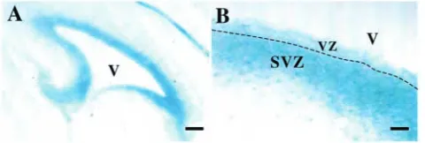
shown that TNC is expressed during CNS development from E10 onwards (Sheppard et al., 1991; Kawano et al., 1995; Gotz et al., 1997). To confirm expression in the embryonic SVZ, we performed β-galactosidase staining on sections from TNC null mice, which were created by the insertion of a lacZ cassette within the TNC gene. We found transgene expression within the neuroepithelium at stages during which neurogenesis occurs (E14.5, not shown) and also within the SVZ at later stages during which gliogenesis occurs (E17.5) (Fig. 1A,B).
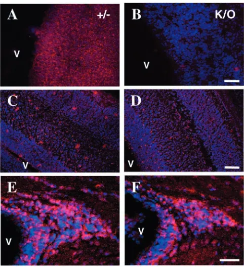
To examine the hypothesis that TNC within the VZ and SVZ regulates the response of neural stem cells to growth factors we examined the timing of the acquisition of EGF receptors (EGFRs) in the embryonic CNS in vivo by comparing TNC-deficient mice with their heterozygote littermates. Previous work has established that EGFR expression first occurs between E11 and E14 (with the plug date being considered E1) (Burrows et al., 1997; Kornblum et al., 1997; Kalyani et al., 1999; Tropepe et al., 1999) and is promoted by FGF2 and inhibited by BMP4, both of which are present in the developing CNS (Lillien and Raphael, 2000). Using immunohistochemistry, we found that EGFR expression was delayed in TNC null mice as compared with their normal littermates. At E12.5 (with the plug date being considered E0 in all our experiments) EGFR immunostaining was evenly distributed in the developing cortex of heterozygous mice (Fig. 2) whereas no labelling was observed in TNC null littermates (Fig. 2). EGFR immunoreactivity was, however, observed in the TNC null mice at later time points from E17.5 to postnatal day (P)17. The distribution was similar in both heterozygous and null littermates (Fig. 2), becoming restricted to the dorsolateral part of the SVZ (Fig. 2) where TNC mRNA and protein is also expressed.
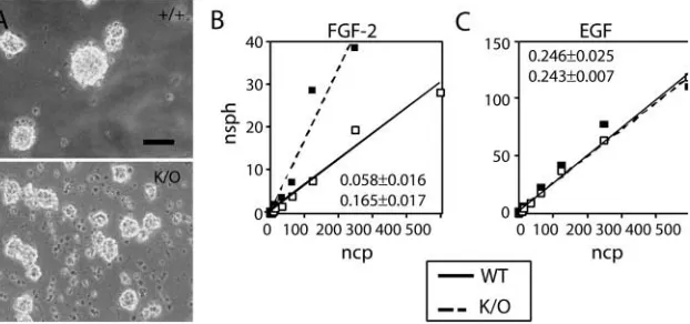
[image:3.612.323.563.584.665.2]To confirm the differences in the timing of EGFR expression in neural stem cells suggested by the expression data in the TNC-deficient mice, we performed cell culture experiments in which telencephalic cells from E10.5 mice were grown at clonal density to generate neurospheres. These are aggregates of cells comprising a mixture of the stem and precursor cells found in the germinal regions of the CNS (Reynolds and Weiss, 1992) that derive from single neural stem cells when dissociated cells are grown at clonal density (as described in Materials and methods). As a consequence, the number of neurospheres obtained in these clonal density cultures provides a measure of neural stem cell number in the cell population.
Fig. 1. Expression of tenascin C (TNC) in subventricular zone (SVZ) cells revealed by lacZ expression from the transgene in TNC heterozygous mice. This was observed within the regions
We observed that neurospheres grew in FGF2 in similar numbers from wild type and TNC null mice (Fig. 3A). In the presence of EGF small numbers of neurospheres could also be obtained from wild-type cells, consistent with previous work showing that the EGF-responsive neural stem cell population is present at this stage of embryonic development, but is much smaller than the FGF-responsive population. However, no neurospheres formed from TNC-deficient cells at this stage (Fig. 3A), confirming that the EGF-responsive neural stem cell population is not yet present at this stage in the TNC-deficient mice.
To show that the failure to express the EGFR and respond to EGF at E10.5 was caused directly by the loss of TNC, we performed rescue experiments by adding back exogenous TNC to cultures of telencephalic cells. To detect the earliest responses in the TNC-deficient cells we used a biochemical endpoint for these experiments (expression of the EGFR detected by Western blotting) rather than subsequent neurosphere formation in the presence of EGF. Heterozygous E10.5 cells expressed EGFR in response to FGF2 (Fig. 3B), as described previously for wild-type cells (Ciccolini and Svendsen, 1998; Lillien and Raphael, 2000). TNC-deficient cells did not express the EGFR after 5 days in FGF2, but the addition of exogenous TNC to the culture over this time rescued EGFR expression, although levels were still lower than in the heterozygous cells (Fig. 3B). This result confirms that
TNC is essential for the timely expression of the EGFR in neural stem cells, and is sufficient to rescue the abnormality seen in neural stem cell development in the TNC null mice.
Taken together, the experiments above show that the expression of the EGFR in neural stem cells is delayed by the absence of TNC. Previous work has shown that EGF receptor expression is stimulated by FGF2 and inhibited by BMP4 (Lillien and Raphael, 2000). If, as we have hypothesised, TNC regulates neural stem cell development by modulating growth factor signalling then we would predict changes in the response to the effects of FGF2 and/or BMP4 in the TNC null cells. We therefore examined neural stem cell proliferation and EGFR expression in response to FGF2 and BMP4, respectively in wild-type and TNC-deficient cells. As shown in Fig. 4A-B, E12.5 and P0 cells from wild-type mice proliferate more than TNC null cells in response to FGF2, showing that TNC enhances FGF2 signalling in neural stem cells.
[image:4.612.47.288.71.335.2]We investigated the effect of TNC deficiency on BMP4 signalling by examining EGF receptor expression in response to FGF2 in the presence or absence of exogenous BMP4. With E12.5 cells (as opposed to the E10.5 cells examined earlier), EGFR expression was detected in both wild-type and TNC-deficient cells after 3 days of culture in response to 20 ng/ml FGF2 and after 7 days of culture in response to 2 ng/ml FGF2 (Fig. 4C). Similar results were obtained with P0 neural stem cells (Fig. 4C). Unlike the experiments performed with E10.5 neural stem cells, no differences in the expression of EGF receptors were found in response to FGF2 between wild type and TNC null cells in E12.5 and P0 cells. This is consistent with our immunocytochemical experiments showing normal EGFR expression at later embryonic stages in TNC null mice
Fig. 2. Acquisition of the epidermal growth factor receptor (EGFR – shown in red with nuclei counterstained in blue) by NSCs is delayed in tenascin C (TNC) null brains. At E12.5, note the absence of EGFR immunostaining in the TNC null mice (K/O, B), whereas EGFR is present in their heterozygous littermates (+/–, A). At later stages (E17.5, C,D; P17, E,F), similar patterns of EGFR immunolabelling are observed in homozygous and heterozygous animals. Similar results were observed in three sets of littermates. Scale bars: 100 µm in A,B; 200 µm in C,D; 80 µm in E,F.
Fig. 3. (A) Numbers of neurospheres from E10.5 rostral
[image:4.612.317.560.428.543.2]and that the lack of TNC therefore delays but does not prevent EGFR expression. However, at both ages we observed enhanced potency of inhibitory BMP4 signalling in the TNC null cells. For E12.5 cells grown for 7 days in 2 ng/ml FGF2, BMP4 inhibited EGFR expression in both wild-type and null cells. However, for E12.5 or P0 cells grown for 7 days in 20 ng/ml FGF2, inhibition of EGFR expression by BMP4 was only seen in the absence of TNC (Fig. 4C), from which we conclude that TNC normally functions as an inhibitor of BMP4 signalling and so enhances EGFR expression.
Regulation of neural stem cell differentiation by TNC Having established a role for TNC in the acquisition of EGFR expression by neural stem cells, we next
examined the consequences of TNC deficiency on neural stem cell development and differentiation. To do this, we examined in embryonic and newborn TNC-deficient mice the numbers of FGF2- and EGF-responsive neural stem cells, the morphology and numbers of radial glial cells and the ability of the neural stem cells to form neurones and glia.
To count neural stem cells in the mice we used neurosphere-forming assays. In initial experiments plating cells at clonal density in the presence of FGF2, we observed more neurospheres in cultures prepared from the newborn TNC-deficient mice although the spheres from the TNC mice were smaller then wild-type spheres (Fig. 5A). The same increase in sphere number and reduction in size was also seen in the populations of secondary neurospheres that grew from single cells replated at clonal density following dissociation of the primary neurospheres (not shown). This excludes the possibility that the initial smaller spheres seen with the TNC-deficient cells represented clusters of precursor cells, as these would not generate secondary neurospheres following passaging. While the smaller size of the spheres could reflect a reduced number of precursor cells, whose proliferation rate is
reduced in the absence of TNC, the consistent increase in sphere numbers suggests that the TNC-deficient CNS (and TNC-deficient neurospheres) may contain a higher number of neural stem cells. To quantify this we used a serial dilution assay based on that described originally by Bellows and Aubin (Bellows and Aubin, 1989) and modified by Tropepe et al. (Tropepe et al., 1999) to assay the neurosphere-forming potential of newborn cell populations. These assays were performed in two ways. First, we used as the starting population a fixed number of cells (4000) obtained from dissociated newborn cortex and striatum. The use of both regions ensured that all the SVZ was included. Cells were grown in either FGF2 or EGF, and the number of neurospheres
Fig. 4. (A) BrdU uptake in E12.5 neurosphere cells in response to fibroblast growth factor (FGF2) (20 ng/ml). (B) BrdU uptake in P0 neurosphere cells to increasing concentration of FGF2. Note the reduction in cells stained for BrdU in the tenascin C (TNC) null neurospheres at both ages. Results represent mean±s.e.m. of three independent experiments. *P<0.05, ***P<0.001, using Student’s t-test. (C) Expression of epidermal growth factor receptor (EGFR) in neurospheres grown in FGF2 assessed by Western blotting. One of three independent experiments is presented, with all three giving the result shown. Cells lacking TNC show enhanced sensitivity to the inhibitory effects of bone morphogenic protein 4 (BMP4), as discussed in the main text.
[image:5.612.51.368.73.246.2] [image:5.612.257.568.437.583.2]formed at different dilutions counted as described in Materials and methods. As shown in Fig. 5B, the percentage of cells with the ability to form neurospheres (indicated by the slope of the line when the number of spheres formed is plotted against the number of cells plated) was significantly greater in the TNC-deficient cells when cells were grown in FGF2. In contrast, no differences between TNC-deficient and wild-type cells were observed in EGF, showing that the differences in the numbers of EGF-responsive neural stem cells seen at E10.5 are no longer present at birth.
The results from the clonal density and serial dilution experiments suggest that the TNC-deficient CNS contains more FGF2-responsive neural stem cells than wild-type CNS. However, interpretation is complicated by the possibility that the TNC-deficient CNS, or TNC-deficient neurospheres, might contain fewer precursor cells in addition to any changes in the stem cell population. This could result from the reduced proliferation observed in the precursor population secondary to the loss of TNC (Garcion et al., 2001). Consequently, the fixed number of TNC-deficient cells used in these experiments might contain relatively more stem cells simply because there were fewer precursors in the starting population. The result would then be that stem cell numbers in both FGF2 and EGF would be artificially elevated. To address this concern, we used a second dilution assay in which we dissociated and plated the entire cortex/striatum from newborn wild-type or TNC-deficient mice, and counted the number of cells able to form spheres at progressively lower plating densities. This assay now estimates the absolute, rather than relative, number of neurosphere-forming cells in the TNC-deficient and wild-type CNS. We observed one or more neurospheres at greater dilutions in the FGF2 experiments with TNC-deficient cells (18±1.7 serial twofold dilutions as compared with 13±1 for wild-type cells, P<0.02, n=3 animals of each genotype). This confirms the presence of a greater number of FGF2-responsive NSCs in the CNS of newborn TNC-deficient mice, as suggested by the experiments above. In contrast, and again in keeping with the previous experiments, no increase in the
number of EGF-responsive NSCs was seen, with the dilution factor being 14.6±0.6 for both TNC-deficient and wild-type cells.
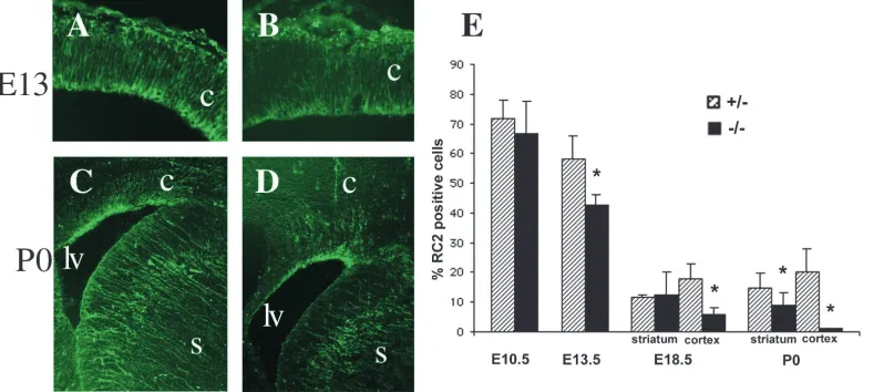
As another method to analyse the effect of TNC deficiency on neural stem cell populations, we also examined radial glial cells. These cells, which represent a subset of the total stem cell population in the embryonic CNS (Hartfuss et al., 2001; Heins et al., 2002), were identified using the RC2 antibody (Edwards et al., 1990). No changes in morphology were seen at either of the ages examined (E13 and P0), with processes remaining straight and lacking the sharp turns as seen, for example, in the Pax6 mutant cortex (Gotz et al., 1998). However, the intensity of RC2 labelling was diminished in the TNC-deficient CNS (Fig. 6A-D). To determine whether this reflected a reduction in the number of radial glia we performed RC2 immunocytochemistry on acutely dissociated cell populations (Fig. 6E). We found a reduction in RC2+ cells at E13.5, E18.5 and P0, demonstrating a reduction in radial glial numbers in the TNC-deficient CNS. At E10.5, at which stage RC2 labels neuroepithelial cells prior to the development of radial glia (Malatesta et al., 2003), no differences were seen between the genotypes.
To measure the formation of neurones and glia in the presence or absence of TNC, spheres were plated onto poly-D-lysine substrates under which conditions they flatten, with adherent cells on the substrate now differentiating into neurones, astrocytes and oligodendrocytes (Reynolds and Weiss, 1992; Zhou and Chiang, 1998). To confirm the presence of TNC in the neurospheres we performed RT-PCR analyses of spheres from wild-type or TNC null mice. We found TNC expression in cells derived from wild-type but not TNC-deficient mice (Fig. 7A). This shows that the TNC producing cells were either part of or derived from the neural stem cell population and that a comparison of the behaviour of neural stem cells within spheres from wild-type and transgenic mice is a valid test of the effects of TNC on neurogenesis and gliogenesis. We observed the generation of all three neural cell types from spheres grown from the cortex or striatum of either
[image:6.612.104.503.506.683.2]wild-type or TNC null animals. No quantitative differences were observed in the relative number of glial cells within the adherent cell population, although qualitative differences were observed as oligodendrocytes from null cells had longer processes than wild-type cells (Fig. 7B,C). However, we observed a significant increase in the relative number of neurones in striatum-derived spheres from TNC-deficient animals (Fig. 7D-F). This increase was seen in spheres grown in either EGF or FGF2 (Fig. 7F). Cortex-derived spheres from the TNC-deficient animals also showed increased neurogenesis, but only when grown in EGF (not shown). Rescue experiments in which exogenous TNC was added to the cultures grown in FGF2 reversed the increased neurogenesis seen in the spheres derived from TNC-deficient animals (Fig. 7G). We conclude, therefore, that TNC can inhibit neurogenesis in cells derived from either EGF- or FGF2-responsive neural stem cells without altering the number of glial cells. The overall effect of TNC is therefore to shift the balance of neural stem cell differentiation towards a glial fate.
Discussion
Although the fundamental properties of self-renewal and multipotency are retained by stem cells within the CNS throughout life, these cells undergo a developmental programme which results in their localization to a specific region of the CNS, the SVZ, and a switch in the major cell type produced by the stem cells from neurons to glia (Kilpatrick and Bartlett, 1995; Qian et al., 2000; Alvarez-Buylla et al., 2001; Sun et al., 2001). This developmental programme of the neural stem cells appears to be regulated by both intrinsic and extrinsic factors. The observation that single stem cells grown in micro culture give rise first to neurons and then later to glia in sequential cell divisions points to intrinsic timing mechanisms regulating the fate of the daughter cells (Qian et al., 1998; Qian et al., 2000). Conversely, the finding that FGF2 and BMP4 will promote and inhibit respectively the expression of the EGF receptor on neural stem cells points to the existence of extrinsic mechanisms (Ciccolini and Svendsen, 1998; Lillien and Raphael, 2000). Together, these mechanisms ensure that gliogenesis follows a period of neurogenesis in CNS development whilst maintaining a population of stem cells in the developing CNS that will subsequently persist throughout adult life (Alvarez-Buylla and Temple, 1998; Alvarez-Buylla et al., 2001). Here we provide evidence that ECM molecules expressed in the SVZ also contribute to this developmental programme by showing that TNC is an extrinsic regulator of neural stem cell behaviour. This conclusion is based on three lines of evidence. First, mice lacking TNC showed a delayed expression of the EGF receptor in cell populations containing neural stem cells both in vivo and in vitro. In the cell culture experiments this failure of normal EGF receptor expression could be rescued by the addition of exogenous TNC. Second, TNC-deficient mice have altered numbers of different neural stem cell populations. Third, neural stem cells from TNC-deficient mice show increased levels of neurogenesis.
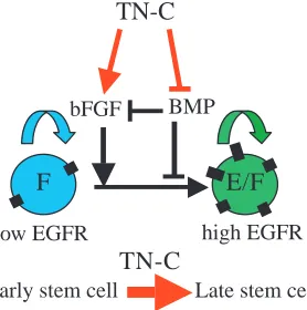
Neural stem cell development is accompanied by changes in response to growth factors such as FGF, BMPs and EGF as embryonic development proceeds (Burrows et al., 1997; Ciccolini and Svendsen, 1998; Kalyani et al., 1999; Tropepe et al., 1999; Lillien and Raphael, 2000; Ciccolini, 2001). We and others have shown previously that TNC can alter the response of cells to mitogenic growth factors, and our results here suggest that this provides a mechanism by which TNC facilitates normal stem cell development. In this model (Fig. 8), TNC modulates the sensitivity to the two growth factors, FGF2 and BMP4, that regulate EGF receptor expression so as to promote EGF receptor acquisition. TNC enhances sensitivity to FGF2, and the reduced sensitivity to FGF2 in the TNC null mice may contribute to the reduced level of proliferation we have previously noted in the SVZ of postnatal TNC-deficient mice (Garcion et al., 2001). However, the fact that we observe an increase rather than a decrease in FGF2-responsive stem cells in the absence of TNC emphasizes that the predominant effect of TNC on cells in the SVZ is
[image:7.612.52.350.77.288.2]the regulation of developmental progression rather than proliferation. In contrast to FGF2, TNC decreases the sensitivity to BMP4, as we observed in the TNC-deficient mice an increase in the inhibitory effects of BMP4 on EGF receptor expression. Several potential mechanisms exist by which these effects of TNC could be mediated. An extracellular interaction of the growth factor with TNC could inhibit or potentiate its effect, as seen with the interaction between FGF and heparan sulphate proteoglycans (Yayon et al., 1991; Nurcombe et al., 1993; Brickman et al., 1995; Caldwell and Svendsen, 1998). Alternatively, TNC could interact with a specific cell surface receptor, and intracellular components of the signalling pathway downstream of this TNC receptor could then interact with growth factor receptor signalling pathways (Jones and Jones, 2000), as we have shown for oligodendrocyte precursor cells in the TNC null mouse (Garcion et al., 2001). Finally, TNC could regulate intracellular signalling by the interaction with cell surface phosphatases such as members of the receptor-like protein tyrosine phosphatase family, which could then in turn regulate the activity of intracellular kinases (Milev et al., 1997). In keeping with this we observed increased levels of Smad1 phosphorylation in cells from TNC-deficient animals (E.G., unpublished). Smad1 is a downstream target of BMP4 (Kretzschmar et al., 1997) and increased phosphorylation (reflecting increased signalling activity in this pathway) in the absence of TNC points to an inhibitory effect of TNC on BMP4 signalling.
Why are the numbers of FGF2-responsive neural stem cells increased in the TNC-deficient mice? We hypothesize that this increase results from the accumulation of neural stem cells in the FGF2-responsive compartment as a result of their delayed exit into the next, EGF-responsive compartment. The delay is consistent with a ‘feed-forward’ model of neural stem cell development (Panchision et al., 2001) in which the transition to successive stem cell developmental stages is a consequence of signalling by the growth factor to which the stem cells within the previous stage are responsive. In this way, BMP and FGF2 have both been shown to promote proliferation of a particular stage of NSC development and the expression of the next growth factor receptor in the developmental progression, the BMPR1B receptor and the EGFR, respectively (Lillien and
Raphael, 2000; Panchision et al., 2001). Once these receptors reach a threshold level, signalling is initiated and the transition to the next developmental stage is made. Neural stem cells from TNC null mice show reduced sensitivity to the effects of FGF2. As we have shown, this reduced sensitivity inhibits EGFR expression and entry into the EGF-responsive stem cell compartment. We propose that this in turn expands the number of FGF2 stem cells left undergoing self-renewing divisions (which require lower levels of FGF2 signalling) in their compartment, although contributions by other mechanisms such as enhanced sensitivity of the FGF2-responsive NSCs to the proliferative as well as the inhibitory effects of BMPs in the absence of TNC, are obviously not excluded. Eventually, the development of the increased numbers of FGF2-responsive cells is sufficient to correct the deficit in the EGF-responsive neural stem cell number, even though the probability of an individual neural stem cell transiting from one compartment to the next may remain diminished. The reduction in the numbers of radial glial cells, a subset of the neural stem cell population that appears within the neuroepithelium at E13 (Hartfuss et al., 2001; Heins et al., 2002), also confirms that alterations of neural stem cell development are present throughout embryogenesis. Although the precise relationships between radial glia, neurosphere-forming cells and EGF-responsive neural stem cells are unknown, radial glia do develop from the original neuroepithelium so their reduction in the TNC-deficient mice would also be consistent with a role for TNC in promoting the developmental progression by which the neural stem cells of the neuroepithelium alter both growth factor sensitivity and cellular identity.
In this model a critical function for TNC is therefore the acceleration of the feed-forward mechanism and this may be one role for the observed maintenance of expression of TNC in the postnatal SVZ (Gates et al., 1995), so providing an environment that facilitates the production and differentiation of precursor cells when required in response to injury or cell loss. In addition, the expression of TNC by the radial glial cells themselves (Gotz et al., 1998) and the observation from gene expression profiling studies that TNC is very highly enriched in 8-day-old neurospheres (Ramalho-Santos et al., 2002) shows that TNC is produced by neural stem cells or their progeny, and will therefore provide an autocrine/paracrine factor in a positive feedback loop for neural stem cell development. A consequence may be the generation of a community effect within the germinal zones of the CNS. This effect was originally defined as a mechanism dependent on cell-cell interactions that ensured coordinated fate specification in developing cell populations (Gurdon, 1988), and cell-cell interactions have been shown to sharpen the boundaries of dose-response thresholds in Xenopus mesoderm fate determination (Green et al., 1994; Wilson and Melton, 1994). Community effects have also been shown to influence cell fate decisions in response to TGFβby neural crest progenitors, with either neural fates or apoptosis obtained within narrow ranges of increasing levels of TGFβ when cells were present in clusters (Hagedorn et al., 2000). Similar effects of cell-cell interactions based on the production of TNC within the germinal zones of the CNS may ensure the coordinated development of neural stem cells at appropriate FGF2 concentrations, so providing the extracellular matrix with a role
low EGFR high EGFR
TN-C
F
E/F
bFGF BMP
Early stem cell Late stem cell
[image:8.612.97.236.76.216.2]TN-C
in both the spatial and temporal regulation of neural stem cell development.
TNC also appears to inhibit neurogenesis, as TNC null neurospheres show a relative increase in the numbers of neurones when grown in conditions that allow differentiation. This result is surprising in light of the reduction observed in radial glia numbers in TNC-deficient animals. Radial glia generate neurones during embryonic development driven, at least in part, by the transcription factor Pax6 (Heins et al., 2002). If the reduction in radial glia was the only abnormality then TNC deficiency might be expected to result in lower, rather than higher, levels of neurogenesis. However, our work also shows an increase in the FGF2-responsive stem cell population in association with TNC deficiency, which could contribute to the increased neurogenesis as previous work has established that these stem cells are likely to differentiate into neurones (Levitt et al., 1983; Vescovi et al., 1993; Kilpatrick and Bartlett, 1995; Johe et al., 1996; Qian et al., 1997; Tropepe et al., 1999; Qian et al., 2000; Raballo et al., 2000). Moreover, this is unlikely to be the only mechanism as spheres grown in EGF also generate more neurones, and previous studies have suggested that the EGF-responsive neural stem cell population is distinct from the FGF2-responsive population (Tropepe et al., 1999). Although we cannot determine to what extent the increased neurogenesis in the absence of TNC reflects changes either in differentiation or in survival of neuronal and glial precursor populations, we can conclude from the rescue effect of exogenous TNC that prior differences in the balance of different stem cell populations are not solely responsible for the increased neurogenesis observed in the TNC-deficient spheres. TNC is therefore likely to regulate other transitions within the stem cell/precursor/neurone lineage in addition to that between FGF- and EGF-responsive stem cells. Further studies on the relationship between the FGF- and EGF-responsive neural stem cells and radial glia, and on the proliferation and differentiation of radial glial cells in the TNC mice will be required to elucidate the differentiation phenotype we have reported here.
Taken together our results show how TNC contributes to the regulation of the self-renewal and output of a stem cell population. Microenvironments that are able to regulate stem cell self-renewal and output in this way are commonly defined as niches, and are present in many different vertebrate and invertebrate stem cell systems. We conclude, therefore, that TNC is an important component of the neural stem cell niche in the SVZ. TNC is also present in the haematopoetic stem cell microenvironment (Klein et al., 1993), and the observation that mice lacking TNC show reduced haematopoesis (Ohta et al., 1998) confirms a role in the regulation of stem cell output in this example of a stem cell niche. Together with our present results, these observations raise the possibility that TNC may play an important general role in stem cell niches as a modulator of growth factor signalling. If so, however, why have TNC-deficient mice been shown to have a normal morphological phenotype in many different systems (Saga et al., 1992; Forsberg et al., 1996)? This may reflect the compensatory abilities of many developmental processes. We have described how reduced levels of apoptosis observed in newly differentiated oligodendrocytes in TNC-deficient mice could compensate for the reduced proliferation of oligodendrocyte precursor cells seen earlier in development
(Garcion et al., 2001). The ability to correct final cell numbers in this way could also correct any consequences of alterations in precursor numbers resulting from any changes in stem cell development. We would predict, however, that the ability of the CNS to respond to injury or perturbations of development would be compromised by these abnormalities in the stem cell compartment.
E.G. was supported by a grant from GlaxoSmithKline, and A.H. by a grant from Merck, Sharpe and Dohme. The work was also supported by the Multiple Sclerosis Society of Great Britain and Northern Ireland, and by the Wellcome Trust through a research leave fellowship to C.ff.-C, and by the German Research Council (DFG SPP-1109) to A.F.. We are grateful to Perry Bartlett, Siddarthan Chandran and Maeve Caldwell, in addition to members of the ffrench-Constant Laboratory for many helpful discussions.
References
Altman, J. and Bayer, S. A. (1990a). Horizontal compartmentation in the germinal matrices and intermediate zone of the embryonic rat cerebral cortex. Exp. Neurol. 107, 36-47.
Altman, J. and Bayer, S. A. (1990b). Vertical compartmentation and cellular transformations in the germinal matrices of the embryonic rat cerebral cortex. Exp. Neurol. 107, 23-35.
Alvarez-Buylla, A., Garcia-Verdugo, J. M. and Tramontin, A. D. (2001). A unified hypothesis on the lineage of neural stem cells. Nat. Rev. Neurosci. 2, 287-293.
Alvarez-Buylla, A. and Temple, S. (1998). Stem cells in the developing and adult nervous system. J. Neurobiol. 36, 105-110.
Bellows, C. G. and Aubin, J. E. (1989). Determination of numbers of osteoprogenitors present in isolated fetal rat calvaria cells in vitro. Dev. Biol. 133, 8-13.
Benoit, B. O., Savarese, T., Joly, M., Engstrom, C. M., Pang, L., Reilly, J., Recht, L. D., Ross, A. H. and Quesenberry, P. J. (2001). Neurotrophin channeling of neural progenitor cell differentiation. J. Neurobiol. 46, 265-280.
Brickman, Y. G., Ford, M. D., Small, D. H., Bartlett, P. F. and Nurcombe, V. (1995). Heparan sulfates mediate the binding of basic fibroblast growth factor to a specific receptor on neural precursor cells. J. Biol. Chem. 270, 24941-24948.
Burrows, R. C., Wancio, D., Levitt, P. and Lillien, L. (1997). Response diversity and the timing of progenitor cell maturation are regulated by developmental changes in EGFR expression in the cortex. Neuron 19, 251-267.
Caldwell, M. A. and Svendsen, C. N. (1998). Heparin, but not other proteoglycans potentiates the mitogenic effects of FGF-2 on mesencephalic precursor cells. Exp. Neurol. 152, 1-10.
Ciccolini, F. (2001). Identification of two distinct types of multipotent neural precursors that appear sequentially during CNS development. Mol. Cell. Neurosci. 17, 895-907.
Ciccolini, F. and Svendsen, C. N. (1998). Fibroblast growth factor 2 (FGF-2) promotes acquisition of epidermal growth factor (EGF) responsiveness in mouse striatal precursor cells: identification of neural precursors responding to both EGF and FGF-2. J. Neurosci. 18, 7869-7880.
Doetsch, F. (2003). The glial identity of neural stem cells. Nat. Neurosci. 6, 1127-1134.
Doetsch, F., Garcia-Verdugo, J. M. and Alvarez-Buylla, A. (1997). Cellular composition and three-dimensional organization of the subventricular germinal zone in the adult mammalian brain. J. Neurosci. 17, 5046-5061. Edwards, M. A., Yamamoto, M. and Caviness, V. S., Jr (1990). Organization
of radial glia and related cells in the developing murine CNS. An analysis based upon a new monoclonal antibody marker. Neuroscience 36, 121-144. Faissner, A. and Kruse, J. (1990). J1/tenascin is a repulsive substrate for central
nervous system neurons. Neuron 5, 627-637.
Forsberg, E., Hirsch, E., Frohlich, L., Meyer, M., Ekblom, P., Aszodi, A., Werner, S. and Fassler, R. (1996). Skin wounds and severed nerves heal normally in mice lacking tenascin-C. Proc. Natl. Acad. Sci. USA 93, 6594-6599.
Furuta, Y., Piston, D. W. and Hogan, B. L. (1997). Bone morphogenetic proteins (BMPs) as regulators of dorsal forebrain development. Development 124, 2203-2212.
reveal a contribution of the extracellular matrix molecule tenascin-C to neural precursor proliferation and migration. Development 128, 2485-2496. Gates, M. A., Thomas, L. B., Howard, E. M., Laywell, E. D., Sajin, B.,
Faissner, A., Gotz, B., Silver, J. and Steindler, D. A. (1995). Cell and molecular analysis of the developing and adult mouse subventricular zone of the cerebral hemispheres. J. Comp. Neurol. 361, 249-266.
Gotz, M., Bolz, J., Joester, A. and Faissner, A. (1997). Tenascin-C synthesis and influence on axonal growth during rat cortical development. Eur. J. Neurosci. 9, 496-506.
Gotz, M., Stoykova, A. and Gruss, P. (1998). Pax6 controls radial glia differentiation in the cerebral cortex. Neuron 21, 1031-1044.
Graham, A., Koentges, G. and Lumsden, A. (1996). Neural crest apoptosis and the establishment of craniofacial pattern: an honorable death. Mol. Cell. Neurosci. 8, 76-83.
Green, J. B., Smith, J. C. and Gerhart, J. C. (1994). Slow emergence of a multithreshold response to activin requires cell-contact-dependent sharpening but not prepattern. Development 120, 2271-2278.
Gritti, A., Frolichsthal-Schoeller, P., Galli, R., Parati, E. A., Cova, L., Pagano, S. F., Bjornson, C. R. and Vescovi, A. L. (1999). Epidermal and fibroblast growth factors behave as mitogenic regulators for a single multipotent stem cell-like population from the subventricular region of the adult mouse forebrain. J. Neurosci. 19, 3287-3297.
Gritti, A., Parati, E. A., Cova, L., Frolichsthal, P., Galli, R., Wanke, E., Faravelli, L., Morassutti, D. J., Roisen, F., Nickel, D. D. et al. (1996). Multipotential stem cells from the adult mouse brain proliferate and self-renew in response to basic fibroblast growth factor. J. Neurosci. 16, 1091-1100. Gross, R. E., Mehler, M. F., Mabie, P. C., Zang, Z., Santschi, L. and Kessler,
J. A. (1996). Bone morphogenetic proteins promote astroglial lineage commitment by mammalian subventricular zone progenitor cells. Neuron 17, 595-606.
Gurdon, J. B. (1988). A community effect in animal development. Nature 336, 772-774.
Hagedorn, L., Floris, J., Suter, U. and Sommer, L. (2000). Autonomic neurogenesis and apoptosis are alternative fates of progenitor cell communities induced by TGFbeta. Dev. Biol. 228, 57-72.
Hartfuss, E., Galli, R., Heins, N. and Gotz, M. (2001). Characterization of CNS precursor subtypes and radial glia. Dev. Biol. 229, 15-30.
Heins, N., Malatesta, P., Cecconi, F., Nakafuku, M., Tucker, K. L., Hack, M. A., Chapouton, P., Barde, Y. A. and Gotz, M. (2002). Glial cells generate neurons: the role of the transcription factor Pax6. Nat. Neurosci. 5, 308-315. Johe, K. K., Hazel, T. G., Muller, T., Dugich-Djordjevic, M. M. and McKay, R. D. (1996). Single factors direct the differentiation of stem cells from the fetal and adult central nervous system. Genes Dev. 10, 3129-3140. Jones, F. S. and Jones, P. L. (2000). The tenascin family of ECM glycoproteins:
structure, function, and regulation during embryonic development and tissue remodeling. Dev. Dyn. 218, 235-259.
Kalyani, A. J., Mujtaba, T. and Rao, M. S. (1999). Expression of EGF receptor and FGF receptor isoforms during neuroepithelial stem cell differentiation. J. Neurobiol. 38, 207-224.
Kawano, H., Ohyama, K., Kawamura, K. and Nagatsu, I. (1995). Migration of dopaminergic neurons in the embryonic mesencephalon of mice. Brain Res. Dev. Brain Res. 86, 101-113.
Kiernan, B. W., Garcion, E., Ferguson, J., Frost, E. E., Torres, E. M., Dunnett, S. B., Saga, Y., Aizawa, S., Faissner, A., Kaur, R. et al. (1999). Myelination and behaviour of tenascin-C null transgenic mice. Eur. J. Neurosci. 11, 3082-3092.
Kilpatrick, T. J. and Bartlett, P. F. (1993). Cloning and growth of multipotential neural precursors: requirements for proliferation and differentiation. Neuron 10, 255-265.
Kilpatrick, T. J. and Bartlett, P. F. (1995). Cloned multipotential precursors from the mouse cerebrum require FGF-2, whereas glial restricted precursors are stimulated with either FGF-2 or EGF. J. Neurosci. 15, 3653-3661. Klein, G., Beck, S. and Muller, C. A. (1993). Tenascin is a cytoadhesive
extracellular matrix component of the human hematopoietic microenvironment. J. Cell Biol. 123, 1027-1035.
Kornblum, H. I., Hussain, R. J., Bronstein, J. M., Gall, C. M., Lee, D. C. and Seroogy, K. B. (1997). Prenatal ontogeny of the epidermal growth factor receptor and its ligand, transforming growth factor alpha, in the rat brain. J. Comp. Neurol. 380, 243-261.
Kretzschmar, M., Liu, F., Hata, A., Doody, J. and Massague, J. (1997). The TGF-beta family mediator Smad1 is phosphorylated directly and activated functionally by the BMP receptor kinase. Genes Dev. 11, 984-995. Levitt, P., Cooper, M. L. and Rakic, P. (1983). Early divergence and changing
proportions of neuronal and glial precursor cells in the primate cerebral ventricular zone. Dev. Biol. 96, 472-484.
Li, W., Cogswell, C. A. and LoTurco, J. J. (1998). Neuronal differentiation of precursors in the neocortical ventricular zone is triggered by BMP. J. Neurosci. 18, 8853-8862.
Lillien, L. and Raphael, H. (2000). BMP and FGF regulate the development of EGF-responsive neural progenitor cells. Development 127, 4993-5005. Lim, D. A., Tramontin, A. D., Trevejo, J. M., Herrera, D. G.,
Garcia-Verdugo, J. M. and Alvarez-Buylla, A. (2000). Noggin antagonizes BMP signaling to create a niche for adult neurogenesis. Neuron 28, 713-726. Malatesta, P., Hack, M. A., Hartfuss, E., Kettenmann, H., Klinkert, W.,
Kirchhoff, F. and Gotz, M. (2003). Neuronal or glial progeny: regional differences in radial glia fate. Neuron 37, 751-764.
Milev, P., Fischer, D., Haring, M., Schulthess, T., Margolis, R. K., Chiquet-Ehrismann, R. and Margolis, R. U. (1997). The fibrinogen-like globe of tenascin-C mediates its interactions with neurocan and phosphacan/protein-tyrosine phosphatase-zeta/beta. J. Biol. Chem. 272, 15501-15509.
Nurcombe, V., Ford, M. D., Wildschut, J. A. and Bartlett, P. F. (1993). Developmental regulation of neural response to FGF-1 and FGF-2 by heparan sulfate proteoglycan. Science 260, 103-106.
Ohta, M., Sakai, T., Saga, Y., Aizawa, S. and Saito, M. (1998). Suppression of hematopoietic activity in tenascin-C-deficient mice. Blood 91, 4074-4083. Panchision, D. M., Pickel, J. M., Studer, L., Lee, S. H., Turner, P. A., Hazel, T. G. and McKay, R. D. (2001). Sequential actions of BMP receptors control neural precursor cell production and fate. Genes Dev. 15, 2094-2110. Qian, X., Davis, A. A., Goderie, S. K. and Temple, S. (1997). FGF2
concentration regulates the generation of neurons and glia from multipotent cortical stem cells. Neuron 18, 81-93.
Qian, X., Goderie, S. K., Shen, Q., Stern, J. H. and Temple, S. (1998). Intrinsic programs of patterned cell lineages in isolated vertebrate CNS ventricular zone cells. Development 125, 3143-3152.
Qian, X., Shen, Q., Goderie, S. K., He, W., Capela, A., Davis, A. A. and Temple, S. (2000). Timing of CNS cell generation: a programmed sequence of neuron and glial cell production from isolated murine cortical stem cells. Neuron 28, 69-80.
Raballo, R., Rhee, J., Lyn-Cook, R., Leckman, J. F., Schwartz, M. L. and Vaccarino, F. M. (2000). Basic fibroblast growth factor (Fgf2) is necessary for cell proliferation and neurogenesis in the developing cerebral cortex. J. Neurosci. 20, 5012-5023.
Ramalho-Santos, M., Yoon, S., Matsuzaki, Y., Mulligan, R. C. and Melton, D. A. (2002). ‘Stemness’: transcriptional profiling of embryonic and adult stem cells. Science 298, 597-600.
Reynolds, B. A. and Weiss, S. (1992). Generation of neurons and astrocytes from isolated cells of the adult mammalian central nervous system. Science 255, 1707-1710.
Richards, L. J., Kilpatrick, T. J. and Bartlett, P. F. (1992). De novo generation of neuronal cells from the adult mouse brain. Proc. Natl. Acad. Sci. USA 89, 8591-8595.
Saga, Y., Yagi, T., Ikawa, Y., Sakakura, T. and Aizawa, S. (1992). Mice develop normally without tenascin. Genes Dev. 6, 1821-1831.
Sheppard, A. M., Hamilton, S. K. and Pearlman, A. L. (1991). Changes in the distribution of extracellular matrix components accompany early morphogenetic events of mammalian cortical development. J. Neurosci. 11, 3928-3942.
Sun, Y., Nadal-Vicens, M., Misono, S., Lin, M. Z., Zubiaga, A., Hua, X., Fan, G. and Greenberg, M. E. (2001). Neurogenin promotes neurogenesis and inhibits glial differentiation by independent mechanisms. Cell 104, 365-376. Temple, S. (2001). The development of neural stem cells. Nature 414, 112-117. Tropepe, V., Sibilia, M., Ciruna, B. G., Rossant, J., Wagner, E. F. and van der Kooy, D. (1999). Distinct neural stem cells proliferate in response to EGF and FGF in the developing mouse telencephalon. Dev. Biol. 208, 166-188. Vescovi, A. L., Reynolds, B. A., Fraser, D. D. and Weiss, S. (1993). bFGF
regulates the proliferative fate of unipotent (neuronal) and bipotent (neuronal/astroglial) EGF-generated CNS progenitor cells. Neuron 11, 951-966.
Watt, F. M. and Hogan, B. L. (2000). Out of Eden: stem cells and their niches. Science 287, 1427-1430.
Whetton, A. D. and Graham, G. J. (1999). Homing and mobilization in the stem cell niche. Trends Cell Biol. 9, 233-238.
Wilson, P. A. and Melton, D. A. (1994). Mesodermal patterning by an inducer gradient depends on secondary cell-cell communication. Curr. Biol. 4, 676-686.
Yayon, A., Klagsbrun, M., Esko, J. D., Leder, P. and Ornitz, D. M. (1991). Cell surface, heparin-like molecules are required for binding of basic fibroblast growth factor to its high affinity receptor. Cell 64, 841-848. Zhou, F. C. and Chiang, Y. H. (1998). Long-term nonpassaged EGF-responsive