The Rockefeller University Press, 0021-9525/2002/09/863/10 $5.00
The Journal of Cell Biology, Volume 158, Number 5, September 2, 2002 863–872 http://www.jcb.org/cgi/doi/10.1083/jcb.200204127
JCB
Article
863
DNA replication is required for the checkpoint
response to damaged DNA in
Xenopus
egg extracts
Matthew P. Stokes,1 Ruth Van Hatten,1 Howard D. Lindsay,2 and W. Matthew Michael1
1The Biological Laboratories, Department of Molecular and Cellular Biology, Harvard University, Cambridge, MA 02138 2Genome Damage and Stability Center, University of Sussex Falmer, Brighton BN1 9QG, UK
lkylating agents, such as methyl methanesulfonate (MMS), damage DNA and activate the DNA damage checkpoint. Although many of the checkpoint proteins that transduce damage signals have been identified and characterized, the mechanism that senses the damage and activates the checkpoint is not yet understood. To address this issue for alkylation damage, we have reconstituted the
checkpoint response to MMS in Xenopus egg extracts.
Using four different indicators for checkpoint activation (delay on entrance into mitosis, slowing of DNA replication,
A
phosphorylation of the Chk1 protein, and physical associationof the Rad17 checkpoint protein with damaged DNA), we report that MMS-induced checkpoint activation is dependent upon entrance into S phase. Additionally, we show that the replication of damaged double-stranded DNA, and not replication of damaged single-stranded DNA, is the molecular event that activates the checkpoint. There-fore, these data provide direct evidence that replication forks are an obligate intermediate in the activation of the DNA damage checkpoint.
Introduction
Damaged chromosomal DNA activates a signaling path-way (checkpoint) that serves to protect the cell against the genotoxic consequences of continuation through the cell cycle in the presence of damage (Weinert and Hartwell, 1988). When DNA damage is encountered during the S phase of the cell cycle, two of the major checkpoint-controlled responses are the active prevention of entrance into mitosis and suppression of further DNA replication (for reviews see O’Connell et al., 2000; Zhou and Elledge, 2000). Ge-netic experiments in both budding and fission yeast have provided insight into how the damage checkpoint con-trols cell cycle progression. In fission yeast, DNA damage activates the Rad3 protein, which is a large, phosphatidyl-inositol-like protein kinase (for review see O’Connell et al., 2000). Activation of Rad3 leads to phosphorylation and activation of the effector kinases Chk1 and Cds1. Both Chk1 and Cds1 directly phosphorylate and
inacti-vate the Cdc25 protein phosphatase, thus preventing en-trance into mitosis.
Vertebrates contain two Rad3-like kinases, ataxia telangi-ectasia-mutated (ATM)* and the ATM- and Rad3-related protein kinase (ATR; for review see Abraham, 2001). ATM is activated by double-stranded DNA breaks (DSB), and promotes cell cycle arrest through phosphorylation of vertebrate Cds1 (also known as Chk2; for reviews see Zhou and Elledge, 2000; Abraham, 2001). ATR is activated by a wide range of DNA structural aberrations, including UV light–induced damage and DNA replication blocks. Upon activation, ATR directly phosphorylates vertebrate Chk1 (Abraham, 2001). Thus, unlike fission yeast, where the single Rad3 kinase phosphorylates both Chk1 and Cds1, in vertebrate cells ATM and ATR appear to specifically act upon Cds1 and Chk1, respectively. After activation by ATM/ATR, both Chk1 and Cds1 phosphorylate and inac-tivate the vertebrate Cdc25 orthologue Cdc25C (Zhou and Elledge, 2000).
In addition to ATM/ATR, another group of checkpoint proteins (Rad17, Rad9, Hus1, and Rad1) is required to transmit the presence of damage to Chk1/Cds1. Rad17 is homologous to the large subunit of the replication factor C (RFC) complex (Griffiths et al., 1995). RFC is a five-subunit complex (comprised of the RFC1–5 proteins) required during DNA replication to recognize primer ends and to load the PCNA clamp protein onto the primed templates. Rad17 has been shown to assemble into complexes with the
Address correspondence to W. Matthew Michael, The Biological Labora-tories, Dept. of Molecular and Cellular Biology, 16 Divinity Ave., Harvard University, Cambridge, MA 02138. Tel.: (617) 496-2940. Fax: (617) 384-7423. E-mail: [email protected]
*Abbreviations used in this paper: ATM, ataxia telangiectasia–mutated; ATR, ATM and Rad3-related; bio-dUTP, biotinylated dUTP; DSB, dsDNA break; dsDNA, double-stranded DNA; HSS, high speed superna-tant; MMS, methyl methanesulfonate; NPE, nucleoplasmic extract; preRC, prereplicative complex; ssDNA, single-stranded DNA; XB, extract buffer. Key words: cell cycle; Rad17; DNA damage; DNA replication; S phase
on June 10, 2008
www.jcb.org
Downloaded from
864 The Journal of Cell Biology|Volume 158, Number 5, 2002
RFC2–5 subunits, to form what might be a damaged DNA-specific clamp-loading protein (Shimomura et al., 1998; Shimada et al., 1999; Green et al., 2000; Lindsey-Boltz et al., 2001). Consistent with this idea, the Rad9, Hus1, and Rad1 proteins display homology to PCNA, and interact with one another to form a complex (the 9–1-1 complex; for review see Melo and Toczyski, 2002). Both Rad17 and 9–1-1 associate with damaged chromatin (Burtelow et al., 2000; Kai et al., 2001; Kondo et al., 2001; Melo et al., 2001; Zou et al., 2002), and chromatin association of 9–1-1 has been shown to be dependent on Rad17 (Kondo et al., 2001; Melo et al., 2001; Zou et al., 2002).
A major question in the cell cycle field is how checkpoint proteins sense damaged DNA to activate the checkpoint re-sponse. This problem is especially vexing for recognition of damage induced by either alkylating agents or UV light, as the lesions created by these particular genotoxic agents are not as overt as DSBs, and often exhibit modest chemical dif-ferences relative to undamaged DNA. One possibility is that the lesions themselves are not recognized by the checkpoint, rather the checkpoint senses a consequence of the presence of the lesion. For example, both alkylation and UV light–induced lesions are known to block replication fork progression in prokaryotic systems (Friedberg et al., 1995). Therefore, in eu-karyotes, it is possible that the checkpoint actually senses the stalled replication fork, and is otherwise incapable of sensing the damage. Although this model is appealing, direct experi-mental evidence that the replication of double-stranded DNA (dsDNA) is essential for checkpoint activation in re-sponse to alkylation or UV light–induced damage has not yet been provided. Additionally, any model that assumes that replication forks are essential components of the damage sensing activity that activates the checkpoint must account for how, or if, these lesions are detected outside of S phase.
Most of what is known about the cell cycle phase depen-dence of damage checkpoint activation has come from ge-netic analysis in budding yeast. For UV light–induced dam-age, checkpoint activation occurs in all phases of the cell cycle (Siede et al., 1993, Vialard et al., 1998) when repair is proficient, but is restricted to S phase when repair is defi-cient (Neecke et al., 1999). One interpretation of these find-ings is that the act of repairing the damage is sufficient to ac-tivate the checkpoint, perhaps through generation of nucleic acid intermediates such as single-stranded DNA (ssDNA), but when repair is prevented, the checkpoint is then depen-dent on stalled replication forks for activation. Thus, in this scenario, repair and replication serve redundant functions in checkpoint activation.
In the case of alkylation damage, the work in budding yeast has yielded less conclusive results. Paulovich and Hartwell showed that when budding yeast cells are synchro-nized in G1 with factor mating pheromone, and then re-leased back into the cell cycle in the presence of methyl methanesulfonate (MMS), the cell cycle is not significantly slowed until cells enter S phase (Paulovich and Hartwell, 1995; Paulovich et al., 1997). This suggests that budding yeast cells do not mount a rigorous G1 response to MMS. Consistent with this, it has been shown that MMS-induced attenuation of the utilization of origins of replication in budding yeast is restricted to late-firing origins, even if
dam-age is induced during G1 (Shirahige et al., 1998). The fact that only late-firing origins are negatively regulated by MMS suggests that cells must enter S phase in order to generate the signal that blocks origin firing, which is consistent with the lack of a G1 response to alkylation damage. In contrast to these findings, however, are studies that used a different cri-terion for checkpoint activation, that being phosphorylation of the Rad53 checkpoint protein (a substrate of the budding yeast ATR homologue Mec1). In these experiments, it was shown that when cells harboring temperature-sensitive mu-tations in any number of genes that are required for S phase entry were incubated at the restrictive temperature, then Rad53 was still phosphorylated in response to MMS (Pellici-oli et al., 1999). Similarly, in another study, when cells were blocked in G1 with factor, they still induced Rad53 phos-phorylation in response to MMS (Vialard et al., 1998). Thus, cells arrested in G1 have been shown to be capable of detecting the alkylation damage, and activating Rad53. Therefore, these data are difficult to reconcile with the stud-ies on the regulation of origin firing in response to MMS, which is known to require Rad53 (Shirahige et al., 1998). If Rad53 activation can occur in G1 in response to MMS, why aren’t early firing origins prevented from firing when MMS is added before S phase entry?
To address the question of whether detection of alkylation damage by the checkpoint is restricted to S phase, and to ex-plore the mechanism for checkpoint activation during S phase, we have taken a biochemical approach using Xenopus
egg extracts. We report here that the mechanism that senses alkylation damage operates exclusively during S phase, and is dependent on the replication of dsDNA molecules. These results indicate that replication forks represent the major damage-sensing activity for activation of the DNA damage checkpoint in response to alkylation damage.
Results
Alkylation damage of DNA activates the DNA damage checkpoint in Xenopus egg extracts
To address how alkylation damage of DNA generates a checkpoint signal in Xenopus egg extracts, we first sought to establish that the egg extract system induces a bona fide checkpoint response to MMS-induced damage. Therefore, demembranated sperm chromatin was treated with MMS to alkylate the DNA, and the MMS was then removed from the chromatin by multiple washings. To determine if the alkyl-ated sperm chromatin triggers the DNA damage checkpoint in vitro, we used cycling egg extracts (Murray, 1991). These extracts are produced by low speed centrifugation of freshly laid eggs. Upon supplementation with an energy regenerat-ing system and sperm chromatin, the extracts assemble a nu-cleus around the chromatin, the DNA is replicated, and, af-ter replication, the extracts activate the cdc2-cyclin B kinase and enter mitosis. It has been shown that damaged DNA in the form of DSBs or UV light–induced lesions, generates a signal that prevents mitosis in cycling egg extracts (Kumagai et al., 1998; Michael and Newport, 1998; Guo and Dunphy, 2000). To ask if alkylation damage generates such a signal, either control or alkylated chromatin was mixed with cycling extracts and entrance into mitosis was determined
on June 10, 2008
www.jcb.org
Downloaded from
Replication-dependent checkpoint activation | Stokes et al. 865
scopically through visualization of nuclear envelope break-down (Murray, 1991). We found that the alkylated chroma-tin induced a substantial delay on entrance into mitosis (Fig. 1 A), as extracts containing MMS-treated chromatin entered mitosis an average of 75 min after a control reaction contain-ing undamaged DNA entered mitosis. As expected, DSBs also generated a signal that delayed mitosis in these extracts. To determine if the lag on entrance into mitosis was due to activation of the DNA damage checkpoint, we also per-formed the experiments in the presence of 5 mM caffeine. Caffeine is an inhibitor of both the ATM and ATR check-point kinases (Sarkaria et al., 1999), and inclusion of caffeine completely reversed the mitotic delay imposed by MMS-treated chromatin and DSBs (Fig. 1 A).
In budding yeast, MMS-induced damage activates the Mec1 protein kinase. The metazoan Mec1 homologue is ATR, and it was therefore of interest to determine if ATR activation occurs in response to alkylation damage in Xeno-pus. To do so, we used a previously published Chk1 phos-phorylation assay (Michael et al., 2000). Chk1 is an ATR substrate, and removal of ATR from egg extract by
immu-nodepletion prevents Chk1 phosphorylation in response to stalled replication forks or UV light–induced damage (Guo et al., 2000). Therefore, Chk1 phosphorylation is a reliable indicator of activation of ATR kinase in Xenopus extracts. To assay for ATR activation, cycling extracts were prepared and supplemented with a radio-labeled fragment of Xenopus
Chk1 (Chk1 KD; for review see Michael et al., 2000). Af-ter a 60-min incubation period, a sample of the extract was removed and analyzed by SDS-PAGE for slower migrating forms of Chk1 KD, which we have previously shown to re-sult from checkpoint-mediated phosphorylation (Michael et al., 2000). As shown in Fig. 1 B, the DNA replication inhib-itor (aphidicolin) induced phosphorylation of Chk1 KD, as expected (Michael et al., 2000), as did MMS treatment of the chromatin sample. Chk1 KD phosphorylation was not observed in an extract containing control, undamaged sperm chromatin, nor in an extract containing DSBs, as expected from published data showing that DSBs do not induce Chk1 phosphorylation (Guo and Dunphy, 2000). Taken together, the data in Fig. 1 show that MMS treatment of sperm chromatin activates the ATR checkpoint kinase and delays entrance into mitosis in frog egg extracts.
MMS treatment has been shown to slow progression through S phase in human (Painter, 1977) and yeast (Pau-lovich and Hartwell, 1995) cells. Therefore, we asked if DNA replication in frog egg extracts is affected by alkyla-tion. To study replication, we turned to a soluble system for DNA replication analysis that makes use of concentrated nucleoplasmic extracts (NPEs; Walter and Newport, 1999). To prepare NPE, nuclei are formed in the conventional ex-tract system. The nuclei are then purified, and the soluble, nucleoplasmic fraction from the purified nuclei is extracted. Although it is devoid of membranes, this soluble fraction contains all of the components required for rapid, complete, and cell cycle–regulated chromosomal replication. To mea-sure DNA replication in this system, DNA templates are first incubated in the high speed supernatant (HSS) of egg extract. This incubation allows prereplicative complex (preRC) formation to occur before the addition of NPE, and is necessary because NPE contains activities that are in-hibitory toward preRC assembly. Upon NPE addition, DNA replication initiates synchronously from all active ori-gins, and rapidly goes to completion (Walter et al., 1998).
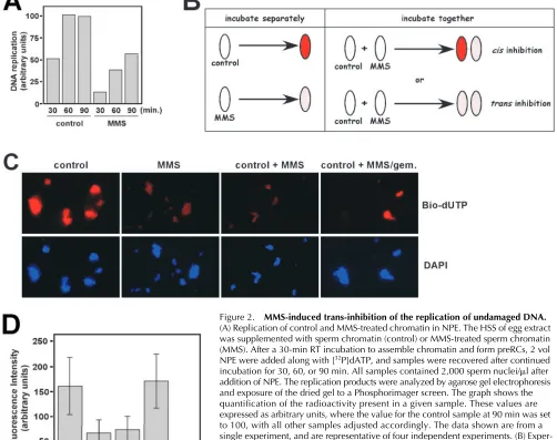
[image:3.612.81.263.55.262.2]To compare the replication of MMS-treated chromatin with undamaged chromatin in NPE, we incubated both types of chromatin first in HSS, to assemble the preRCs, and then NPE, along with radio-labeled dATP, was added to initiate replication. Aliquots of the reaction were taken every 30 min for 90 min, and the amount of replication was determined by agarose gel electrophoresis of the radio-labeled replication products. As shown in Fig. 2 A, MMS treatment of the chro-matin induced a substantial delay in the kinetics of DNA rep-lication. After 30 min, the alkylated sample had replicated to just 26% of the control; after 60 min, the control sample had completed replication, whereas the MMS-treated sample had replicated to 38% of the level of the control sample. Even after 90 min, the MMS-treated sample had not finished replication, and was just 57% of the final value for the control. From this experiment, we conclude that MMS treatment slows DNA replication in Xenopus, just as it does in yeast and human cells.
Figure 1. Alkylation damage of DNA activates the DNA damage checkpoint in Xenopus egg extracts. (A) Cycling extracts were prepared and supplemented with sperm chromatin (control), MMS-treated sperm chromatin (MMS), or sperm chromatin and linearized plasmid DNA at 25 ng/l (DSB). Additionally, where indicated, 5 mM caffeine was also included. Extracts were incubated at RT and examined for entrance into mitosis by DAPI staining of nuclei to visualize nuclear envelope breakdown. An extract was scored as mitotic when 50% of the nuclei had undergone nuclear envelope breakdown. The data are plotted as mitotic delay, which is the difference in time between entrance into mitosis for the control extract and entrance into mitosis for the experimental extracts. Control extracts typically required 55–70 min to enter mitosis. All samples contained 2,000 sperm nuclei/l. (B) 35S-labeled Chk1 KD protein (Michael et al., 2000) was added to cycling egg extracts along with the following: sperm chromatin and no further additions (-), or sperm chromatin and aphidicolin (100 g/ml; aphid.), MMS-treated chromatin (MMS), or sperm chromatin and plasmid DNA that had been linearized by restriction enzyme digestion (25 ng/l; DSB). After a 60-min incubation, samples were recovered and analyzed by SDS-PAGE for mobility shifts of the labeled proteins. The input lane shows the Chk1 KD protein before incubation with Xenopus egg extract.
on June 10, 2008
www.jcb.org
Downloaded from
866 The Journal of Cell Biology|Volume 158, Number 5, 2002
In budding yeast, MMS treatment causes a checkpoint-dependent inhibition of further DNA replication through generation of a diffusible signal that inhibits late origin fir-ing (Shirahige et al., 1998), and a checkpoint-independent inhibition of chain elongation (Tercero and Diffley, 2001). To ask if alkylation damage generates a diffusible signal that blocks further DNA replication in the frog egg extract
[image:4.612.62.564.69.466.2]sys-tem, we next asked if the MMS-induced replication delay was limited to the alkylated DNA, or if it also extended to undamaged DNA. To do so, replication of individual sperm chromatin templates within mixed populations of damaged and undamaged DNA was assessed by fluorescence micros-copy in NPE. Because NPE does not contain membranes, nuclear envelopes do not form around sperm chromatin
Figure 2. MMS-induced trans-inhibition of the replication of undamaged DNA.
(A) Replication of control and MMS-treated chromatin in NPE. The HSS of egg extract was supplemented with sperm chromatin (control) or MMS-treated sperm chromatin (MMS). After a 30-min RT incubation to assemble chromatin and form preRCs, 2 vol NPE were added along with [32P]dATP, and samples were recovered after continued
incubation for 30, 60, or 90 min. All samples contained 2,000 sperm nuclei/l after addition of NPE. The replication products were analyzed by agarose gel electrophoresis and exposure of the dried gel to a Phosphorimager screen. The graph shows the quantification of the radioactivity present in a given sample. These values are expressed as arbitrary units, where the value for the control sample at 90 min was set to 100, with all other samples adjusted accordingly. The data shown are from a single experiment, and are representative of four independent experiments. (B) Exper-imental strategy. When control (undamaged) or MMS-treated chromatin are incubated separately in NPE containing fluorescent nucleotides, the control sample will appear brighter due to its enhanced ability to undergo DNA replication. To ask if the MMS-treated sample inhibits DNA replication in trans, control and MMS-MMS-treated chromatin are incubated together in the same NPE. If the inhibition works in cis only, then two populations of fluorescent chromatin will result: a bright population (corresponding to the control template) and a dim population (corresponding to the MMS-treated sample). However, if inhibition can also work in trans, then all chromatin templates in the coincubation are expected to exhibit reduced fluorescence, relative to the control sample alone. (C) Either control, undamaged sperm chromatin (control), or MMS-treated sperm chromatin (MMS), or a 50:50 mixture of both (control MMS) were incubated for 30 min in HSS. After the 30-min incubation, NPE containing bio-dUTP was added to the reactions. Incubation was performed for an additional 30 min before processing of the samples for detection of bio-dUTP incorporation with fluorescent streptavidin. In the panel labeled control MMS/gem., MMS-treated chromatin was incubated in HSS with recombinant geminin for 30 min, then combined with control, undamaged chromatin that had been incubated separately in HSS-lacking geminin, also for 30 min. NPE-containing bio-dUTP was then added to the combined sample. Panels labeled Bio-dUTP display signal obtained from staining of the samples with Texas red–conjugated streptavidin, to detect the bio-dUTP, and panels labeled DAPI correspond to DAPI staining of the samples to visualize the DNA. (D) Quantification of the data presented in C. The fluorescent intensity of a minimum of 50 nuclei for each sample from each of two independent experiments was determined using the Scion Image software package from images obtained from a fluorescence microscope (BX51; Olympus) attached to a Spot camera (Diagnostic Instruments, Inc.). The control MMS/gem. sample is represented by two bars (labeled dim and bright) to provide individual quantification for the two classes of nuclei present in this particular sample.
on June 10, 2008
www.jcb.org
Downloaded from
Replication-dependent checkpoint activation | Stokes et al. 867
templates when they are incubated in NPE (Walter et al., 1998). Thus, there are no physical barriers separating dam-aged and undamdam-aged DNA if they are coincubated in the same NPE. Given this, we reasoned that if replication of un-damaged DNA was affected by the presence of alkylated DNA, then this could be uncovered through the visualiza-tion of the replicavisualiza-tion of individual sperm chromatin tem-plates in NPEs containing a mixture of damaged and un-damaged samples (see Fig. 2 B). To visualize replication of individual nuclei within a population, biotinylated dUTP (bio-dUTP) was added to NPE during the replication reac-tion, and, after incubareac-tion, the samples were fixed and stained with fluorescent streptavidin to monitor uptake of the bio-dUTP within individual nuclei.
As shown in Fig. 2 C (control), when undamaged sperm chromatin alone was used in the experiment, the templates were highly fluorescent, indicating that the bio-dUTP was efficiently being incorporated into the replication templates. In contrast, when the MMS-treated sperm chromatin was used in the reaction, the fixed samples were not nearly as bright (Fig. 2 C, MMS). Quantification of the fluorescence intensity of 100 representative nuclei demonstrates that rep-lication in the MMS-treated sample was 41% that of the control sample, which is consistent with the reduced level of DNA synthesis in NPE containing alkylated templates, as measured by uptake of radio-labeled nucleotides (see Fig. 2 A). To ask if the presence of alkylated chromatin affected replication of undamaged chromatin, we analyzed a sample containing 50% control and 50% MMS-treated chromatin (Fig. 2 C, control MMS). We found the level of fluores-cence in this mixed sample was uniform, indicating that all templates had replicated to the same relative level, but that this level was less than that observed in the sample contain-ing undamaged sperm chromatin alone. Indeed, quantifica-tion showed that the level of replicaquantifica-tion in the mixed sample was very similar to that observed in the sample containing MMS-treated chromatin alone (see Fig. 2 D). From this ex-periment, we conclude that alkylation damage of DNA gen-erates a diffusible inhibitor that negatively regulates the rep-lication of undamaged DNA when coincubated in NPE. Taken together with the data in Fig. 1, these experiments also show that the canonical features of the DNA damage re-sponse (delay on entrance into mitosis, slowing of DNA rep-lication, and activation of ATR) are recapitulated in the cell-free egg extract system.
DNA replication is required to generate the MMS-induced DNA damage signal
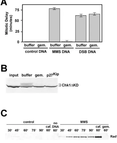
Having established that MMS treatment of sperm chroma-tin activates a bona fide checkpoint response in this egg ex-tract system, we next turned to the question of the S phase dependence of activation of this checkpoint. To do so, we first examined the ability of MMS-treated chromatin to in-hibit mitosis in cycling extracts containing the DNA replica-tion inhibitor, geminin (McGarry and Kirschner, 1998). Geminin targets Cdt1, an essential replication factor that is a component of the preRCs that form on origins of replica-tion before S phase entry (Wohlschlegel et al., 2000; Tada et al., 2001). Therefore, chromatin that is formed in geminin-containing extracts does not contain preRCs, and cannot
initiate DNA replication. As shown in Fig. 3 A, and as ex-pected (for review see Michael et al., 2000), geminin had no influence on the kinetics of entrance into mitosis of the con-trol sample. In contrast, in extracts containing MMS-treated chromatin, the addition of geminin completely abrogated the mitotic delay normally seen in the alkylated DNA–con-taining samples (Fig. 3 A). Geminin itself is not a global checkpoint inhibitor, as it had no effect at all on the ability of DSBs to induce a mitotic delay in cycling extracts (Fig. 3 A), and its ability to block the alkylation damage checkpoint was lost if geminin were added to the reaction after preRCs had assembled on the chromatin (unpublished data).
Con-Figure 3. The initiation of DNA replication is required to generate the MMS damage signal. (A) Cycling extracts were prepared and supplemented with either PBS (buffer) or recombinant geminin (gem.). The samples were then further supplemented with either sperm chromatin (control DNA), MMS-treated sperm chromatin (MMS DNA) or sperm chromatin and plasmid DNA that had been linearized by restriction enzyme digestion (25 ng/l; DSB DNA). Entrance into mitosis was determined as in Fig. 1 A, and the data are plotted as in Fig. 1 A. (B) Cycling egg extracts were prepared and supplemented with cyclohexamide and either PBS (buffer), recom-binant geminin (250 nM; gem.), or recomrecom-binant p27Kip (500 nM).
Extracts were further supplemented with MMS-treated chromatin and 35S-labeled Chk1 KD protein. After a 60-min incubation, samples were recovered for SDS-PAGE. The input lane shows the Chk1 KD protein before incubation with Xenopus egg extract. (C) Cycling extracts were prepared and supplemented with cyclohexamide and either control, undamaged sperm chromatin, or MMS-treated sperm chromatin. Chromatin was isolated from these extracts at the indicated times (in minutes), and probed for the presence of Rad17 using anti–Xenopus Rad17 antibodies. Where indicated, the samples also included caffeine (caf., at 5 mM) or recombinant geminin (gem., at 250 nM). The sample labeled no DNA refers to a sample that was processed in the absence of any sperm chromatin addition, showing that Rad17 recovery is dependent on the addition of chro-matin. Sperm chromatin was added to 2,000/l in all samples.
on June 10, 2008
www.jcb.org
Downloaded from
[image:5.612.307.542.64.351.2]868 The Journal of Cell Biology|Volume 158, Number 5, 2002
sistent with the loss of mitotic delay in extracts containing alkylated chromatin and geminin, we also found that gemi-nin prevented the ability of alkylated chromatin to induce phosphorylation of Chk1 KD (Fig. 3 B). It was important to ensure that damage checkpoint activation in response to MMS was dependent on DNA replication, and was not blocked due to an unknown activity of geminin. Therefore, we used p27Kip (Toyoshima and Hunter, 1994), an inhibitor
of the cdk2-cyclin E kinase that is required to initiate DNA replication. Treatment of extracts with p27Kip allows preRC
assembly, but prevents replication forks from assembling at origins of replication (Michael et al., 2000). As shown in Fig. 3 B, treatment of extracts with p27Kip also prevented
Chk1 KD phosphorylation in response to the MMS-treated chromatin. Thus, two independently acting inhibi-tors of entrance into S phase, geminin and p27Kip, both
block Chk1 phosphorylation in response to alkylation dam-age. From the data in Fig. 3, we conclude that Xenopus egg extracts must enter S phase in order to generate the DNA damage checkpoint signal that activates ATR, and delays en-trance into mitosis.
Next, we asked if DNA replication was required for the MMS-treated sperm chromatin to inhibit, in trans, the rep-lication of undamaged DNA (Fig. 2). To do so, we estab-lished conditions where preRC assembly was selectively in-hibited on alkylated chromatin, and then coincubated this chromatin, in NPE, with undamaged chromatin that con-tained intact preRCs. To block preRC assembly, alkylated chromatin was incubated in HSS containing geminin. Con-trol, undamaged chromatin was incubated in HSS lacking geminin, and consequently contained intact preRCs. Once preRCs are formed, geminin can no longer inhibit DNA replication (McGarry and Kirschner, 1998). After separate incubations in HSS, the two samples were combined in the same NPE, and replication was measured by uptake of bio-dUTP. Unlike the experiment shown in Fig. 2 C (control MMS), where the fluorescence intensity was low and uni-form from sample to sample, the experiment containing un-damaged chromatin with geminin-treated un-damaged chroma-tin (Fig. 2 C, control MMS/gem.) showed two distinct classes of chromatin samples, one very bright (105% of the control sample after quantification of the fluorescence inten-sity) and the other exhibiting low levels of fluorescence (13% after quantification of the fluorescence intensity). Thus, the geminin-treated damaged chromatin, unlike the damaged chromatin lacking geminin, could not reduce the fluorescence intensity of the undamaged sample. From this data, we conclude that the alkylated chromatin must initiate replication in order to generate the trans-acting signal that blocks replication on undamaged replicons.
The Rad17 checkpoint protein associates specifically with damaged DNA in a DNA replication–dependent manner
Rad17 is thought to be part of a complex that loads the 9–1-1 complex onto damaged DNA (for review see Melo and Toczyski, 2002). Consistent with this, Rad17 itself has been shown to physically associate with chromatin in both human (Zou et al., 2002) and fission (Kai et al., 2001) yeast cells. Because Rad17 is required for checkpoint activation in all
systems tested, we determined its chromatin binding proper-ties in Xenopus egg extracts. Egg extracts were prepared and supplemented with either control, undamaged sperm chro-matin, or with sperm chromatin that had been treated with MMS. Chromatin was isolated after 30, 45, 60, 75, and 90 min of incubation by sucrose density centrifugation, and probed by immunoblotting for the presence of Xenopus
Rad17. As shown in Fig. 3 C, little if any Rad17 could be detected in any of the undamaged chromatin samples except for the 45-min time point. This time point corresponds to maximal DNA replication activity in these extracts, and it is therefore tempting to speculate that this low level associa-tion of Rad17 with actively replicating chromatin is func-tionally significant. In contrast to the undamaged chroma-tin, when MMS-treated chromatin was added to the extract, we found a robust and time-dependent association of Rad17 with the chromatin. This association peaked by 60 min, and showed a modest decrease at 90 min. Interestingly, Rad17 chromatin association was markedly enhanced when the ATR inhibitor caffeine was included in the extract. To ask if DNA replication was required for Rad17 chromatin associa-tion with MMS-treated DNA, the experiment was per-formed in the presence of geminin. As shown in Fig. 3 C, no Rad17 could be detected in the MMS-treated chromatin fraction purified from extract containing geminin. Taken to-gether, the data in Fig. 3 C show that Rad17 specifically in-teracts with damaged DNA, and that this interaction re-quires ongoing DNA replication. If Rad17 association with damaged chromatin were a prerequisite for checkpoint acti-vation, as seems likely (for review see Kai et al., 2001), then this would explain the requirement for DNA replication in activation of the DNA damage checkpoint.
DNA synthesis, per se, is not sufficient for damage checkpoint activation
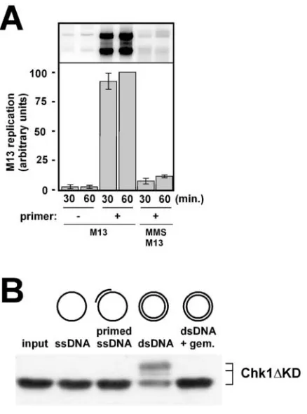
The data in Figs. 2 and 3 show a clear S phase dependence for multiple aspects of checkpoint signaling, including the blocks to mitosis and further DNA replication, activation of ATR kinase, and physical association of Rad17 with chroma-tin. There are many possible reasons for this requirement for S phase entry. For example, unwinding of the DNA duplex before replication may be the critical event, as unwinding generates ssDNA and/or exposes lesions to the checkpoint sensors. Indeed, during DSB repair, it is has been shown that the amount of ssDNA that is produced during the repair re-action influences the intensity of the checkpoint response (Lee et al., 1998). Therefore, it is possible that alkylated ssDNA is sufficient to activate the checkpoint. Alternatively, physical contact between DNA polymerase and the lesion may be the event that triggers checkpoint activation. If this were so, then primer extension on alkylated ssDNA tem-plates could, in principle, activate the checkpoint. It is also possible that some aspect of dsDNA replication, such as physical contact between the replicative helicase and lesions, is the signal that activates the checkpoint. To begin to ad-dress which aspect of DNA replication is required to gener-ate the MMS damage signal, we compared the ability of alkylated M13 circular ssDNA templates to alkylated plas-mid dsDNA templates to activate the checkpoint in the NPE system. We chose NPE because this system, unlike
on June 10, 2008
www.jcb.org
Downloaded from
Replication-dependent checkpoint activation | Stokes et al. 869
tional egg extracts, is capable of efficient and cell cycle–regu-lated replication of simple DNA substrates such as plasmid DNAs (Walter et al., 1998). To prepare for this analysis, rep-lication of undamaged M13 ssDNA was first assessed in NPE. We found that M13 ssDNA replication in NPE re-quires a preannealed primer, as samples that lack a primer are not replicated (Fig. 4 A). This was surprising, as it is well es-tablished that M13 ssDNA replication in conventional egg
extracts is not dependent on a primer. At present, we do not understand why M13 ssDNA replication is primer-depen-dent in NPE. If the M13 ssDNA was treated with MMS be-fore incubation in NPE, then replication was blocked despite the presence of a preannealed primer (Fig. 4 A). This shows that alkylation blocks even simple primer extension reac-tions, and supports the idea that MMS-induced lesions form a physical blockade to polymerase movement in eukaryotes, as has been shown in prokaryotes (Friedberg et al., 1995).
Having shown that alkylation blocks the primer-depen-dent replication of M13 ssDNA in NPE, we next assessed the ability of the M13 templates to activate the damage checkpoint in NPE. As a positive control, we determined that alkylation of closed circular dsDNA induced phosphor-ylation of Chk1 in NPE (Fig. 4 B). Furthermore, we found that checkpoint activation by the dsDNA molecules re-quired DNA replication, as the Chk1 phosphorylation did not occur in extracts containing geminin. In contrast to the dsDNA, we found that neither the primed nor unprimed alkylated M13 ssDNA templates were capable of activating the checkpoint (Fig. 4 B). The inability of the ssDNA to trigger a checkpoint response was not due to alkylation, as unalkylated M13 ssDNA also failed to trigger the check-point (unpublished data). From these data, we conclude that large amounts of ssDNA are not sufficient to activate the checkpoint. Therefore, the involvement of DNA replication in activation of the checkpoint is likely to extend beyond simple unwinding of the template strands. The experiment also shows that the attempt at DNA synthesis, per se, is also not sufficient to activate the checkpoint. If so, then the in-clusion of the primer on the MMS-treated M13 ssDNA templates would be expected to activate the checkpoint. Based on these results, we conclude that replication of dam-aged dsDNA molecules is the S phase activity that generates the alkylation damage signal for checkpoint activation.
Discussion
In this article, we have used Xenopus egg extracts to address an important area in cell cycle checkpoint research, how damaged DNA is sensed to trigger the checkpoint response. In particular, we examined the checkpoint response to al-kylation damage, as induced by the commonly used geno-toxic agent MMS. The findings reported here make three important points with regard to how alkylation damage is sensed by the checkpoint in Xenopus. First, MMS-induced checkpoint activation can be recapitulated in the in vitro egg extract system. Second, the ability to activate the checkpoint in response to alkylation damage requires entrance into S phase. Third, DNA synthesis, per se, is not sufficient for checkpoint activation, and neither is the accumulation of ssDNA. Rather, a structure that forms when replication is attempted on damaged dsDNA constitutes the signal for ac-tivation of the checkpoint in response to alkylation damage.
Recapitulation of the alkylation damage checkpoint in Xenopus egg extracts
[image:7.612.64.283.58.353.2]To apply biochemical approaches to the question of damage checkpoint activation, it was first necessary to establish that key features of the checkpoint response to alkylation damage
Figure 4. Replication structures specific for dsDNA replication are required to generate the MMS damage signal. (A) M13 ssDNA was annealed to an oligonucleotide primer, or not, and then incubated in NPE containing [32P]dATP (final concentration of M13 ssDNA
was 5 ng/l). Additionally, M13 ssDNA was alkylated, annealed to a primer, and incubated in NPE at 5 g/ml. After either 30 or 60 min, aliquots were removed from the reaction and processed for DNA replication analysis on agarose gels. The top portion of the figure shows the gel, and beneath it is a bar graph showing the values obtained after analysis of the scanned gel on a Phosphorimager. After quantification of the data by Phosphorimager analysis, the value for the 60-min time point for M13 ssDNA plus primer was set to 100, and all other data were normalized accordingly. (B) NPE was supplemented with recombinant, bacterially expressed Chk1KD at 5 ng/l. The reactions were further supplemented with either alkylated M13 ssDNA (ssDNA), alkylated M13 ssDNA containing a preannealed oligonucleotide primer (primed ssDNA), or plasmid DNA that had been incubated previously in 0.5 vol of HSS either in the absence (dsDNA) or presence (dsDNA gem.) of recombinant geminin (at 250 nM). After 60 min of incubation, samples were withdrawn and analyzed by SDS-PAGE, and immunoblotting for the phosphor-ylation status of Chk1KD was performed. Chk1KD was detected using a T7 mAb that recognizes the epitope tag supplied by the expression vector used to produce the recombinant Chk1KD. Plasmid DNA was present at 25 ng/l final concentration, whereas M13 DNA was present at 75 ng/l final concentration. The input lane shows the Chkl1 KD protein before incubation with
Xenopus egg extract.
on June 10, 2008
www.jcb.org
Downloaded from
870 The Journal of Cell Biology|Volume 158, Number 5, 2002
could be reconstituted in the cell-free egg extract system. For this, we demonstrated that MMS treatment of sperm chro-matin templates resulted in the induction of a delay on en-trance into mitosis in cycling extracts, and that the alkylated chromatin induced phosphorylation of the ATR substrate Chk1. Additionally, we found that the ATM substrate Cds1/Chk2 was not phosphorylated in response to MMS treatment (unpublished data). These data strongly suggest that alkylation damage activates the ATR, and not the ATM, checkpoint kinase in Xenopus.
To explore other aspects of damage checkpoint activa-tion, we examined binding of the Rad17 protein to dam-aged and undamdam-aged DNA, and found that Rad17 specifi-cally binds to damaged chromatin in Xenopus. Thus, Rad17 association with DNA is induced by the presence of damage in this system, and this association likely repre-sents a very early event in activation of the checkpoint. This finding is consistent with work from fission yeast (Kai et al., 2001), but unlike the findings in a recent re-port in which human Rad17 was found to constitutively associate with chromatin (Zou et al., 2002). This discrep-ancy may be due to species differences or to differences in the chromatin isolation protocols used in frog egg extracts relative to intact human tissue culture cells. Additionally, we note that inhibition of ATR kinase activity, by caffeine, seems to induce an enhanced association of Rad17 with damaged DNA. The enhancement of Rad17 association chromatin in response to caffeine in our experiment might reflect a requirement for ATR in release of Rad17 from damaged chromatin. Although this is an attractive hypoth-esis, especially in light of recent reports showing that Rad17 is a direct substrate of the ATM/ATR kinase family (Bao et al., 2001; Post et al., 2001), further work will be required to verify this.
When DNA replication of MMS-treated chromatin was assessed, we found that replication was significantly delayed, as has been shown in vivo in both yeast and human cells. Furthermore, this replication slow down was not restricted to the damaged DNA, as we found that replication of un-damaged DNA was also negatively affected when coincu-bated with damaged DNA. This is consistent with genera-tion of a diffusible inhibitor that can negatively regulate DNA replication in trans. In budding yeast, a diffusible in-hibitor in the form of Rad53-mediated attenuation of Cdc7 activity has been shown to negatively regulate late origin fir-ing in response to MMS treatment (for review see Jares et al., 2000). At present, we do not know if the block to repli-cation of undamaged DNA in our experiments acts at the level of origin utilization, chain elongation, or both.
Detection of alkylation damage requires S phase entry As detailed in the Introduction, genetic experiments in bud-ding yeast have produced conflicting results as to whether alkylation damage can activate a checkpoint response out-side of S phase. In this work, we use four indicators of dam-age checkpoint activation to determine whether S phase en-try is required for checkpoint activation in response to MMS-induced DNA damage. These indicators include mi-totic delay, the block to replication of undamaged DNA, and the molecular markers Chk1 phosphorylation and
phys-ical association of the Rad17 checkpoint protein with dam-aged DNA. In all four cases, we find that checkpoint activa-tion requires entrance into S phase. If entrance into S phase in extracts containing alkylated chromatin is blocked by pre-venting preRC formation with the Cdt1 inhibitor geminin, then the extracts will enter mitosis, they will fail to phos-phorylate the Chk1 protein, and Rad17 does not associate with the damaged DNA. Additionally, the diffusible signal that damaged DNA generates to prevent replication of undamaged DNA requires that replication be initiated on the damaged DNA. Thus, in Xenopus egg extracts, alkylation damage can only be sensed by the checkpoint after entrance into S phase has occurred.
Replication of dsDNA is required to generate the DNA damage signal
To define the S phase event that is critical for checkpoint ac-tivation, we used simplified DNA templates and found that replication on dsDNA, and not ssDNA, is required to gen-erate the damage signal. This implies that, at least for alkyla-tion damage detecalkyla-tion in Xenopus, ssDNA alone is not the signal that activates the checkpoint. The finding that repli-cation on primed ssDNA templates also fails to activate the checkpoint suggests that stalled DNA polymerases are insuf-ficient to generate the signal, and indicates that the replica-tion structures that do generate the signal are specific for the replication of dsDNA.
What might these structures correspond to? In Esche-richia coli, replication forks that are stalled by base lesions are processed in a manner that allows lesion bypass and replication restart, and the biochemistry of these compli-cated reactions is becoming understood (for review see Cox et al., 2000; Michel, 2000). Many of the processes that promote lesion bypass involve formation of Holliday junc-tions. Holliday junctions are four-stranded, cross-shaped DNA structures that are produced after replication fork re-gression and subsequent unwinding of the newly synthe-sized strands, which allows the nascent strands to anneal to one another. Holliday junction formation allows the na-scent chain that was blocked by the lesion to be extended using the nascent strand on its sister chromatid as a tem-plate. If such DNA rearrangements also occur in eukary-otes, as seems likely, then one possibility is that the check-point is activated by a nucleic acid structure that is present during either formation, or resolution, of the Holliday junctions. This is consistent with our finding that check-point activation requires replication of dsDNA, and not ssDNA. A top priority for future work on the problem of damage checkpoint activation will involve testing this hy-pothesis by asking if synthetic nucleic acid structures that mimic Holliday junctions are capable of activating the DNA damage checkpoint in Xenopus egg extracts.
Materials and methods
Xenopus egg extract preparation
Cycling extracts were prepared exactly as described by Murray (1991), ex-cept that calcium ionophore, at 0.5 g/ml, was used to activate the eggs. 100 g/ml cycloheximide was added to the extracts to prevent protein syn-thesis, when noted. HSS and NPE were prepared exactly as described pre-viously (Walter et al., 1998).
on June 10, 2008
www.jcb.org
Downloaded from
Replication-dependent checkpoint activation | Stokes et al. 871
Sperm chromatin preparation and alkylation
Sperm chromatin was purified from the testes of male frogs as described pre-viously (Walter and Newport, 1999). To prepare MMS-treated sperm chro-matin, purified sperm chromatin was soaked in buffer X (10 mM Hepes, pH 7.4, 80 mM KCl, 5 mM MgCl2, 1 mM EDTA, 200 mM sucrose, 3% BSA, 1
mM DTT, 10 g/ml aprotinin, 10 g/ml leupeptin) containing 100 mM MMS (Sigma-Aldrich) for 30 min at RT with mild agitation. After incubation, the MMS was removed by three rounds of washing with buffer X, and the sperm chromatin preparations were flash frozen and stored at 80 C.
Replication assays
To measure DNA replication in NPE, sperm chromatin templates were in-cubated in HSS for 30 min to assemble chromatin and to allow preRCs to form on the DNA (Walter et al., 1998). After this incubation, 2 vol NPE were added to initiate DNA synthesis. To analyze bulk DNA synthesis, the reaction products were electrophoresed on agarose gels exactly as de-scribed previously (Walter and Newport, 1999). Quantification was per-formed through exposure of the dried gels to Phosphorimager screens, and subsequent analysis of the scanned images using the Molecular Dynamics software package.
Chk1 phosphorylation assays
Chk1 phosphorylation assays, using the Chk1 KD construct, were per-formed exactly as described previously (Michael et al., 2000). The assay performed in Fig. 1 B and Fig. 3 B used 35S-labeled Chk1 KD produced in rabbit reticulocyte lysate. The assay presented in Fig. 4 B used recombi-nant Chk1 KD produced in bacteria as described previously (Michael et al., 2000).
Chromatin isolation
Chromatin isolations were performed as follows: 50 l of cycling extract containing cycloheximide and sperm chromatin was incubated for 60 min, diluted with 200 l of extract buffer (XB) (Murray, 1991), and layered over 800 l of a sucrose cushion consisting of XB plus 870 mM sucrose in 1.5-ml Eppendorf tubes. The samples were centrifuged in a Super T21 centri-fuge equipped with an ST-H50 rotor (Sorval) for 10 min at 6,000 rpm. After centrifugation, the supernatants were removed, and the chromatin pellet was resuspended in XB plus 0.6% Triton and recentrifuged through an identical sucrose cushion. The supernatant was again removed and the pellet was resuspended in SDS-PAGE sample buffer.
Fluorescence-based DNA replication assays
NPE replication assays were assembled as described above except that bio-dUTP (Boehringer) was added, to 10 uM, along with the NPE, and [32
P]dATP was omitted. After 30 min of incubation, the samples were fixed, and spun through a sucrose cushion onto poly-L-lysine–coated glass
coverslips as described previously (Walter and Newport, 1999). The sam-ples were stained with Texas red–conjugated streptavidin (Pierce Biotech-nology, Inc.) as described previously (Walter and Newport, 1999).
Recombinant proteins and antibodies
Recombinant geminin and p27Kip
were produced as described previously (Michael et al., 2000). To produce antibodies against Xenopus Rad17, a 1 kb-COOH-terminal fragment of Xenopus Rad17 was generated by PCR and cloned into pET16b (Novagen). The protein was expressed in E. coli
and purified on nickel-NTA agarose (QIAGEN) under denaturing condi-tions according to the manufacturer’s instruccondi-tions. Purified protein was sent to Eurogentec for injection into rabbits. Serum was affinity-purified us-ing the antigen peptide coupled to AminoLink Plus Resin (Pierce Biotech-nology, Inc.). Eluted antibodies were diluted into storage buffer (20 mM Hepes, pH 7.6, 100 mM KCl, 5 mM EDTA) and concentrated to a protein concentration of 2 mg/ml in a Vivaspin 6 centrifugal concentrator (30K MWCO; Vivascience).
We thank Andrew Murray, Greg Verdine, and Johannes Walter for helpful discussions.
This work was supported by start up funds from Harvard University, a Searle Scholar Award, and a National Science Foundation CAREER Award (MCB 0133774) to W.M. Michael. M.P. Stokes was supported by a Na-tional Institutes of Health training grant (T32GM07598). H.D. Lindsay was supported by a Medical Research Council grant (G9901480).
Submitted: 24 April 2002 Revised: 15 July 2002 Accepted: 16 July 2002
References
Abraham, R.T. 2001. Cell cycle checkpoint signaling through the ATM and ATR kinases. Genes Dev. 15:2177–2196.
Bao, S., R.S. Tibbetts, K.M. Brumbaugh, Y. Fang, D.A. Richardson, A. Ali, S.M. Chen, R.T. Abraham, and X.F. Wang. 2001. ATR/ATM-mediated phos-phorylation of human Rad17 is required for genotoxic stress responses. Na-ture. 411:969–974.
Burtelow, M.A., S.H. Kaufmann, and L.M. Karnitz. 2000. Retention of the hu-man Rad9 checkpoint complex in extraction-resistant nuclear complexes af-ter DNA damage. J. Biol. Chem. 275:26343–26348.
Cox, M.M., M.F. Goodman, K.N. Kreuzer, D.J. Sherratt, S.J. Sandler, and K.J. Marians. 2000. The importance of repairing stalled replication forks. Na-ture. 404:37–41.
Friedberg, E.C., G.C. Walker, and W. Siede. 1995. DNA Repair and Mutagenesis. American Society for Microbiology Press, Washington, D.C. 698 pp. Green, C.M., H. Erdjument-Bromage, P. Tempst, and N.F. Lowndes. 2000. A
novel Rad24 checkpoint protein complex closely related to replication factor C. Curr. Biol. 10:39–42.
Griffiths, D.J., N.C. Barbet, S. McCready, A.R. Lehmann, and A.M. Carr. 1995. Fission yeast rad17: a homologue of budding yeast RAD24 that shares re-gions of sequence similarity with DNA polymerase accessory proteins.
EMBO J. 14:5812–5823.
Guo, Z., and W.G. Dunphy. 2000. Response of Xenopus Cds1 in cell-free extracts to DNA templates with double-stranded ends. Mol. Biol. Cell. 11:1535– 1546.
Guo, Z., A. Kumagai, S.X. Wang, and W.G. Dunphy. 2000. Requirement for Atr in phosphorylation of Chk1 and cell cycle regulation in response to DNA replication blocks and UV-damaged DNA in Xenopus egg extracts. Genes Dev. 14:2745–2756.
Jares, P., A. Donaldson, and J.J. Blow. 2000. The Cdc7/Dbf4 protein kinase: tar-get of the S phase checkpoint? EMBO Rep. 1:319–322.
Kai, M., H. Tanaka, and T.S. Wang. 2001. Fission yeast Rad17 associates with chromatin in response to aberrant genomic structures. Mol. Cell. Biol. 21: 3289–3301.
Kondo, T., T. Wakayama, T. Naiki, K. Matsumoto, and K. Sugimoto. 2001. Re-cruitment of Mec1 and Ddc1 checkpoint proteins to double-strand breaks through distinct mechanisms. Science. 294:867–870.
Kumagai, A., Z. Guo, K.H. Emami, S.X. Wang, and W.G. Dunphy. 1998. The
Xenopus Chk1 protein kinase mediates a caffeine-sensitive pathway of check-point control in cell-free extracts. J. Cell Biol. 142:1559–1569.
Lee, S.E., J.K. Moore, A. Holmes, K. Umezu, R.D. Kolodner, and J.E. Haber. 1998. Saccharomyces Ku70, mre11/rad50 and RPA proteins regulate adapta-tion to G2/M arrest after DNA damage. Cell. 94:399–409.
Lindsey-Boltz, L.A., V.P. Bermudez, J. Hurwitz, and A. Sancar. 2001. Purification and characterization of human DNA damage checkpoint Rad complexes.
Proc. Natl. Acad. Sci. USA. 98:11236–11241.
McGarry, T.J., and M.W. Kirschner. 1998. Geminin, an inhibitor of DNA repli-cation, is degraded during mitosis. Cell. 93:1043–1053.
Melo, J., and D. Toczyski. 2002. A unified view of the DNA-damage checkpoint.
Curr. Opin. Cell Biol. 14:237–245.
Melo, J.A., J. Cohen, and D.P. Toczyski. 2001. Two checkpoint complexes are in-dependently recruited to sites of DNA damage in vivo. Genes Dev. 15:2809– 2821.
Michael, W.M., and J. Newport. 1998. Coupling of mitosis to the completion of S phase through Cdc34-mediated degradation of Wee1. Science. 282:1886– 1889.
Michael, W.M., R. Ott, E. Fanning, and J. Newport. 2000. Activation of the DNA replication checkpoint through RNA synthesis by primase. Science. 289: 2133–2137.
Michel, B. 2000. Replication fork arrest and DNA recombination. Trends Biochem. Sci. 25:173–178.
Murray, A.W. 1991. Cell cycle extracts. Methods Cell Biol. 36:581–605. Neecke, H., G. Lucchini, and M.P. Longhese. 1999. Cell cycle progression in the
presence of irreparable DNA damage is controlled by a Mec1- and Rad53-dependent checkpoint in budding yeast. EMBO J. 18:4485–4497. O’Connell, M.J., N.C. Walworth, and A.M. Carr. 2000. The G2-phase
DNA-damage checkpoint. Trends Cell Biol. 10:296–303.
Painter, R.B. 1977. Inhibition of initiation of HeLa cell replicons by methyl meth-anesulfonate. Mutat. Res. 42:299–303.
Paulovich, A.G., and L.H. Hartwell. 1995. A checkpoint regulates the rate of pro-gression through S phase in S. cerevisiae in response to DNA damage. Cell.
82:841–847.
on June 10, 2008
www.jcb.org
Downloaded from
872 The Journal of Cell Biology|Volume 158, Number 5, 2002
Paulovich, A.G., R.U. Margulies, B.M. Garvik, and L.H. Hartwell. 1997. RAD9, RAD17, and RAD24 are required for S phase regulation in Saccharomyces cerevisiae in response to DNA damage. Genetics. 145:45–62.
Pellicioli, A., C. Lucca, G. Liberi, F. Marini, M. Lopes, P. Plevani, A. Romano, P.P. Di Fiore, and M. Foiani. 1999. Activation of Rad53 kinase in response to DNA damage and its effect in modulating phosphorylation of the lagging strand DNA polymerase. EMBO J. 18:6561–6572.
Post, S., Y.C. Weng, K. Cimprich, L.B. Chen, Y. Xu, and E.Y. Lee. 2001. Phos-phorylation of serines 635 and 645 of human Rad17 is cell cycle regulated and is required for G(1)/S checkpoint activation in response to DNA dam-age. Proc. Natl. Acad. Sci. USA. 98:13102–13107.
Sarkaria, J.N., E.C. Busby, R.S. Tibbetts, P. Roos, Y. Taya, L.M. Karnitz, and R.T. Abraham. 1999. Inhibition of ATM and ATR kinase activities by the ra-diosensitizing agent, caffeine. Cancer Res. 59:4375–4382.
Shimada, M., D. Okuzaki, S. Tanaka, T. Tougan, K.K. Tamai, C. Shimoda, and H. Nojima. 1999. Replication factor C3 of Schizosaccharomyces pombe, a small subunit of replication factor C complex, plays a role in both replica-tion and damage checkpoints. Mol. Biol. Cell. 10:3991–4003.
Shimomura, T., S. Ando, K. Matsumoto, and K. Sugimoto. 1998. Functional and physical interaction between Rad24 and Rfc5 in the yeast checkpoint path-ways. Mol. Cell. Biol. 18:5485–5491.
Shirahige, K., Y. Hori, K. Shiraishi, M. Yamashita, K. Takahashi, C. Obuse, T. Tsurimoto, and H. Yoshikawa. 1998. Regulation of DNA-replication ori-gins during cell-cycle progression. Nature. 395:618–621.
Siede, W., A.S. Friedberg, and E.C. Friedberg. 1993. RAD9-dependent G1 arrest defines a second checkpoint for damaged DNA in the cell cycle of Saccharo-myces cerevisiae. Proc. Natl. Acad. Sci. USA. 90:7985–7989.
Tada, S., A. Li, D. Maiorano, M. Mechali, and J.J. Blow. 2001. Repression of ori-gin assembly in metaphase depends on inhibition of RLF-B/Cdt1 by gemi-nin. Nat. Cell Biol. 3:107–113.
Tercero, J.A., and J.F. Diffley. 2001. Regulation of DNA replication fork progres-sion through damaged DNA by the Mec1/Rad53 checkpoint. Nature. 412: 553–557.
Toyoshima, H., and T. Hunter. 1994. p27, a novel inhibitor of G1 cyclin-Cdk protein kinase activity, is related to p21. Cell. 78:67–74.
Vialard, J.E., C.S. Gilbert, C.M. Green, and N.F. Lowndes. 1998. The budding yeast Rad9 checkpoint protein is subjected to Mec1/Tel1-dependent hyper-phosphorylation and interacts with Rad53 after DNA damage. EMBO J. 17: 5679–5688.
Walter, J., L. Sun, and J. Newport. 1998. Regulated chromosomal DNA replica-tion in the absence of a nucleus. Mol. Cell. 1:519–529.
Walter, J., and J. Newport. 1999. The use of Xenopus laevis interphase egg extracts to study genomic DNA replication. In Eukaryotic DNA Replication. S. Cot-terill, editor. Oxford Press, Oxford, UK. 201–222.
Weinert, T.A., and L.H. Hartwell. 1988. The RAD9 gene controls the cell cycle re-sponse to DNA damage in Saccharomyces cerevisiae. Science. 241:317–322. Wohlschlegel, J.A., B.T. Dwyer, S.K. Dhar, C. Cvetic, J.C. Walter, and A. Dutta.
2000. Inhibition of eukaryotic DNA replication by geminin binding to Cdt1. Science. 290:2309–2312.
Zhou, B.B., and S.J. Elledge. 2000. The DNA damage response: putting check-points in perspective. Nature. 408:433–439.
Zou, L., D. Cortez, and S.J. Elledge. 2002. Regulation of ATR substrate selection by Rad17-dependent loading of Rad9 complexes onto chromatin. Genes Dev. 16:198–208.
on June 10, 2008
www.jcb.org
Downloaded from