Introduction
Th e infl ammatory myopathies – including dermatomyo-sitis (DM), inclusion body myodermatomyo-sitis, and polymyodermatomyo-sitis (PM) – are poorly understood autoimmune diseases aff ect ing skeletal muscle. Evidence regarding the signifi -cance of type 1 interferons to these diseases, especially DM, is reviewed in the present article, with much of this material recently discussed elsewhere [1,2].
Th e type 1 interferons are a class of molecules that include IFNα and IFNβ. After binding to the type 1 inter-feron receptor (IFNAR) on target cells, these cytokines can stimulate the transcription of a set of genes, the type 1 interferon-inducible genes. Proteins abundantly produced from these genes’ transcripts – such as myxovirus resis-tance protein A, interferon-stimulated gene 15 (ISG15), and 2΄,5΄-oligoadenylate synthetase 1 – remain inside cells. Th ey normally function as defenses against viral infections through a variety of means, such as inhibiting viral transcription, translation, or assembly of viral nucleocapsids. It is possible that the chronic intracellular overproduction of these transcripts and proteins might
be directly harmful to cells, such as muscle fi bers in myositis [3].
Myositis, pathology, and mechanisms
Th e varied forms of myositis have distinct clinical and pathological features (Figure 1), and probably involve distinct mechanisms of tissue injury. DM, in addition to clinical skin involvement, has two unique pathological features (perifascicular atrophy and endothelial cell tubuloreticular inclusions) that distinguish it from other muscle diseases. Inclusion body myositis has a unique clinical distribution of involvement, with substantial weak ness of the quadriceps and wrist and fi nger fl exors, as well as specifi c suggestive pathological features includ-ing rimmed vacuoles. Th e broad category of PM is mainly distinguished by a collection of otherwise individually non specifi c features. Although frequently lumped together, DM and PM probably involve entirely diff erent mecha-nisms of tissue injury.
Because type 1 interferons may have many eff ects on cells of the immune system, they may have roles in the varied immune responses present across multiple myo-sitis subtypes. Yet studies to date suggest that only in DM are these molecules strongly and directly infl uencing molecular events in muscle.
Why focus on type 1 interferons in dermatomyositis mechanism?
Recognition that cytokines are present in myositis muscle biopsy samples began with immunohistochemical studies for cytokine proteins [4]. Th is approach is confounded by a number of technical and biological limitations [5],
includ ing nonspecifi c immunoreactivity, transient
expres sion of cytokines, and their low concentration. For these reasons, some investigators turned to examining cytokine mRNA transcripts in muscle homogenates [6]. Initial PCR-based studies of cytokine transcripts including IFNα and IFNγ generally found no myositis subtype-specifi c diff erences compared with nonmyositis muscle – except for granulocyte–macrophage colony-stimulating factor, which was detected in 12 out of 15 myositis samples but in none out of 10 controls [6]. Many subsequent studies of cytokine transcripts and proteins (discussed in [7-9]) have reported variable and often confl icting results.
Abstract
Recent studies suggest a mechanistic role for molecules induced by type 1 interferons in the pathogenesis of some forms of myositis. For
dermatomyositis, evidence that these molecules injure myofi bers seems especially strong. In the group of disorders known as polymyositis, the study of blood samples suggests a potential role. It is unknown what drives the sustained presence of type 1 interferon-inducible molecules in these diseases, as the type 1 interferons themselves have not been specifi cally detected along with their downstream biomarkers. Therapeutic development for blockade of IFNα is in progress aided by the identifi cation of blood genomic biomarkers.
© 2010 BioMed Central Ltd
Type 1 interferons and myositis
Steven A Greenberg*
R E V I E W
*Correspondence: [email protected]
Department of Neurology, Division of Neuromuscular Disease, Brigham and Women’s Hospital, 75 Francis Street, Boston, MA 02115, USA
Because of the potential for transient expression and the low concentration of cytokines, downstream persist-ing eff ects of cytokines have been sought. Two types of biomarkers (macromolecular and molecular) provide strong evidence that DM muscle has experienced strong signaling of the type 1 interferon receptor. More than 25 years ago, tubuloreticular inclusions (also known as lupus inclusions) – macromolecular structures commonly visible with electron microscopy in DM muscle endothelial cells [10,11] and rarely seen in other forms of myositis – were recognized as downstream markers of type 1 interferon signaling. Tubuloreticular inclusions in circulating blood cells develop in patients treated with IFNα[12,13] and those in cultured endothelial cells and other cells develop directly in response to IFNα and IFNβ [14-16], but not IFNγ [13]. For uncertain reasons, no PubMed indexed publication made a connection between this tubulo-reticular inclusion litera ture and DM over 20 years [17].
Over the past 8 years the marked overproduction of type 1 interferon-inducible transcripts and proteins in muscle has been found to be remarkably unique to DM in comparison with all other muscle diseases studied [2,18,19]. Microarray gene expression studies of muscle biopsy specimens measuring approximately 18,000 transcripts in each of 113 muscle biopsy samples from patients with a wide range of myopathies showed that only DM samples with perifascicular atrophy have marked
elevation of type 1 interferon-inducible trans cripts (Figure 2a) [2]. In analyses combining publicly available data, the remarkable specifi city of these trans cript abundances for DM is impressive. For example, the transcript for the type 1 interferon-inducible gene ISG15 was higher in muscle in all 28 biopsies from adults with DM and perifascicular atrophy and from children with juvenile DM than in every one of 199 non-DM biopsy samples from a wide range of neuromuscular diseases (Figure 2b).
Two type 1 interferon-inducible proteins have similarly been shown to be highly specifi c biomarkers of DM muscle. Myxovirus resistance protein A is impressively and uniquely (in comparison with other muscle diseases) abundant in DM myofi bers with perifascicular atrophy and in DM capillaries (Figure 3) [18]. ISG15, a ubiquitin-like modifi er, is furthermore attached to many other proteins in DM muscle, the identities of which have not been determined (Figure 2c). Exposure of human skeletal muscle cell cultures to IFNα or IFNβ produces a similar picture of ISG15 conjugation present in human DM samples (Figure 2c) [2].
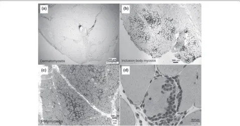
[image:2.612.67.546.89.341.2]DM is a systemic disease, involving muscle, skin, and, variably, other tissues. Skin gene expression profi ling, reported only in abstract format to date [20], has similarly shown marked abundance of type 1 interferon-inducible transcripts. Th e topology of keratinocyte injury in DM skin is similar to that of myofi ber injury in DM muscle [21]. Figure 1. Diff ering pathologies in myositis subtypes. The distribution of immune system cells diff ers among myositis subtypes. (a) In dermatomyositis, immune system cells are predominantly in the regions of connective tissue that lie between muscle fascicles and include medium-sized and large blood vessels. In (b)inclusion body myositis and (c)polymyositis, immune cells surround myofi bers. (d)Especially in inclusion body myositis, these may sometimes invade myofi bers.
Greenberg Arthritis Research & Therapy 2010, 12(Suppl 1):S4 http://arthritis-research.com/content/12/S1/S4
Patients with dermatological features of DM who lack signifi cant clinical evidence of muscle involvement have been classifi ed as clinically amyopathic DM. Auto-antibodies to a classic type 1 interferon-inducible protein IFIH1 (inter feron induced with helicase C domain; also called MDA-5) have been recently identifi ed [22]. Using optimized cutoff values in an ELISA assay, the presence of anti-IFIH1 antibodies in clinically amyopathic DM among 262 patients with a range of connective tissue diseases was 69% sensitive and 99.6% specifi c. Signifi cant
[image:3.612.66.549.89.485.2]clinically amyopathic DM, and evokes an autoantibody response.
A blood type 1 interferon-inducible signature
Blood gene expression profi ling has also demonstrated marked abundance of these transcripts in patients with active DM, such as untreated patients, but also in PM (see below) [19]. One study that did not fi nd marked type 1 interferon-inducible transcript abundance in DM blood samples had included almost only treated patients (11 out of 12 patients receiving prednisone; eight of these patients receiving an additional second immuno sup pressant agent) [23]. Microarray experiments measure the abundance of 10,000s of transcripts simultaneously; any set of transcripts may be called a signature, but what is impressive about such experiments in DM and PM blood samples is the dominance of these gene expression patterns by type 1 interferon-inducible genes. In a study of 23 patients with DM and PM, at least 24 of the highest expressed 25 genes among approximately 38,000 genes studied are all known to be highly inducible by type 1 interferons [19].
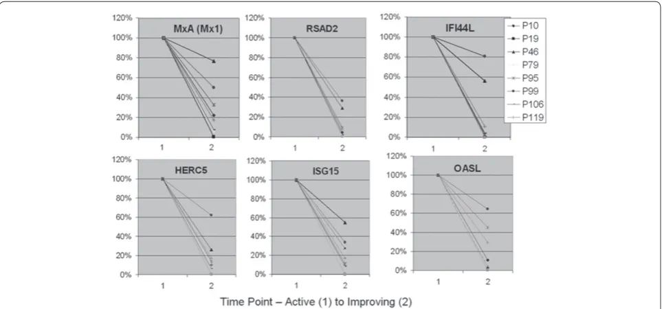
Th e type 1 interferon-inducible transcript overproduc-tion in DM and PM is highly correlated, within individual patients, with clinical measures of disease activity [19] (Figure 4 and unpublished data). Although the absolute magnitude of the upregulation of this signature does not highly correlate with disease severity across patients, within individual patients the signature does track disease activity – a situation similar to the commonly used clinical biomarker creatine kinase.
What drives the production of these downstream biomarkers of type 1 interferon signaling in DM muscle?
A fundamental question that has not been answered is what drives the production of type 1 interferon-inducible
molecules in DM muscle. Specifi c elevation of type 1 inter feron transcripts or proteins has not been demon-strated in DM muscle. Both microarray and real-time quantitative PCR do not show impressive diff er ences, compared with other myositis samples, in transcripts encoding a range of IFNα subtypes or IFNβ in the same DM muscle samples that have marked upregulation (10-fold to 100-fold) of downstream type 1 interferon-inducible transcripts (unpublished data). Immunoblots from myositis samples similarly do not appear to show diff erential presence of IFNα or IFNβ protein in DM muscle (preliminary unpublished data).
Th is situation parallels that seen in systemic lupus erythematosus. Although studies performed almost 30 years ago detected molecules believed to be IFNα in 60 to 76% of systemic lupus erythematosus blood samples using functional antiviral assays sometimes in combina-tion with neutralizing antibodies [24-27], the literature has been notable for the absence of detection of IFNα in systemic lupus erythematosus blood or tissue samples by direct methods such as ELISA, immunoblot, or mass spectrometry. For example, one study found measurable levels of IFNα protein by ELISA in only two out of 38 patients, while most of these same 38 samples showed marked increases in type 1 interferon-inducible trans-cripts [28]. Th e lack of direct detection of IFNα protein has been attributed to potential technical limitations, although unexpected results in science have often been assumed to be erroneous. A more recent functional assay looking at type 1 interferon-inducible transcription has similarly detected activity in systemic lupus erythema-tosus plasma [29].
Th e interpretation of the results of functional assays is complicated by potential type 1 interferon autocrine mechanisms. In mouse cells, autostimulation of the IFNAR by early secreted type 1 interferons results in marked amplifi cation of IFNαproduction [30-33]. Anti-IFNα antibodies used in functional assays to neutralize sample IFNα could potentially diminish type 1 inter feron-inducible gene transcription through neutralizing early secreted reporter cell IFNα, although this possibility is speculative.
For juvenile DM, the functional assay for gene trans-cription has been used for detection of serum type 1 interferon-inducing activity and similarly interpreted as indicating the presence of IFNα in some blood samples [34]. What is particularly remarkable about this study was the marked range of interferon-inducing activity of serum from healthy children and adults, which varies by over 100-fold and includes many healthy people whose activity exceeded the mean value for the juvenile DM population.
Th ese data suggest that the marked production of
[image:4.612.66.300.89.230.2]type 1 interferon-inducible transcripts and proteins in Figure 3. Myxovirus resistance protein A expression in
dermatomyositis muscle. Image of whole muscle section stained for myxovirus resistance protein A (MxA) showing abundant myofi ber protein expression (brown) preferentially in a perifascicular distribution. Adapted from [3] with permission.
Greenberg Arthritis Research & Therapy 2010, 12(Suppl 1):S4 http://arthritis-research.com/content/12/S1/S4
DM muscle, probably by myofi bers, might result from sustained activation of the type 1 interferon receptor IFNAR in the absence of excessive (compared with the wide range of normal) type 1 interferons, or through mechanisms even further downstream that bypass IFNAR. Th e most natural interpretation of the data to date suggests that what may turn out to be most crucial with regard to DM myofi ber injury is not the abundance of a type 1 interferon, but rather sustained abnormal function of the IFNAR or a further downstream process.
Potential role for type 1 interferons in polymyositis and inclusion body myositis
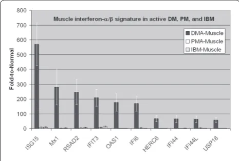
PM is an umbrella term for patients with various forms of myositis that are diffi cult to classify. DM and PM have substantial diff erences with regard to abundance of type 1 interferon-inducible molecules in muscle biopsy samples (Figure 5), yet marked overexpression of type 1 interferon-inducible genes has also been found in blood in PM [19]. Within PM muscle, infl ammatory cells typically surround, displace, and sometimes invade muscle fi bers. Th ese cells include T cells, myeloid dendritic cells, macrophages, and plasma cells (reviewed in [3]). Type 1 interferons have multiple eff ects on these cell types, and it is possible that through these eff ects the type 1 interferon system is contributing to PM myofi ber injury.
Patients with inclusion body myositis – a highly infl ammatory disorder of muscle, as judged by abundance of immune system cells and transcripts in muscle – do not have high levels of muscle or blood type 1
interferon-inducible transcripts (Figure 5), although a small propor tion of patients may have modest elevation of such transcripts in blood alone. As in PM, the mechanistic interpretation of blood expression of these transcripts is uncertain, and could refl ect less-specifi c eff ects driving immune cell development.
Predicted exacerbations with TNFα inhibition
TNFα appears to have an antagonistic relationship with type 1 interferons [35]. It may directly inhibit the genera-tion of plasmacytoid dendritic cells from progenitor cells and may inhibit plasmacytoid dendritic cell production of type 1 interferons. Studies of etanercept in Sjogren’s syndrome indeed showed that this drug increased type 1 interferon activity [36]. Accordingly, models that propose a signifi cant role for type 1 interferons in the pathogenesis of myositis predict that TNFα inhibition might exacer-bate myositis. Published experience with TNFα inhibition in patients with myositis appears to support this model. Two open-label studies of infl iximab have been termi-nated before completion or had substantial dropout rates for reasons that included disease progression [37,38]. Although this class of drugs may prove useful in the management of some patients, currently it appears unlikely to be of more general use for myositis.
Conclusion: diagnostics and therapeutic development
Th e presence of marked overproduction of type 1
[image:5.612.66.548.89.314.2]patients with active DM and PM has potential for diagnostic use [19]. Th ese biomarkers may be able to distinguish these disorders from inclusion body myositis and other muscle diseases that sometimes present diag-nostic uncertainty. Furthermore, they may be useful for therapeutic development. A phase 1b trial of anti-IFNα therapy has been initiated in DM and PM [39]. Entry criteria into this study include the presence of suffi ciently high type 1 interferon-inducible gene expression in blood. Future studies targeting the IFNAR or further down stream events have strong rationale in DM, and perhaps in PM.
Abbreviations
DM = dermatomyositis; ELISA = enzyme-linked immunosorbent assay; IFN = interferon; IFNAR = type 1 interferon receptor; ISG15 = interferon-stimulated gene 15; PCR = polymerase chain reaction; PM = polymyositis; TNF = tumor necrosis factor.
Competing interests
SAG has worked as a consultant regarding clinical trial planning for MedImmune, LLC and has a Sponsored Research Agreement with MedImmune, LLC. SAG is an inventor of intellectual property pertaining to myositis diagnostics. SAG has National Institutes of Health funding and the publication of articles constitutes evidence of productivity that may be used to support future requests for National Institutes of Health funding.
Acknowledgements
The present work was supported by grants to SAG from the National Institutes of Health (R01NS43471 and R21NS057225), the Muscular Dystrophy Association (MDA3878), and MedImmune, LLC.
This article is part of Arthritis Research & Therapy Volume 12 Supplement 1: The role of IFN alpha in autoimmune disease. The full contents of the supplement are available online at http://arthritis-research.com/supplements/12/S1. Publication of the supplement has been supported with funding from MedImmune, LLC.
Published: 14 April 2010
References
1. Greenberg SA: Infl ammatory myopathies: disease mechanisms. Curr Opin Neurol 2009, 22:516-523.
2. Salajegheh M, Kong SW, Pinkus JL, Walsh RJ, Liao A, Nazareno R, Amato AA, Krastins B, Morehouse C, Higgs BW, Jallal B, Yao Y, Sarracino DA, Parker KC, Greenberg SA: Interferon-stimulated gene 15 (ISG15) conjugates proteins in dermatomyositis muscle with perifascicular atrophy.Ann Neurol 2009, 67:53-63
3. Greenberg SA: Proposed immunologic models of the infl ammatory myopathies and potential therapeutic implications.Neurology 2007, 69:2008-2019.
4. Isenberg DA, Rowe D, Shearer M, Novick D, Beverley PC: Localization of interferons and interleukin 2 in polymyositis and muscular dystrophy.Clin Exp Immunol 1986, 63:450-458.
5. Emslie-Smith AM, Arahata K, Engel AG: Major histocompatibility complex class I antigen expression, immunolocalization of interferon subtypes, and T cell-mediated cytotoxicity in myopathies. Hum Pathol 1989, 20:224-231. 6. Lundberg I, Brengman JM, Engel AG: Analysis of cytokine expression in
muscle in infl ammatory myopathies, Duchenne dystrophy, and non-weak controls.J Neuroimmunol 1995, 63:9-16.
7. Figarella-Branger D, Civatte M, Bartoli C, Pellissier JF: Cytokines, chemokines, and cell adhesion molecules in infl ammatory myopathies. Muscle Nerve 2003, 28:659-682.
8. Salomonsson S, Lundberg IE: Cytokines in idiopathic infl ammatory myopathies.Autoimmunity 2006, 39:177-190.
9. Schmidt J, Barthel K, Wrede A, Salajegheh M, Bahr M, Dalakas MC: Interrelation of infl ammation and APP in sIBM: IL-1 beta induces accumulation of beta-amyloid in skeletal muscle.Brain 2008, 131(Pt 5):1228-1240.
10. Banker BQ: Dermatomyostis of childhood, ultrastructural alteratious of muscle and intramuscular blood vessels.J Neuropathol Exp Neurol 1975, 34:46-75.
11. Norton WL, Velayos E, Robison L: Endothelial inclusions in dermatomyositis.
Ann Rheum Dis 1970, 29:67-72.
12. Grimley PM, Davis GL, Kang YH, Dooley JS, Strohmaier J, Hoofnagle JH: Tubuloreticular inclusions in peripheral blood mononuclear cells related to systemic therapy with alpha-interferon.Lab Invest 1985, 52:638-649. 13. Rich SA, Owens TR, Bartholomew LE, Gutterman JU: Immune interferon does
not stimulate formation of alpha and beta interferon induced human lupus-type inclusions.Lancet 1983, 1:127-128.
14. Grimley PM, Rutherford MN, Kang YH, Williams T, Woody JN, Silverman RH: Formation of tubuloreticular inclusions in human lymphoma cells compared to the induction of 2΄-5΄-oligoadenylate synthetase by leucocyte interferon in dose-eff ect and kinetic studies. Cancer Res 1984, 44:3480-3488.
15. Kuyama J, Kanayama Y, Mizutani H, Katagiri S, Tamaki T, Yonezawa T, Tarui S, Morise H, Arimura H, Suyama T: Formation of tubuloreticular inclusions in mitogen-stimulated human lymphocyte cultures by endogenous or exogenous alpha-interferon.Ultrastruct Pathol 1986, 10:77-85.
16. Feldman D, Goldstein AL, Cox DC, Grimley PM: Cultured human endothelial cells treated with recombinant leukocyte A interferon. Tubuloreticular inclusion formation, antiproliferative eff ect, and 2΄,5΄ oligoadenylate synthetase induction. Lab Invest 1988, 58:584-589.
17. Greenberg SA, Amato AA: Uncertainties in the pathogenesis of adult dermatomyositis.Curr Opin Neurol 2004, 17:359-364.
18. Greenberg SA, Pinkus JL, Pinkus GS, Burleson T, Sanoudou D, Tawil R, Barohn RJ, Saperstein DS, Briemberg HR, Ericsson M, Park P, Amato AA: Interferon-alpha/beta-mediated innate immune mechanisms in dermatomyositis.
Ann Neurol 2005, 57:664-678.
19. Walsh RJ, Kong SW, Yao Y, Jallal B, Kiener PA, Pinkus JL, Beggs AH, Amato AA, Greenberg SA: Type I interferon-inducible gene expression in blood is present and refl ects disease activity in dermatomyositis and polymyositis.
Arthritis Rheum 2007, 56:3784-3792.
[image:6.612.65.298.90.247.2]20. Kea B, Pesich R, Chung LS, Brown PO, Fiorentino D: Genomic analyses identify lipid metabolism abnormalities in dermatomyositis patients [abstract].J Invest Dermatol 2007, 127(Suppl 1):S12.
21. Greenberg SA, Fiorentino D: Similar topology of injury to keratinocytes and myofi bres in dermatomyositis skin and muscle. Br J Dermatol 2009, 160:464-465.
Figure 5. Distinct muscle expression of type 1 interferon-inducible genes in infl ammatory myopathies. Distinct muscle expression of type 1 interferon-inducible genes in dermatomyositis (DM) compared with polymyositis (PM) and inclusion body myositis (IBM). Muscle microarray data shown for 20 patients (fi ve each with active dermatomyositis (DMA), active polymyositis (PMA), untreated IBM, and normal) with plotted mean values and error bars for mean ± standard error for each group. Highly expressed genes in DM muscle are orders of magnitude greater than in PM and IBM. Adapted from [19] with permission.
Greenberg Arthritis Research & Therapy 2010, 12(Suppl 1):S4 http://arthritis-research.com/content/12/S1/S4
22. Sato S, Hoshino K, Satoh T, Fujita T, Kawakami Y, Fujita T, Kuwana M: RNA helicase encoded by melanoma diff erentiation-associated gene 5 is a major autoantigen in patients with clinically amyopathic
dermatomyositis: association with rapidly progressive interstitial lung disease. Arthritis Rheum 2009, 60:2193-2200.
23. Baechler EC, Bauer JW, Slattery CA, Ortmann WA, Espe KJ, Novitzke J, Ytterberg SR, Gregersen PK, Behrens TW, Reed AM: An interferon signature in the peripheral blood of dermatomyositis patients is associated with disease activity.Mol Med 2007, 13:59-68.
24. Hooks JJ, Jordan GW, Cupps T, Moutsopoulos HM, Fauci AS, Notkins AL: Multiple interferons in the circulation of patients with systemic lupus erythematosus and vasculitis.Arthritis Rheum 1982, 25:396-400. 25. Hooks JJ, Moutsopoulos HM, Geis SA, Stahl NI, Decker JL, Notkins AL:
Immune interferon in the circulation of patients with autoimmune disease. N Engl J Med 1979, 301:5-8.
26. Preble OT, Black RJ, Friedman RM, Klippel JH, Vilcek J: Systemic lupus erythematosus: presence in human serum of an unusual acid-labile leukocyte interferon.Science 1982, 216:429-431.
27. Ytterberg SR, Schnitzer TJ: Serum interferon levels in patients with systemic lupus erythematosus. Arthritis Rheum 1982, 25:401-406.
28. Baechler EC, Batliwalla FM, Karypis G, Gaff ney PM, Ortmann WA, Espe KJ, Shark KB, Grande WJ, Hughes KM, Kapur V, Gregersen PK, Behrens TW: Interferon-inducible gene expression signature in peripheral blood cells of patients with severe lupus.Proc Natl Acad Sci U S A 2003, 100:2610-2615. 29. Hua J, Kirou K, Lee C, Crow MK: Functional assay of type I interferon in
systemic lupus erythematosus plasma and association with anti-RNA binding protein autoantibodies.Arthritis Rheum 2006, 54:1906-1916. 30. Sato M, Suemori H, Hata N, Asagiri M, Ogasawara K, Nakao K, Nakaya T,
Katsuki M, Noguchi S, Tanaka N, Taniguchi T: Distinct and essential roles of transcription factors IRF-3 and IRF-7 in response to viruses for IFN-alpha/ beta gene induction.Immunity 2000, 13:539-548.
31. Sato M, Tanaka N, Hata N, Oda E, Taniguchi T: Involvement of the IRF family transcription factor IRF-3 in virus-induced activation of the IFN-beta gene.
FEBS Lett 1998, 425:112-116.
32. Marie I, Durbin JE, Levy DE: Diff erential viral induction of distinct interferon-alpha genes by positive feedback through interferon regulatory factor-7.
Embo J 1998, 17:6660-6669.
33. Sato M, Hata N, Asagiri M, Nakaya T, Taniguchi T, Tanaka N: Positive feedback regulation of type I IFN genes by the IFN-inducible transcription factor IRF-7.FEBS Lett 1998, 441:106-110.
34. Niewold TB, Kariuki SN, Morgan GA, Shrestha S, Pachman LM: Elevated serum interferon-alpha activity in juvenile dermatomyositis: associations with disease activity at diagnosis and after thirty-six months of therapy. Arthritis Rheum 2009, 60:1815-1824.
35. Palucka AK, Blanck JP, Bennett L, Pascual V, Banchereau J: Cross-regulation of TNF and IFN-alpha in autoimmune diseases. Proc Natl Acad Sci U S A 2005, 102:3372-3377.
36. Mavragani CP, Niewold TB, Moutsopoulos NM, Pillemer SR, Wahl SM, Crow MK: Augmented interferon-alpha pathway activation in patients with Sjogren’s syndrome treated with etanercept.Arthritis Rheum 2007, 56:3995-4004.
37. Hengstman GJ, De Bleecker JL, Feist E, Vissing J, Denton CP, Manoussakis MN, Slott Jensen H, van Engelen BG, van den Hoogen FH: Open-label trial of anti-TNF-alpha in dermato- and polymyositis treated concomitantly with methotrexate.Eur Neurol 2008, 59:159-163.
38. Dastmalchi M, Grundtman C, Alexanderson H, Mavragani CP, Einarsdottir H, Helmers SB, Elvin K, Crow MK, Nennesmo I, Lundberg IE: A high incidence of disease fl ares in an open pilot study of infl iximab in patients with refractory infl ammatory myopathies.Ann Rheum Dis 2008, 67:1670-1677. 39. A study to evaluate safety of multi-dose MEDI-545 in adult patients with
dermatomyositis or polymyositis [http://clinicaltrials.gov/ct2/show/ NCT00533091]
doi:10.1186/ar2885