N A N O E X P R E S S
Enzymatic Digestion of Single DNA Molecules Anchored
on Nanogold-Modified Surfaces
Junhong Lu¨Æ Ming YeÆNa DuanÆ Bin Li
Received: 9 January 2009 / Accepted: 14 May 2009 / Published online: 31 May 2009
Óto the authors 2009
Abstract To study enzyme–DNA interactions at single molecular level, both the attachment points and the immediate surroundings of surfaces must be carefully considered such that they do not compromise the structural information and biological properties of the sample under investigation. The present work demonstrates the feasibil-ity of enzymatic digestion of single DNA molecules attached to nanoparticle-modified surfaces. With Nanogold linking DNA to the mica surface by electrostatic interac-tions, advantageous conditions with fewer effects on the length and topography of DNA are obtained, and an appropriate environment for the activities of DNA is cre-ated. We demonstrate that by using Dip-Pen Nanolithog-raphy, individual DNA molecules attached to modified mica surfaces can be efficiently digested by DNase I.
Keywords Gold nanoparticlesMicaDNA Atomic force microscopy Dip-Pen Nanolithography
Introduction
Advances in single-molecule techniques make it possible to explore new phenomena and unravel novel mechanisms in biology that were largely inaccessible by traditional bulk measurements [1]. For example, studies of DNA–protein interaction at single molecular level could characterize the
distributions of molecular properties and observe the tem-poral evolution of complicated reaction pathways [2]. It is generally understood that single-molecule measurements require adsorption and fixation of single DNA molecules on a solid support surface [1,3] before the protein motion along the DNA can be tracked. Among the many kinds of substrate surfaces, mica is ideal because of its atomic smoothness. Since newly cleaved mica is negatively charged at basic pH [4], an advisable surface modification is critical to bind the negatively charged phosphate back-bone of DNA. Typically, poly-L-lysine [5,6], silane [7,8], and divalent cations, such as Ni2? and Mg2?, have been used to provide positively charged sites and/or hydrophobic surfaces for enhancing the interactions between DNA and surfaces [4,9,10]. However, these modification methods usually compromise the inherent surface roughness of mica, making it more difficult to gain structural insight into biomolecules with nanometer resolution. Also such modi-fied surfaces are not well suited for dynamic measurements of protein or DNA molecules, because the entire DNA molecule is often fixed tightly on the surface, leading to little or tardy response of the molecule to environmental changes.
To fix DNA on a surface for investigation into its interaction with other reactants, one strategy is to modify the terminal of the DNA strands, so that they specifically bind to surfaces [11–13]. For instance, van Oijen et al. used biotin–avidin system to fix only one end and allow the rest of the single DNA molecule to interact with exonuclease [3]. Medalia et al. demonstrated a method that anchors two ends of a DNA fragment with a thiol group on a gold film-modified mica surface [14]. Recently, a novel strategy named ‘‘protein-assisted DNA immobilization’’ was pro-posed by Dukkipati et al. in which DNA binding proteins such as restriction enzymes or RNA polymerases are used
Electronic supplementary material The online version of this article (doi:10.1007/s11671-009-9350-6) contains supplementary material, which is available to authorized users.
J. Lu¨M. YeN. DuanB. Li (&)
Shanghai Institute of Applied Physics, Chinese Academy of Sciences, P.O. Box 800-204, Shanghai 201800, China e-mail: [email protected]
as attachment points to adsorb DNA on surfaces [15]. Although this method can maintain the biological activity of the immobilized DNA molecules, it is not suitable for higher resolution imaging at nanometer scale by atomic force microscopy (AFM), because hydrophobic polymeth-ylmethacrylate (PMMA) surfaces have to absorb proteins. We are working on single-molecule enzymatic reactions on mica surfaces by controlled dipping of a nonspecific endonuclease over the DNA molecules based on nanoma-nipulation [16]. To simultaneously realize the goals of obtaining structural insights into biomolecules with nano-meter resolution and providing an appropriate condition for their biological processes, we investigated enzymatic reactions (DNase I) at single DNA molecules attached and immobilized on mica surfaces functioned by gold nano-particles (GNPs), 1.4 nm-diameter nanonano-particles (Nano-gold). We demonstrate that Nanogold-modified mica surfaces (Nanogold-mica) have less effect on the length and topography of DNA molecules and provide a suitable environment for higher efficiency of enzymatic reactions on DNA.
Materials and Methods
The original DNA solutions (Shanghai Sangon Biological Engineering Technology and Services Co., Ltd) were diluted to final concentrations of 1 ng/lL fork DNA and 0.1 ng/lL for pBR322, in TE buffer (10 mM TE–HCl, pH 8.0). Nanogold-mica was produced by treating freshly cleaved mica with 1–50 fM Nanogold (Nanoprobes, Stony Brook, NY) in water for 1 min. After being dried with nitrogen gas, the ‘‘spin-stretching’’ technique was used to stretch and fix DNA [17]. Briefly, 2–5lL DNA was put on a Nanogold-mica, which was adhered firmly on a centri-fuge. The spin speed was limited to\3,000 rpm to extend DNA for 30 s. Samples were washed twice with 10lL deionized water and dried for imaging.
AFM imaging was conducted using the tapping mode of a MultiMode Scanning Probe microscope (NanoScope IIIa, Digital Instruments, Santa Barbara, CA) with a J Scanner. Noncontact cantilevers (NSC11, MikroMasch) with a res-onance frequency of*300 kHz and a spring constant of *40 Nm-1were used for imaging at room temperature (in an ambient situation). All AFM images were flattened and analyzed with the microscope’s software system. The contour lengths of single DNA molecules and percentage of DNA occupied on surfaces were determined using METAMORPH software (MDS, Inc.) (see supporting information on the method of calculating DNA length and coverage).
For enzymatic digestion of DNA molecules, Dip-Pen Nanolithography (DPN) [16, 18–20] was used to deposit
DNase I on DNA. Briefly, an AFM tip coated with 0.01– 0.05 unit/lL DNase I (Sigma) in 20 mM Tris–HCl, pH 8.3, 2 mM MgCl2, and 2 mM CaCl2 was mounted on the sample stage. After the first DNA image was obtained by tapping mode, lift mode was turned on to move the AFM tip closer to the surface by setting a negative lift height value. The tip remained for a moment once it touched the surface to induce a meniscus between the tip and the sur-face. Then, the first image was scanned again with tapping mode but this time by depositing DNase I on the surface and the DNA. Afterwards, several images were recorded in situ to observe the process of DNA digestion. The digestion experiments were conducted in a relative humidity of 30– 40% and a temperature of 20–25°C.
Results and Discussion
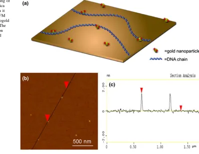
Nanogold is generally used as a contrast agent in electron microscopy [21]. In our experiments, we utilize the unique properties of the positively charged Nanogold to act as cross-linker between negatively charged DNA and mica through electrostatic interactions (Fig. 1a). We expect that most parts of DNA are free except for the binding sites to Nanogold. Due to the fact that only bare mica is used and no other additional surface modification is needed, the inherent surface properties of mica such as its atomic flatness and hydrophilicity are less affected. So the features of DNA can be clearly observed, and a suitable surface for observing the biological activities of proteins can be provided.
As shown in Fig.1b and c, after the modification pro-cess, the Nanogold, 1.4 nm in height, is randomly dis-persed on the mica surface. The roughness of the mica surface is changed a little by the sparse distribution of small size nanoparticles. The root mean square (RMS) roughness measured on the 1.75 lm91.75lm area of the mica surface was *0.06 nm. Although there is a slight increase in this value compared with a freshly cleaved mica surface of*0.05 nm, it is sufficient for imaging DNA and studying the interaction between protein and DNA.
However, a higher density of Nanogold would influence the topography of DNA, thus it is important to control the numbers of Nanogold on mica surface to achieve a better DNA topography. In Fig.2b, there are a few nanoparticles that are used to attach lambda DNA molecules on the surface, and the lower DNA molecule is anchored only by a single Nanogold. From the cross-section profile of Fig.2c as shown in Fig.2d, the measured height of the binding site is 1.8 nm (arrow 1), equaling the value of DNA height of 0.4 nm (the measured height of most parts of DNA, arrow 2), plus a Nanogold height of 1.4 nm (arrow 3). In addition, there is the measured height of 0.8 nm (three thin arrows in Fig.2c) along DNA strands, implying other structures of DNA existing on the surface.
We have also explored the general applicability of Nanogold to deposit circular and linear DNA on mica. Circular pBR322 DNA andPst1linearized pBR322 were chosen for this purpose. It has been reported that the enzyme sometimes shows limited catalytic activity on overstretched DNA molecules. Although it is possible to avoid overstretching by reducing the hydrophobic effects during the DNA-stretching processes [22], the problem of controlling this effect persists. However, in our experi-ments, DNA molecules are easily attached but not over-stretched. As shown in Fig.3, the measured lengths of DNA range from 1.31 to 1.48lm regardless of linear or
circular molecules, which is very close to the actual length,
1.48lm. The preserved conformation of DNA would be a
potential advantage for reactions of DNA with other mol-ecules like proteins and enzymes.
[image:3.595.141.541.61.370.2]After being able to reproducibly deposit linear and circular DNA molecules on mica without overstretching them, it would be very interesting to explore whether DNA molecules attached on Nanogold-mica are beneficial for the investigation into enzymatic reactions along a single DNA molecule. To this end, a digestion reaction with DNase I was carried out. DNase I is a paradigm endonuclease used routinely for nonspecific cleavage of DNA in molecular biology. Figure4 shows the process of the enzymatic reaction. The uniform linear DNA (Fig.4a) was digested into several fragments immediately (Fig.4b) after DNase I ink (bright spots in image) was transferred from the coated tip to the surface and DNA. The size of spots changed along with the time passed. About half an hour later, the volume of the ink spots decreased greatly (Fig.4c). To observe DNA clearly, the sample was imaged again after 10 h. All bright spots and most parts of DNA disappeared, but tracks of DNA still remained (Fig.4d). This phenomenon is interesting, its mechanism however is unclear so far. We think the disappearance of ink (Fig.4b–d) may be caused by the tip’s effects, such as tip-induced diffusion and/or adsorption, during
scanning processes. Other factors, such as liquid evapo-ration and liquid diffusion may also play a rule. To exclude any chance that the observed gaps could have been caused by mechanical force applied by the AFM tip, control experiments with denatured enzyme were
performed, and no such digestion phenomenon occurred. The results imply that the flat, hydrophilic Nanogold-mica surface is suitable for the detection of enzymatic diges-tions of DNA by AFM. We note that no additional sample washing steps were needed; therefore, this technique not
Fig. 2 Typical AFM images of lambda DNA anchored on Nanogold-mica modified witha
50 fM andb5 fM Nanogold. Height bar=5 nmcAn enlarged image from the mini square in Fig.2b. Height bar=2 nmdA height profile of DNA indicated by alinein Fig.2c
[image:4.595.181.545.57.636.2]only completely eliminates any possible artifacts caused by the water flow, but also has the potential to be developed into a method for recording digestion reactions in a time-lapse manner. It should be noted that although the cleavage of DNA can be observed on other modified surfaces, such as APTES-mica [16] and Ni-mica [23], using Nanogold-mica facilitates the detection of small gaps in the DNA due to the relatively free state of the molecule. Most of the DNA has weak interaction with the surface except at the points that are anchored by Nano-gold. Once the phosphodiester linkages are broken, the ends of the DNA fragments have a tendency to adjust their positions because of their entropic property, so a larger gap appears. Additionally, the modified surface is flat, providing a unique platform to probe the topography of DNA. Moreover, the entire smooth surface is hydro-philic because of the hydrohydro-philic mica surface and the water soluble Nanogold. The flat, hydrophilic surface facilitates ink and small DNA fragments to diffuse on the substrate, leading to an enlarged gap and a clear view field. So a digestion reaction of DNA can be probed clearly, even without washing steps.
Conclusions
We have demonstrated that we are able to facilely deposit and anchor DNA molecules on a mica surface using Nanogold for single-molecule enzymatic reactions. The immobilization of DNA on Nanogold-modified surfaces does not require time-consuming steps, and the fixed DNA strands on the surface can easily be observed on AFM images. Because the Nanogold distribution largely deter-mines the interaction forces between mica and the adsorbed DNA molecules, we could minimize any possible influence of the surface on the native properties of DNA molecules by adjusting the concentrations of nanoparticles, thus providing conditions in which distinct conformations of DNA molecules and their interactions with proteins or other materials can be studied better. By using Dip-Pen Nanolithography to dip DNase I over DNA molecules, we have realized to digest single DNA molecules with higher efficiency. Further research toward more careful control over the deposited density of the Nanogold on surfaces for fixing DNA in solution and probe the structure-related properties of DNA with various kinds of restriction
[image:5.595.182.545.58.423.2]endonucleases needs to be conducted. Some of this research is currently under way in our research group.
Acknowledgment This work was supported by grants from NSFC (10675160, 10604061, and 10874198).
References
1. M.-L. Visnapuu, D. Duzdevich, E.C. Greene, Mol. Biosyst.4, 394 (2008). doi:10.1039/b800444g
2. E. Rhoades, E. Gussakoysky, G. Haran, Proc. Natl Acad. Sci. USA100, 3197–3202 (2003). doi:10.1073/pnas.2628068100 3. A.M. van Oijen, P.C. Blainey, D.J. Crampton, C.C. Richardson,
T. Ellenberger, X.S. Xie, Science301, 1235 (2003). doi:10.1126/ science.1084387
4. H.G. Hansma, D.E. Laney, Biophys. J. 70, 1933 (1996). doi: 10.1016/S0006-3495(96)79757-6
5. M. Bussiek, K. Toth, N. Muecke, N. Brun, J. Langowski, Bio-phys. J.88, 58a (2005)
6. S.J.T. van Noort, O.H. Willemsen, K.O. van der Werf, B.G. de Grooth, J. Greve, Langmuir 15, 7101 (1999). doi:10.1021/ la990459a
7. Y.L. Lyubchenko, A.A. Gall, L.S. Shlyakhtenko, R.E. Harring-ton, B.L. Jacobs, P.I. Oden, S.M. Lindsay, J. Biomol. Struct. Dyn.
10, 589 (1992)
8. M. Sasou, S. Sugiyama, T. Yoshino, T. Ohtani, Langmuir 19, 9845 (2003). doi:10.1021/la035054b
9. T. Thundat, D.P. Allison, R.J. Warmack, G.M. Brown, K.B. Jacobson, J.J. Schrick, T.L. Ferrell, Scanning Microsc. 6, 911 (1992)
10. T.F. Chan, C. Ha, A. Phong, D. Cai, E. Wan, L. Leung, P.Y. Kwok, M. Xiao, Nucleic Acids Res. 34, e113 (2006). doi: 10.1093/nar/gkl593
11. S. Matsuura, J. Komatsu, K. Hirano, H. Yasuda, K. Takashima, S. Katsura, A. Mizuno, Nucleic Acids Res. 29, e79 (2001). doi: 10.1093/nar/29.16.e79
12. J.H. Kim, R.G. Larson, Nucleic Acids Res.35, 3848 (2007). doi: 10.1093/nar/gkm332
13. A. Crut, B. Geron-Landre, I. Bonnet, S. Bonneau, P. Desbiolles, C. Escude, Nucleic Acids Res.33, e98 (2005). doi:10.1093/nar/ gni097
14. O. Medalia, J. Englander, R. Guckenberger, J. Sperling, Ultra-microscopy90, 103 (2002). doi:10.1016/S0304-3991(01)00141-3 15. V.R. Dukkipati, J.H. Kim, S.W. Pang, R.G. Larson, Nano Lett.6,
2499 (2006). doi:10.1021/nl0617484
16. B. Li, Y. Zhang, S.H. Yan, J.H. Lu, M. Ye, M.Q. Li, J. Hu, J. Am. Chem. Soc.129, 6668 (2007). doi:10.1021/ja0687015
17. H. Yokota, J. Sunwoo, M. Sarikaya, G. van den Engh, R. Aebersold, Anal. Chem.71, 4418 (1999). doi:10.1021/ac9902695 18. R.D. Piner, J. Zhu, F. Xu, S.H. Hong, C.A. Mirkin, Science283,
661 (1999). doi:10.1126/science.283.5402.661
19. B. Li, Y. Zhang, J. Hu, M.Q. Li, Ultramicroscopy 105, 312 (2005). doi:10.1016/j.ultramic.2005.06.056
20. J. Hyun, J. Kim, S.L. Craig, A. Chilkoti, J. Am. Chem. Soc.126, 4770–4771 (2004). doi:10.1021/ja049956q
21. J.F. Hainfeld, R.D. Powell, J. Histochem. Cytochem. 48, 471 (2000)
22. Z. Gueroui, C. Place, E. Freyssingeas, B. Berge, Proc. Natl Acad. Sci. USA99, 6005 (2002). doi:10.1073/pnas.092561399 23. O. Pietrement, D. Pastre, F. Landousy, M.O. David, S. Fusil, L.