R E S E A R C H A R T I C L E
Open Access
Altered lipoproteins in patients with
systemic lupus erythematosus are
associated with augmented oxidative
stress: a potential role in atherosclerosis
Jin Kyun Park
1,2, Jae-Yong Kim
3, Jin Young Moon
2, Eun Young Ahn
2, Eun Young Lee
2, Eun Bong Lee
2,
Kyung-Hyun Cho
3and Yeong Wook Song
1,2,4*Abstract
Background:To examine the structural and oxidative properties of lipoproteins from patients with systemic lupus erythematosus (SLE).
Methods:The lipid profiles of 35 SLE patients and 15 healthy controls (HCs) were compared. Oxidation status, susceptibility to oxidation, and structural integrity of low-density lipoprotein (LDL) were determined by measuring malondialdehyde (MDA), de novo formation of conjugated dienes in the presence of CuSO4, and mobility on gel electrophoresis, respectively. In vitro foam cell formation and the oxidative potential in zebrafish embryos were examined.
Results:LDL levels in SLE patients and HCs were similar (p= 0.277). LDL from SLE patients was more fragmented than that from HCs. In addition, LDL from SLE patients was more oxidized than LDL from HCs (p< 0.001) and more susceptible to de novo oxidation (p< 0.001) in vitro. THP-1 cells engulfed more LDL from SLE patients than LDL from HCs (p< 0.001). LDL from SLE patients, which was injected into zebrafish embryos, induced a higher degree of oxidation and a higher mortality than LDL from HCs (bothp< 0.001). The survival of embryos treated with oxidized LDL was significantly better in the presence of HDL3from HCs than that from SLE patients (allp< 0.001).
Conclusions:Lipoproteins from SLE patients exhibited greater oxidative potential, which might contribute to accelerated atherosclerosis in SLE.
Keywords:Atherosclerosis, Oxidation, Lipoproteins, LDL, Systemic lupus erythematosus
Background
Systemic lupus erythematosus (SLE) is a chronic inflam-matory multisystem disease mediated by immune cell acti-vation and autoantibody production [1]. Patients with SLE carry an increased risk (up to 17-fold) of developing a car-diovascular (CV) disease [2, 3]. Although traditional risk factors for CV disease are more prevalent in patients with
SLE than in the general population [4–7], they do not solely account for the increased CV risk observed in these patients [8, 9]. As an example, a significant reduction of total cholesterol levels with atorvastatin failed to halt pro-gression of atherosclerosis or to decrease inflammatory markers such as C-reactive protein (CRP) in SLE patients [10, 11]. Therefore, SLE with chronic inflammation increases CV risk by influencing traditional and non-traditional pro- and anti-atherogenic factors.
High-density lipoprotein (HDL) is crucial in the pre-vention of the atherosclerosis. It prevents oxidation of low-density lipoprotein (LDL) and removes reactive oxy-gen species from LDL [12, 13]. Dysfunctional HDL has been linked to an increased risk of atherosclerosis during
* Correspondence:ysong@snu.ac.kr
1Department of Molecular Medicine and Biopharmaceutical Sciences,
Graduate School of Convergence Science and Technology, and College of Medicine, Medical Research Center, Seoul National University, Seoul, Republic of Korea
2Division of Rheumatology, Department of Internal Medicine, Seoul National
University Hospital, Seoul, Republic of Korea
Full list of author information is available at the end of the article
chronic inflammation [14, 15]. Indeed, half of women with SLE have high levels of pro-inflammatory HDL, which fails to protect LDL from damaging oxidation [16, 17]. This oxidation of lipoproteins might be further potenti-ated by reactive oxygen species, which are generpotenti-ated ex-cessively within the inflamed tissue of SLE patients. Subsequent accumulation of oxidized LDL (oxLDL) induces apoptosis of vascular smooth muscle cells and accelerates cellular senescence [18]. In addition, oxLDL is engulfed by monocytes, which then produce inflammatory cytokines and transform into foam cells, thereby contrib-uting to the development of atherosclerosis [19, 20]. Taken together, the altered structural and functional prop-erties of lipoproteins might contribute to accelerated atherosclerosis associated with SLE, possibly via inter-action with immune cells [5].
Since oxidation of lipoproteins is a crucial step for atherosclerosis, this study aimed to investigate the oxida-tive properties of lipoproteins from SLE patients both in vitro and in vivo.
Methods Patients
A total of 35 patients fulfilling the 1997 American College of Rheumatology classification criteria for SLE [21] were recruited at Seoul National University Hospital. Disease activity at the time of blood sampling was determined using the SLE disease activity index 2000 (SLEDAI-2 K) [22]. Fifteen individuals without other comorbidities were included as healthy controls (HCs).
Sample preparation and lipoprotein isolation
Blood was obtained after overnight fasting, and serum was separated by low-speed centrifugation and stored at -80 °C until analysis. The storage time did not differ between SLE and HC samples (109.6 ± 69.2 days vs. 88.4 ± 8.7 days, p= 0.40). Lipoproteins, including very low-density lipoprotein (low-density <1.019 g/mL), LDL (low-density 1.019–1.063 g/mL), HDL2 (density 1.064–1.125 g/mL), and HDL3 (density 1.126–1.225 g/mL) were isolated from serum by sequential ultracentrifugation as previ-ously described [23]. Briefly, the density was adjusted by addition of NaCl and NaBr and samples were centri-fuged for 24 hours at 10 °C at 100,000 g using a Himac CP-90α(Hitachi, Tokyo, Japan).
To generate oxLDL, 300 μg of LDL that had been purified from healthy controls was incubated with 10μM CuSO4for 4 hours at 37 °C.
Analysis of lipoproteins
Total cholesterol and triglyceride (TG) levels were mea-sured using commercially available kits (Diagnostics; Osaka, Japan). LDL cholesterol levels were calculated using the Friedewald formula. The protein content of
the lipoproteins was measured using the Lowry protein assay as previously described [24].
Copper-mediated oxidation of lipoproteins
The amount of oxidized species in lipoproteins was quantified by measuring malondialdehyde (MDA) levels using the thiobarbituric acid reactive substance method as previously described [25].
To investigate the susceptibility to copper-mediated de novo oxidation, 300 μg of LDL, which was isolated from SLE or HC, was incubated with 5μM CuSO4for 3 hours. During the incubation, the formation of conjugated dienes was determined by measuring the absorbance at 234 nm at 37 °C using a Beckman DU 800 spectrophotometer (Beckman Coulter, Fullerton, CA, USA), equipped with a MultiTemp III thermocirculator (Amersham Biosciences, Uppsala, Sweden) [24].
Electrophoresis and Western blot analysis
Apolipoprotein/lipoprotein composition was compared by sodium dodecyl sulfate-polyacrylamide gel electro-phoresis (SDS-PAGE). Identical amounts of HDL2, HDL3, and LDL (3μg of total protein), which had been pooled from 19 SLE patients or 8 HCs, were loaded into the lanes (Additional file 1: Table S1 and Figure S1).
For Western blot analysis, proteins were transferred onto a nitrocellulose membrane and the blots were incu-bated with goat anti-human apoA-I antibody (clone# ab7613, Abcam, Cambridge, UK) and then with anti-goat IgG horseradish peroxidase conjugate (Sigma-Aldrich, St. Louis, MO, USA). The detection was performed by a chemiluminescent method (ECL, Amersham Biosciences, Uppsala, Sweden).
For semi-quantitative analysis of lipoproteins, images of SDS-PAGE gels and films were scanned using Gel Doc® XR (Bio-Rad, Hercules, CA, USA) and the intensity of bands was analyzed using Quantity One software (version 4.5.2; Bio-Rad, Hercules, CA, USA).
Cholesteryl ester (CE) transfer assay
Recombinant HDL (rHDL) containing apoA-I was synthe-sized in the presence of [3H]-cholesteryl oleate (TRK886; 3.5μCi/mg of apoA-I; GE Healthcare, Chicago, IL, USA). HDL3 (20 μL, 2 mg/mL), [3H]-rHDL, and human LDL served as a cholesteryl ester (CE) transfer protein, a CE donor, and a CE acceptor, respectively. After incubation at 37 °C, the amount of CE acceptor was measured using scintillation counting and the percentage transfer of [3 H]-CE from rHDL to LDL was calculated as previously described [26, 27].
Paraoxonase assay
diethyl phosphates in the presence of paraoxonase as a catalyst. Briefly, 10 μL of diluted serum was added to 200μL of paraoxon-ethyl (Sigma D-9286; Sigma-Aldrich, St. Louis, MO, USA) in a buffer containing 90 mM Tris-HCl, 3.6 mM NaCl, and 2 mM CaCl2(pH 8.5). The production of p-nitrophenol at 37 °C was determined by measuring the absorbance at 405 nm using a Microplate reader (Bio-Rad, Hercules, CA, USA). A paraoxonase activity of 1 U/L was defined as the formation of 1μmol of p-nitrophenol per minute [28].
Phagocytosis of LDL by macrophages
LDL isolated from SLE patients or HCs was incubated with a fluorescent cholesterol derivative (22-(N-7-nitrobenz-2-oxa-1,3-diazol-4-yl) amino-23, 24-bisnor-5-cholen-3-ol [NBD-cholesterol], Molecular Probes, Eugene, OR, USA, N-1148; 70 μg of NBD-cholesterol/mg of apoA-I). THP-1 cells were then differentiated into macrophages in the pres-ence of phorbol myristate acetate and incubated with 50μL of labeled LDL (1 mg of protein/mL in PBS) for 48 hours at 37 °C in a humidified incubator. After washing with PBS, cells were fixed for 10 min in 4% paraformaldehyde and photographed under a Nikon Eclipse TE2000 microscope (Tokyo, Japan) at × 600 magnification (excitation wave-length = 488 nm; emission wavewave-length = 535 nm). NBD positive area was measured.
Senescence-associated (SA)-β-galactosidase activity
Cultured fibroblasts were used at passages 11–15 (approximately 40% confluence). Cells were incubated with lipoprotein fractions (0.1 mg/mL), fixed with 3% paraformaldehyde for 5 min, washed with PBS, and in-cubated with senescence-associated (SA)-β-galactosidase staining solution (40 mM citric acid, phosphate [pH 6.0], 5 mM potassium ferrocyanide, 5 mM potassium ferri-cyanide, 150 mM NaCl, 2 mM MgCl2, and 1 mg/mL 5-bromo-4-chloro-3-indolyl-X-galactosidase) for 16 hours at 37 °C. The cells were then observed under a light microscope, and the percentage of blue cells was calculated.
Microinjection of zebrafish embryos
All experimental procedures and maintenance of zebrafish (Linebrass, AB strain) were approved by the Committee of Animal Care and Use at Yeungnam University (Gyeongsan, Korea). Embryos (obtained 4 hours after fertilization) were microinjected with PBS or lipoproteins using a pneumatic picopump (PV820, World Precision Instruments; Sarasota, FL, USA). The embryos were then observed for 48 hours under a stereomicroscope (Motic SM 168; Hong Kong) and imaged using a Moticam 2300 CCD camera.
Measurement of oxidation in vivo
After injection, the fluorescence intensity (excitation = 588 nm and emission = 605 nm) of oxidized dihydroethi-dium (DHE; Sigma-Aldrich, St. Louis, MO, USA) in the embryos was examined under a Nikon Eclipse TE2000 microscope (Tokyo, Japan) and quantified using Image Proplus software (version 4.5.1.22; Media Cybernetics, Bethesda, MD, USA).
Statistical analysis
Results are expressed as the mean ± standard deviation (SD). Differences between the two groups were assessed using the Mann-WhitneyUtest orttest as appropriate. All reportedpvalues were two-sided, andpvalues < 0.05 were considered significant. All statistical analyses were performed using GraphPad Prism 5.01 (GraphPad Software Inc.; La Jolla, CA, USA).
Results
Patient characteristics
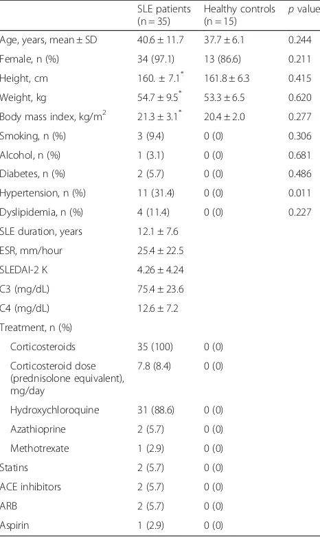
The mean age of the SLE patients was 40.6 ± 11.7 years, and the majority were female (97.1%). Mean disease duration was 12.1 ± 7.6 years, and the mean SLEDAI-2 K was 4.SLEDAI-26 ± 4.SLEDAI-24. The majority of patients were taking glucocorticoids (mean prednisolone equivalent dose, 7.8 mg/d) and hydroxychloroquine at the time of blood sampling. Only few patients were taking an add-itional immunosuppressant such as azathioprine or methotrexate (Table 1).
Comparison of serum lipid profiles between SLE patients and HCs
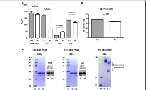
There was no difference in total cholesterol levels between SLE patients and HCs (190.1 ± 54.1 mg/dL vs. 178.3 ± 25.5 mg/dL,p= 0.43). However, TG levels were significantly higher in SLE patients than in HCs (168.9 ± 70.1 mg/dL vs. 69.5 ± 18.8 mg/dL,p< 0.001). There was no difference be-tween SLE patients and HCs with respect to LDL levels (134.7 ± 54.5 mg/dL vs. 118.3 ± 29.9 mg/dL, p= 0.28). However, HDL levels in SLE patients were significantly lower than those in HCs (21.7 ± 9.3 mg/dL vs. 46.2 ± 9.3 mg/dL, p< 0.001) (Fig. 1a). Serum cholesterol ester transfer protein (CETP) activity, which decreases HDL levels by preferentially transferring cholesterol esters from HDL to apoB-containing LDL, was higher in SLE patients than in HCs (38.4% ± 6.8% vs. 34.2% ± 2.6%,
p= 0.03) (Fig. 1b).
Lipoproteins from SLE patients show increased fragmentation
confirmed as apoA-I on Western blot analysis. The intensity of the lipoprotein bands in HC samples was stronger than that from SLE (Fig. 1c; left and middle panels). LDL proteins isolated from SLE patients were more fragmented than those from HCs (Fig. 1c; right panel, short arrows).
SLE-associated lipoproteins show increased oxidation
The increased fragility of SLE-associated lipoproteins sug-gests that they might have undergone additional structural modifications, such as oxidation. Therefore, we isolated HDL2, HDL3, and LDL from SLE patients and HCs and measured the degree of oxidation. HDL2, HDL3, and LDL from SLE patients exhibited higher levels of oxidized spe-cies than those from HCs (HDL2, 22.6 ± 4.7 vs. 11.3 ± 2.0,
p< 0.001; HDL3, 17.5 ± 2.4 vs. 7.8 ± 1.1,p< 0.001; and LDL, 56.2 ± 10.2 vs. 25.4 ± 2.3, p< 0.001) (Fig. 2a). Next, we examined whether lipoproteins from SLE patients were more susceptible to de novo oxidation. The oxidation rate of LDL from SLE patients (SLE-LDL) was significantly higher than LDL from HCs (HC-LDL) (3.6% ± 0.5% vs. 1.9% ± 0.3%, p< 0.001) under conditions of cupric ion-mediated oxidative stress (Fig. 2b). In addition, paraoxonase activity (an HDL-associated enzyme that protects LDL from oxidation) was significantly lower in SLE patients than in HCs (3.53 ± 0.19 vs. 4.04 ± 0.16,p< 0.001) (Fig. 2c).
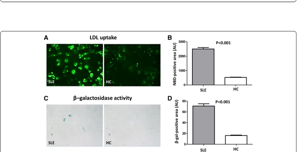
SLE-LDL induces foam cell generation and cellular senescence
THP-1 cells (a human monocytic cell line derived from an acute monocytic leukemia) were incubated with LDL isolated from SLE patients or HCs. THP-1 cells phagocy-tosed significantly more SLE-LDL than HC-LDL (NBD positive area: 2501 ± 401.2 vs. 524.1 ± 59.9 arbitrary units (AUs),p< 0.001) and transformed into foam cells (Fig. 3a and b). Exposure of human fibroblasts to SLE-LDL induced accelerated cellular senescence, as reflected by increasedβ-galactosidase activity (70.9 ± 17.9 vs. 16.3 ± 2.4,
p< 0.001) (Fig. 3c and d).
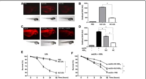
SLE lipoproteins cause oxidative stress in zebrafish embryos
Zebrafish embryos were injected with LDL isolated from SLE patients or HCs. SLE-LDL induced significantly higher levels of DHE oxidation in vivo than HC-LDL (1592.0 ± 58.7 AUs vs. 459.6 ± 66.4 AUs, p< 0.001) (Fig. 4a and b). Next, embryos were injected with oxLDL in the presence of anti-oxidative HDL3 isolated from SLE patients or HCs. Embryos exposed to oxLDL alone showed marked oxidation of DHE (Fig. 4c; left panel). Co-injection of HDL3from HCs or SLE patients reduced DHE oxidation than oxLDL alone (bothp< 0.001). How-ever, HDL3from SLE patients induced significantly more DHE oxidation than that from HCs (3358 ± 208.8 AUs vs. 1299 ± 75.1 AUs,p< 0.001) (Fig. 4d).
[image:4.595.57.290.110.501.2]SLE-LDL was toxic to embryos: 8 hours after injection, 68.3% ± 3.0% of embryos exposed to SLE-LDL remained alive compared with 94.6% ± 1.1% exposed to HC-LDL. After 48 hours, the mean survival rate of SLE-LDL-exposed embryos was 49.3% ± 2.8% whereas that of HC-LDL-exposed embryos was 85.0% ± 2.9% (p< 0.001) (Fig. 4e). The injection of oxidized LDL purified from HCs reduced the embryo survival to 48.6% after 48 hours. The toxicity of oxLDL was partially reversed or neutralized by co-injection of protective HDLs: co-in-jection of HDL3 from HCs improved the embryo sur-vival as compared to HDL3from SLE patients (75.1% ± 2.1% vs. 62.0% ± 3.1%,p< 0.001) (Fig. 4f ).
Table 1Baseline demographic and clinical characteristics of the study participants
SLE patients (n = 35)
Healthy controls (n = 15)
pvalue Age, years, mean ± SD 40.6 ± 11.7 37.7 ± 6.1 0.244 Female, n (%) 34 (97.1) 13 (86.6) 0.211 Height, cm 160. ± 7.1* 161.8 ± 6.3 0.415 Weight, kg 54.7 ± 9.5* 53.3 ± 6.5 0.620 Body mass index, kg/m2 21.3 ± 3.1* 20.4 ± 2.0 0.277 Smoking, n (%) 3 (9.4) 0 (0) 0.306 Alcohol, n (%) 1 (3.1) 0 (0) 0.681 Diabetes, n (%) 2 (5.7) 0 (0) 0.486 Hypertension, n (%) 11 (31.4) 0 (0) 0.011 Dyslipidemia, n (%) 4 (11.4) 0 (0) 0.227 SLE duration, years 12.1 ± 7.6
ESR, mm/hour 25.4 ± 22.5 SLEDAI-2 K 4.26 ± 4.24 C3 (mg/dL) 75.4 ± 23.6 C4 (mg/dL) 12.6 ± 7.2 Treatment, n (%)
Corticosteroids 35 (100) 0 (0) Corticosteroid dose
(prednisolone equivalent), mg/day
7.8 (8.4) 0 (0)
Hydroxychloroquine 31 (88.6) 0 (0) Azathioprine 2 (5.7) 0 (0) Methotrexate 1 (2.9) 0 (0) Statins 2 (5.7) 0 (0) ACE inhibitors 2 (5.7) 0 (0) ARB 2 (5.7) 0 (0) Aspirin 1 (2.9) 0 (0)
*
Available for 32 patients
Discussion
Increased generation of oxLDL is crucial for the patho-genesis of atherosclerosis: oxLDL accumulates in vascu-lar walls and attracts monocytes, which then differentiate into tissue macrophages and release inflam-matory cytokines [29]. Foam cells within the vessel wall form the lipid core of an atherosclerotic plaque [30–32]. Furthermore, oxLDL induces cellular senescence, impairs endothelial cell function, and inhibits the release of protective nitric oxide [33, 34]. Accordingly, because statins make LDL less available for oxidation by reducing the hepatic synthesis of cholesterol (which is the main lipid component of LDL) [35], they lead to a significant reduction in CV-related mortality in the general popula-tion [36]. However, atorvastatin did not inhibit or re-verse the atherosclerosis in patients with SLE, although it reduced LDL levels [10]. This suggests that not only the quantity but also the quality of lipoproteins might, at least in part, account for the non-traditional risk factors for accelerated atherosclerosis in SLE patients.
Here, we provide direct evidence that circulating lipo-proteins in patients with SLE are altered and show specific physicochemical properties. First, SLE-LDL exhibited greater oxidation and fragility than HC-LDL. Second, SLE-LDL was more susceptible to de novo oxidation. Third, SLE-LDL induced foam cells and accelerated cellu-lar senescence. Fourth, injection of SLE-LDL into zebra-fish embryos caused greater oxidative stress and higher embryonic mortality. Finally, HDL from SLE patients had impaired anti-oxidative and protective effects. In short, li-poproteins from SLE patients showed higher oxidative and lower anti-oxidative potential than lipoproteins from HCs with detrimental physiological effects.
Serum lipoproteins are produced by the liver. During acute systemic inflammation, inflammatory cytokines in-crease the hepatic production of acute phase reactants. The levels of serum amyloid protein (SAA), an apolipo-protein associated with HDL [37], increase during active inflammation, as occurs during active SLE [16]. Thus, the SAA content of HDL increases at the expense of
[image:5.595.59.540.88.386.2]Fig. 2Increased oxidation of lipoproteins from systemic lupus erythematosus (SLE) patients.aHDL2, HDL3, and LDL were isolated from SLE patients (n = 19) and healthy controls (HCs) (n = 8), and their oxidation status (i.e., MDA levels) was measured. All lipoprotein fractions from SLE patients showed significantly higher levels of oxidation than those from HCs.bLDL was incubated in the presence of 5μM CuSO4, and the formation of conjugated dienes over time was measured as a marker of de novo oxidation. LDL from SLE patients was significantly more susceptible to oxidation than that from HCs.cSerum paraoxonase activity was significantly lower in SLE samples than in HC samples. Data are expressed as the mean and SEM.HC
healthy controls,MDAmalondialdehyde,SLEsystemic lupus erythematosus
Fig. 3Monocytes show increased uptake of LDL from SLE patients.aandbTHP-1 cells were incubated with LDL isolated from SLE patients (n = 19) and HCs (n = 8). THP-1 cells phagocytosed significantly more LDL from SLE patients and showed greater foam cell formation (NBD-positive area: 2501 ± 401.2 AUs vs. 524.1 ± 59.9 AUs, respectively;p< 0.001).cHuman fibroblast cells were incubated with LDL from SLE patients or HCs, andβ-galactosidase activity (a surrogate marker for cellular senescence) was measured.β-galactosidase activity was higher in fibroblasts treated with SLE-LDL (blue) than in those treated with HC-LDL.dSLE-LDL induced significantly higherβ-galactosidase activity than HC-LDL (β-gal positive area: 70.9 AUs vs. 16.3 AUs, respectively;
p< 0.001). Representative images (×400 magnification) from at least three independent experiments are shown. Data are expressed as the mean and SEM.
[image:6.595.58.537.87.333.2] [image:6.595.56.541.396.643.2]apoA-I; this impairs the reverse cholesterol transport of HDL [38]. Here, we found that the proportion of apoA-I in HDL2 and HDL3 from SLE patients seemed to be lower than that in HDL2and HDL3from HCs (Fig. 1c). Also, LDL from SLE patients was more fragile and more susceptible to oxidation (Fig. 2). Reduced paraoxonase activity, which protects LDL from oxidative modifica-tion, might potentiate the generation of oxLDL in SLE [17]. Taken together, HDL dysfunction (possibly due to altered composition), reduced paraoxonase activity, and increased susceptibility of LDL to oxidation might all contribute to increased generation of oxLDL. Consistent with increased LDL oxidation, we found that THP-1 cells readily engulfed SLE-LDL and transformed into foam cells (Fig. 3). Since monocytes from SLE are the same as those from HCs in terms of their capacity to take up oxLDL and transform into foam cells [39], the increased phagocytosis of LDL is likely due to SLE-specific alterations in structure/function of lipoproteins.
To the best of our knowledge, this study is the first to show that LDL from SLE patients exhibits deleterious oxidative effects in vivo using zebrafish embryos. Zebra-fish embryo is suitable to study in vivo oxidation, since
their optical clarity allows dynamic tracking of the oxi-dation process using a fluorescence probe [40]. Further studies are needed to show whether the alteration of li-poproteins can be translated into accelerated athero-sclerosis in vivo as well.
It is not clear whether the findings observed herein are SLE-specific or are generalizable to other chronic inflam-matory diseases such as rheumatoid arthritis and primary systemic vasculitis, both of which are associated with an increased risk of CV-related morbidity [41]. Since even a slight increase in CRP levels is associated with increased CV-related morbidity, one might speculate that smolder-ing inflammation in general might be associated with alterations in the properties of lipoproteins [42, 43]. The finding that anti-oxidant vitamins did not prevent athero-sclerosis raises the question of whether the detrimental effects of oxidized lipoproteins are irreversible [44]. Tight control of SLE disease activity and the associated systemic inflammation, might reduce CV risk as seen in patients with rheumatoid arthritis [45].
The present study has several limitations. First, the HCs were not matched for all comorbidities. Second, the relatively small number of SLE patients does not allow
[image:7.595.58.539.87.349.2]to assess the effects of medical treatment, particularly those of corticosteroids, hydroxychloroquine, and statins, which could have pleiotropic effects on lipid metabolism [46]. Third, since the lipid oxidation can occur during storage, the possible impact of the storage time on the de-gree of spontaneous lipid oxidation needs further investi-gation. Fourth, due to a technical limitation, the protein fragments in the SLE-LDL could not be unequivocally identified as apolipoproteins. Fifth, it might be of interest to examine changes in the physicochemical properties of lipoproteins in treated SLE patients over time. Ultimately, further studies should determine whether the findings of the present study can be translated into an in vivo model of accelerated atherosclerosis.
Conclusions
Lipoproteins from SLE patients show altered structural and functional properties with higher oxidative potential in vitro and in vivo. Further studies should examine whether alterations in lipoproteins directly contribute to the accelerated atherosclerosis associated with SLE.
Additional file
Additional file 1:Table S1. Baseline demographic and clinical characteristics of the participants included in the pooled samples.Figure S1. Age distribution of 19 systemic lupus erythematosus (SLE) patients and 8 healthy controls (HCs) included in the pooled samples. (PDF 222 kb)
Abbreviations
CE:cholesteryl ester; CEPT: cholesterol ester transfer protein; CRP: C-reactive protein; CV: cardiovascular; DHE: dihydroethidium; HC: healthy control; HDL: high-density lipoprotein; LDL: low-density lipoprotein;
MDA: malondialdehyde; oxLDL: oxidized LDL; rHDL: recombinant HDL; SA: senescence-associated; SAA: serum amyloid protein; SDS-PAGE: sodium dodecyl sulfate-polyacrylamide gel electrophoresis; SLE: systemic lupus erythematosus; SLEDAI: SLE disease activity index; TG: triglyceride
Acknowledgements Not applicable.
Funding
This work was supported by a grant from the Korea Health Technology R&D Project through the Korea Health Industry Development Institute funded by the Ministry of Health and Welfare, Republic of Korea (grant number: HI13C1754) and by a grant from the Mid-carrier Researcher Program (2014-11049455) through the National Research Foundation funded by the Ministry of Science, ICT, and Future Planning, Republic of Korea.
Availability of data and materials Not applicable.
Authors’contributions
JKP and YWS conceived the study, analyzed and interpreted data, and participated in drafting the manuscript. JYK, JYM, EYA, EYL, EBL, and KHC made substantial contributions to data analysis, data interpretation, and drafting of the manuscript. All authors read and approved the final manuscript.
Authors’information
Not applicable.
Competing interests
The authors declare that they have no competing interests.
Consent for publication Not applicable.
Ethical approval and consent to participate
Informed consent was obtained from all participants in accordance with the Declaration of Helsinki. The study was approved by the Institutional Review Board at Seoul National University Hospital.
Author details
1Department of Molecular Medicine and Biopharmaceutical Sciences,
Graduate School of Convergence Science and Technology, and College of Medicine, Medical Research Center, Seoul National University, Seoul, Republic of Korea.2Division of Rheumatology, Department of Internal Medicine, Seoul
National University Hospital, Seoul, Republic of Korea.3Department of
Medical Biotechnology, Yeungnam University, Gyeongsangbuk-Do, Republic of Korea.4Division of Rheumatology, Department of Internal Medicine, Seoul
National University College of Medicine, 101 Daehak-ro, Jongno-gu, Seoul 03080, Republic of Korea.
Received: 5 July 2016 Accepted: 5 December 2016
References
1. Liu Z, Davidson A. Taming lupus-a new understanding of pathogenesis is leading to clinical advances. Nat Med. 2012;18(6):871–82.
2. Manzi S, Meilahn EN, Rairie JE, Conte CG, Medsger Jr TA, Jansen-McWilliams L, D’Agostino RB, Kuller LH. Age-specific incidence rates of myocardial infarction and angina in women with systemic lupus erythematosus: comparison with the Framingham Study. Am J Epidemiol. 1997;145(5):408–15.
3. Bessant R, Hingorani A, Patel L, MacGregor A, Isenberg DA, Rahman A. Risk of coronary heart disease and stroke in a large British cohort of patients with systemic lupus erythematosus. Rheumatology (Oxford). 2004;43(7):924–9. 4. Bruce IN, Urowitz MB, Gladman DD, Ibanez D, Steiner G. Risk factors for
coronary heart disease in women with systemic lupus erythematosus: the Toronto Risk Factor Study. Arthritis Rheum. 2003;48(11):3159–67. 5. Chung CP, Avalos I, Oeser A, Gebretsadik T, Shintani A, Raggi P, Michael SC.
High prevalence of the metabolic syndrome in patients with systemic lupus erythematosus: association with disease characteristics and cardiovascular risk factors. Ann Rheum Dis. 2007;66(2):208–14.
6. Esdaile JM, Abrahamowicz M, Grodzicky T, Li Y, Panaritis C, du Berger R, Cote R, Grover SA, Fortin PR, Clarke AE, et al. Traditional Framingham risk factors fail to fully account for accelerated atherosclerosis in systemic lupus erythematosus. Arthritis Rheum. 2001;44(10):2331–7.
7. Magder LS, Petri M. Incidence of and risk factors for adverse cardiovascular events among patients with systemic lupus erythematosus. Am J Epidemiol. 2012;176(8):708–19.
8. Roman MJ, Shanker BA, Davis A, Lockshin MD, Sammaritano L, Simantov R, Crow MK, Schwartz JE, Paget SA, Devereux RB, et al. Prevalence and correlates of accelerated atherosclerosis in systemic lupus erythematosus. N Engl J Med. 2003;349(25):2399–406.
9. Lee AB, Godfrey T, Rowley KG, Karschimkus CS, Dragicevic G, Romas E, Clemens L, Wilson AM, Nikpour M, Prior DL, et al. Traditional risk factor assessment does not capture the extent of cardiovascular risk in systemic lupus erythematosus. Intern Med J. 2006;36(4):237–43.
10. Petri MA, Kiani AN, Post W, Christopher-Stine L, Magder LS. Lupus Atherosclerosis Prevention Study (LAPS). Ann Rheum Dis. 2011;70(5):760–5. 11. Schanberg LE, Sandborg C, Barnhart HX, Ardoin SP, Yow E, Evans GW,
Mieszkalski KL, Ilowite NT, Eberhard A, Imundo LF, et al. Use of atorvastatin in systemic lupus erythematosus in children and adolescents. Arthritis Rheum. 2012;64(1):285–96.
12. Navab M, Hama SY, Anantharamaiah GM, Hassan K, Hough GP, Watson AD, Reddy ST, Sevanian A, Fonarow GC, Fogelman AM. Normal high density lipoprotein inhibits three steps in the formation of mildly oxidized low density lipoprotein: steps 2 and 3. J Lipid Res. 2000;41(9):1495–508. 13. Navab M, Hama SY, Cooke CJ, Anantharamaiah GM, Chaddha M, Jin L,
14. Navab M, Berliner JA, Watson AD, Hama SY, Territo MC, Lusis AJ, Shih DM, Van Lenten BJ, Frank JS, Demer LL, et al. The Yin and Yang of oxidation in the development of the fatty streak. A review based on the 1994 George Lyman Duff Memorial Lecture. Arterioscler Thromb Vasc Biol.
1996;16(7):831–42.
15. Smith CK, Seto NL, Vivekanandan-Giri A, Yuan W, Playford MP, Manna Z, Hasni SA, Kuai R, Mehta NN, Schwendeman A, et al. Lupus high-density lipoprotein induces proinflammatory responses in macrophages by binding lectin-like oxidised low-density lipoprotein receptor 1 and failing to promote activating transcription factor 3 activity. Ann Rheum Dis. 2016; 209683. [Epub ahead of print].
16. McMahon M, Grossman J, FitzGerald J, Dahlin-Lee E, Wallace DJ, Thong BY, Badsha H, Kalunian K, Charles C, Navab M, et al. Proinflammatory high-density lipoprotein as a biomarker for atherosclerosis in patients with systemic lupus erythematosus and rheumatoid arthritis. Arthritis Rheum. 2006;54(8):2541–9. 17. Van Lenten BJ, Wagner AC, Nayak DP, Hama S, Navab M, Fogelman AM.
High-density lipoprotein loses its anti-inflammatory properties during acute influenza a infection. Circulation. 2001;103(18):2283–8.
18. Okura Y, Brink M, Itabe H, Scheidegger KJ, Kalangos A, Delafontaine P. Oxidized low-density lipoprotein is associated with apoptosis of vascular smooth muscle cells in human atherosclerotic plaques. Circulation. 2000;102(22):2680–6.
19. Podrez EA, Poliakov E, Shen Z, Zhang R, Deng Y, Sun M, Finton PJ, Shan L, Gugiu B, Fox PL, et al. Identification of a novel family of oxidized phospholipids that serve as ligands for the macrophage scavenger receptor CD36. J Biol Chem. 2002;277(41):38503–16.
20. Miller YI, Chang MK, Binder CJ, Shaw PX, Witztum JL. Oxidized low density lipoprotein and innate immune receptors. Curr Opin Lipidol. 2003;14(5):437–45. 21. Hochberg MC. Updating the American College of Rheumatology revised
criteria for the classification of systemic lupus erythematosus. Arthritis Rheum. 1997;40(9):1725.
22. Gladman DD, Ibanez D, Urowitz MB. Systemic lupus erythematosus disease activity index 2000. J Rheumatol. 2002;29(2):288–91.
23. Havel RJ, Eder HA, Bragdon JH. The distribution and chemical composition of ultracentrifugally separated lipoproteins in human serum. J Clin Invest. 1955;34(9):1345–53.
24. Markwell MA, Haas SM, Bieber LL, Tolbert NE. A modification of the Lowry procedure to simplify protein determination in membrane and lipoprotein samples. Anal Biochem. 1978;87(1):206–10.
25. Blois MS. Antioxidant determinations by the use of a stable free radical. Nature. 1958;181:1199–200.
26. Cho KH. Synthesis of reconstituted high density lipoprotein (rHDL) containing apoA-I and apoC-III: the functional role of apoC-III in rHDL. Mol Cells. 2009;27(3):291–7.
27. Cho KH, Shin DG, Baek SH, Kim JR. Myocardial infarction patients show altered lipoprotein properties and functions when compared with stable angina pectoris patients. Exp Mol Med. 2009;41(2):67–76.
28. Park KH, Shin DG, Kim JR, Hong JH, Cho KH. The functional and compositional properties of lipoproteins are altered in patients with metabolic syndrome with increased cholesteryl ester transfer protein activity. Int J Mol Med. 2010;25(1):129–36.
29. Levitan I, Volkov S, Subbaiah PV. Oxidized LDL: diversity, patterns of recognition, and pathophysiology. Antioxid Redox Signal. 2010;13(1):39–75. 30. McMahon M, Skaggs BJ, Grossman JM, Sahakian L, Fitzgerald J, Wong WK,
Lourenco EV, Ragavendra N, Charles-Schoeman C, Gorn A, et al. A panel of biomarkers is associated with increased risk of the presence and progression of atherosclerosis in women with systemic lupus erythematosus. Arthritis Rheumatol. 2014;66(1):130–9.
31. Frostegard J, Svenungsson E, Wu R, Gunnarsson I, Lundberg IE, Klareskog L, Horkko S, Witztum JL. Lipid peroxidation is enhanced in patients with systemic lupus erythematosus and is associated with arterial and renal disease manifestations. Arthritis Rheum. 2005;52(1):192–200.
32. Svenungsson E, Jensen-Urstad K, Heimburger M, Silveira A, Hamsten A, de Faire U, Witztum JL, Frostegard J. Risk factors for cardiovascular disease in systemic lupus erythematosus. Circulation. 2001;104(16):1887–93. 33. Chin JH, Azhar S, Hoffman BB. Inactivation of endothelial derived relaxing
factor by oxidized lipoproteins. J Clin Invest. 1992;89(1):10–8. 34. Vink H, Constantinescu AA, Spaan JA. Oxidized lipoproteins degrade the
endothelial surface layer : implications for platelet-endothelial cell adhesion. Circulation. 2000;101(13):1500–2.
35. Davignon J. Beneficial cardiovascular pleiotropic effects of statins. Circulation. 2004;109(23 Suppl 1):III39–43.
36. Stone NJ, Robinson JG, Lichtenstein AH, Bairey Merz CN, Blum CB, Eckel RH, Goldberg AC, Gordon D, Levy D, Lloyd-Jones DM, et al. 2013 ACC/AHA guideline on the treatment of blood cholesterol to reduce atherosclerotic cardiovascular risk in adults: a report of the American College of Cardiology/ American Heart Association Task Force on Practice Guidelines. Circulation. 2014;129(25 Suppl 2):S1–45.
37. Kisilevsky R, Manley PN. Acute-phase serum amyloid A: perspectives on its physiological and pathological roles. Amyloid. 2012;19(1):5–14.
38. Daynes RA, Jones DC. Emerging roles of PPARs in inflammation and immunity. Nat Rev Immunol. 2002;2(10):748–59.
39. Yassin LM, Londono J, Montoya G, De Sanctis JB, Rojas M, Ramirez LA, Garcia LF, Vasquez G. Atherosclerosis development in SLE patients is not determined by monocytes ability to bind/endocytose Ox-LDL. Autoimmunity. 2011;44(3):201–10.
40. Mugoni V, Camporeale A, Santoro MM. Analysis of oxidative stress in zebrafish embryos. J Vis Exp. 2014;(89).
41. Prasad M, Hermann J, Gabriel SE, Weyand CM, Mulvagh S, Mankad R, Oh JK, Matteson EL, Lerman A. Cardiorheumatology: cardiac involvement in systemic rheumatic disease. Nat Rev Cardiol. 2015;12(3):168–76. 42. Ridker PM, Rifai N, Pfeffer MA, Sacks FM, Moye LA, Goldman S, Flaker GC,
Braunwald E. Inflammation, pravastatin, and the risk of coronary events after myocardial infarction in patients with average cholesterol levels. Cholesterol and Recurrent Events (CARE) Investigators. Circulation. 1998;98(9):839–44. 43. Sattar N, Murray HM, Welsh P, Blauw GJ, Buckley BM, Cobbe S, de Craen AJ,
Lowe GD, Jukema JW, Macfarlane PW, et al. Are markers of inflammation more strongly associated with risk for fatal than for nonfatal vascular events? PLoS Med. 2009;6(6):e1000099.
44. Vivekananthan DP, Penn MS, Sapp SK, Hsu A, Topol EJ. Use of antioxidant vitamins for the prevention of cardiovascular disease: meta-analysis of randomised trials. Lancet. 2003;361(9374):2017–23.
45. Ljung L, Rantapaa-Dahlqvist S, Jacobsson LT, Askling J, Group AS. Response to biological treatment and subsequent risk of coronary events in rheumatoid arthritis. Ann Rheum Dis. 2016;75(12):2087–94.
46. Ansell BJ, Navab M, Hama S, Kamranpour N, Fonarow G, Hough G, Rahmani S, Mottahedeh R, Dave R, Reddy ST, et al. Inflammatory/antiinflammatory properties of high-density lipoprotein distinguish patients from control subjects better than high-density lipoprotein cholesterol levels and are favorably affected by simvastatin treatment. Circulation. 2003;108(22):2751–6.
• We accept pre-submission inquiries
• Our selector tool helps you to find the most relevant journal • We provide round the clock customer support
• Convenient online submission • Thorough peer review
• Inclusion in PubMed and all major indexing services • Maximum visibility for your research
Submit your manuscript at www.biomedcentral.com/submit