sublining macrophages display an IL-10
polarized-like phenotype in chronic synovitis
Ambarus
et al.
R E S E A R C H A R T I C L E
Open Access
Intimal lining layer macrophages but not synovial
sublining macrophages display an IL-10
polarized-like phenotype in chronic synovitis
Carmen A Ambarus
1,2*, Troy Noordenbos
1,2, Maria JH de Hair
1, Paul P Tak
1,3and Dominique LP Baeten
1,2Abstract
Introduction:Synovial tissue macrophages play a key role in chronic inflammatory arthritis, but the contribution of different macrophage subsets in this process remains largely unknown. The mainin vitropolarized macrophage subsets are classically (M1) and alternatively (M2) activated macrophages, the latter comprising interleukin (IL)-4 and IL-10 polarized cells. Here, we aimed to evaluate the polarization status of synovial macrophages in
spondyloarthritis (SpA) and rheumatoid arthritis (RA).
Methods:Expression of polarization markers on synovial macrophages, peripheral blood monocytes, andin vitro polarized monocyte-derived macrophages from SpA versus RA patients was assessed by immunohistochemistry and flow cytometry, respectively. The polarization status of the intimal lining layer and the synovial sublining macrophages was assessed by double immunofluorescence staining.
Results:The expression of the IL-10 polarization marker cluster of differentiation 163 (CD163) was increased in SpA compared with RA intimal lining layer, but no differences were found in other M1 and M2 markers between the diseases. Furthermore, no significant phenotypic differences in monocytes andin vitropolarized monocyte-derived macrophages were seen between SpA, RA, and healthy controls, indicating that the differential CD163 expression does not reflect a preferential M2 polarization in SpA. More detailed analysis of intimal lining layer macrophages revealed a strong co-expression of the IL-10 polarization markers CD163 and cluster of differentiation 32 (CD32) but not any of the other markers in both SpA and RA. In contrast, synovial sublining macrophages had a more
heterogeneous phenotype, with a majority of cells co-expressing M1 and M2 markers.
Conclusions:The intimal lining layer but not synovial sublining macrophages display an IL-10 polarized-like phenotype, with increased CD163 expression in SpA versus RA synovitis. These differences in the distribution of the polarized macrophage subset may contribute to the outcome of chronic synovitis.
Introduction
The normal synovial membrane consists of two distinct structures: the intimal lining layer and the synovial sub-lining. The normal intimal lining layer comprises fibro-blast-like synoviocytes and intimal lining layer macrophages, whereas the synovial sublining consists of connective tissue containing blood vessels, fibroblasts, adipocytes, and a limited number of resident immune cells, such as macrophages and mast cells [1]. Chronic
inflammatory arthritis is histologically characterized by marked hyperplasia of the intimal lining layer and mas-sive infiltration of the synovial sublining with innate and adaptive immune cells, which release inflammatory med-iators, promote neoangiogenesis, and drive the destruc-tion of adjacent cartilage and bone [2]. Macrophages play a major role in this process because they contribute to the intimal lining layer hyperplasia [3] and are the main producers of key inflammatory mediators, such as tumor necrosis factor (TNF) [4]. Accordingly, the num-ber of synovial macrophages correlates with clinical dis-ease activity [2,5] and decrdis-eases after clinically efficient but not placebo treatment in both rheumatoid arthritis (RA) [6-9] and spondyloarthritis (SpA) [10-13]. * Correspondence: c.a.ambarus@amc.nl
1Department of Clinical Immunology and Rheumatology, Academic Medical
Center/University of Amsterdam, Meibergdreef 9, 1105 AZ Amsterdam, The Netherlands
Full list of author information is available at the end of the article
Furthermore, selective macrophage depletion has a strong antiinflammatory effect in animal models of arthritis [14].
Tissue macrophages consist of resident macrophages and monocytes that are recruited in inflammatory con-ditions and differentiate into macrophages on tissue entry. These macrophages represent a heterogeneous population, as local mediators can prime macrophages during their maturation and thereby shape their subse-quent response to various activating stimuli [15,16]. Initially, two main polarized macrophage subsets were described based on their pro- versus antiinflammatory functions: classically activated macrophages (M1), which are specialized in the clearance of intracellular patho-gens, and alternatively activated macrophages (M2), which have immunoregulatory properties and are involved in scavenging debris, angiogenesis, and tissue repair [17]. The M1 subset is induced mainly by inter-feron (IFN)-g(MFIFN-g), whereas M2 can be induced in vitroeither by IL-4 (MFIL-4)/IL-13 or by IL-10 (M
FIL-10) [17-20]. Whereas the macrophage polarization has
been best described in rodents, we recently validated specific phenotypic markers [21] and distinct functional characteristics (unpublished observations) for these three main in vitro polarized macrophage subsets in humans.
The increasing knowledge of the phenotypic and func-tional diversity of human macrophages indicates that not only the overall number of macrophages in the inflamed tissue, but also their specific polarization status may determine the disease outcome in terms of severity, chronicity, and tissue damage. Interestingly, we pre-viously described that, despite similar numbers of macrophages in RA and SpA synovitis, a selective over-representation of CD163+ macrophages was present in SpA [3,22-25]. CD163, the scavenger receptor for hemo-globin/haptoglobin complexes, is a known marker for IL-10 or M-CSF polarized M2 [21,26]. Together with the lower levels of M1-derived proinflammatory cyto-kines in SpA versus RA synovial fluid [27] and the defective IFN-gsignature in SpA monocytes [28], these data suggest a preferential M2 polarization in SpA com-pared with an M1 polarization in RA. The aim of this study was to test this hypothesis by detailed characteri-zation of distinct macrophage subsets in SpA and RA synovial tissue.
Materials and methods
Patients and samples
Peripheral blood samples were obtained from 11 SpA and eight RA patients and nine healthy controls. Syno-vial biopsies were obtained with small-bore arthro-scopy from clinically inflamed knee joints, as previously described [29], and 18 SpA and 20 RA
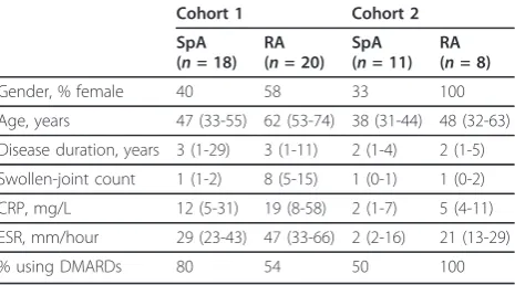
patients were included for the immunohistochemical analysis. All SpA patients fulfilled the criteria of the European Spondyloarthropathy Study Group (ESSG) [30], and all RA patients fulfilled the 1987 ACR classi-fication criteria for RA [31]. Demographic and clinical data of the patients are shown in Table 1. None of the patients was being treated with biologicals. All patients gave written informed consent to participate to the study, as approved by the Medical Ethics Committee of the Academic Medical Centre/University of Amsterdam.
Immunohistochemistry
[image:3.595.306.539.551.680.2]Synovial biopsy samples (six to eight per patient to minimize sampling error) were snap-frozen en blocand mounted in Jung tissue-freezing medium (Leica Instru-ments, Nussloch, Germany). The acetone-fixed sections were stained by using mouse monoclonal antibodies directed toward CD68 (clone EBM-11; Dako, Heverlee, The Netherlands), CD64 (clone 10.1; BioLegend, Uithoorn, The Netherlands), CD14 (clone TUK4; Dako), CD163 (clone 5cFAT; BMA Biomedicals, Augst, Swit-zerland), and CD32 (clone AT10; abcam, Cambridge, UK). Sections were sequentially incubated with a bioti-nylated secondary antibody, a streptavidin-horseradish peroxidase link (LSAB; Dako), aminoethylcarbazole sub-strate as chromogen, and hematoxylin as counterstain [22,25]. Parallel sections were incubated with isotype-and concentration-matched monoclonal antibodies as negative controls. The expression of CD200R was assessed with immunofluorescence, by using a mouse monoclonal antibody (clone OX108; AbD Serotec, Düs-seldorf, Germany). For each cohort, samples were stained in a single run, coded, and scored in a random order on a 4-point semiquantitative scale by two
Table 1 Demographic and clinical features of the patients in the two cohorts
Cohort 1 Cohort 2
SpA
(n= 18)
RA
(n= 20)
SpA
(n= 11)
RA
(n= 8)
Gender, % female 40 58 33 100
Age, years 47 (33-55) 62 (53-74) 38 (31-44) 48 (32-63) Disease duration, years 3 (1-29) 3 (1-11) 2 (1-4) 2 (1-5) Swollen-joint count 1 (1-2) 8 (5-15) 1 (0-1) 1 (0-2) CRP, mg/L 12 (5-31) 19 (8-58) 2 (1-7) 5 (4-11) ESR, mm/hour 29 (23-43) 47 (33-66) 2 (2-16) 21 (13-29)
% using DMARDs 80 54 50 100
independent observers (CAA, TN), as described pre-viously [5].
Monocyte isolation andin vitropolarization
Monocytes were isolated from peripheral blood with gradient centrifugation with Lymphoprep (Axis-Shield PoPAS, Oslo, Norway) and, subsequently, Percoll gradi-ent separation (GE Healthcare, Uppsala, Sweden). Monocytes were analyzed immediately with flow cyto-metry or cultured at a concentration of 0.5 × 10⁶/ml in Iscove Modified Dulbecco Medium (IMDM) (Invitrogen, Breda, The Netherlands) supplemented with 10% fetal calf serum (FCS) (PAA Laboratories, Cölbe, Germany). Cultured monocytes were polarizedin vitrowith human recombinant IFN-g(10 ng/ml; R&D Systems, Abingdon, UK), IL-4 (10 ng/ml; Miltenyi Biotec, Bergisch Glad-bach, Germany), or IL-10 (10 ng/ml; R&D Systems) for 4 days. For further analysis, polarized macrophages were recovered by scraping of the culture plate.
Flow cytometry
Surface-marker expression on monocytes andin vitro polarized macrophages was analyzed with flow cytome-try (BD FACS Calibur Flow Cytometer, Erembodegem, Belgium) by using fluorochrome-labeled monoclonal antibodies against CD80 (clone L307.4; BD Pharmingen, Breda, the Netherlands), CD64 (clone 10.1; BioLegend), CD200R (clone OX108; AbD Serotec), CD14 (clone 61D3; eBioscience, San Diego, CA, USA), CD163 (clone GHI/61; BD Pharmingen), and CD16 (clone DJ130c; AbD Serotec, Düsseldorf, Germany). Equivalent amounts of isotype-matched control antibodies were included in all experiments as negative controls. Before staining, Fc receptors were blocked with 10% human serum (Lonza, Cologne, Germany). Data were analyzed with Flow Jo Flow Cytometry Analysis software (Tree Star, Ashland, OR, USA) after gating on the myeloid population in the FSC/SSC window. Values were expressed as the ratio of the geometric mean fluorescence intensity (gMFI) of the marker of interest over the gMFI of the isotype control.
Double immunofluorescence
Frozen synovial tissue sections were fixed in acetone and blocked with 10% goat serum (Dako, Glostrup, Den-mark), followed by incubation with Biotin blocking sys-tem (Dako). Mouse monoclonal antibodies against CD80 (clone 2D10; BioLegend, Uithoorn, The Nether-lands), CD64 (clone 10.1; BioLegend), CD200R (clone OX108; AbD Serotec, Düsseldorf, Germany), CD14 (clone TUK4; Dako), CD163 (clone 5cFAT; BMA Bio-medicals, Augst, Switzerland), CD16 (clone HI16a; Abbiotec, San Diego, CA, USA), and CD32 (clone AT10; abcam, Cambridge, UK) were added, followed by incubation with Alexa-555-conjugated goat anti-mouse
antibody (Molecular Probes Europe, Leiden, The Neth-erlands). After blocking with 10% mouse serum (Dako), the sections were incubated with biotinylated mouse monoclonal antibodies against CD68 (clone Y1/82A; BioLegend), CD64 (clone 10.1; BioLegend), CD200R (clone OX108; AbD Serotec), and CD163 (clone GHI/ 61; BioLegend). After incubation with streptavidin-Alexa 488 (Molecular Probes Europe, Leiden, The Nether-lands), the slides were mounted with Vectashield con-taining DAPI (Vector Laboratories, Burlingame, CA, USA) and analyzed on a fluorescent imaging microscope (Leica DMRA, Wetzlar, Germany) coupled to a charge-coupled device (CCD) camera and Image-Pro Plus soft-ware (Media Cybernetics, Dutch Vision Components, Breda, The Netherlands). To quantify the coexpression of CD68 with CD64, CD200R, CD14, and CD163, the number of positively stained cells was counted in a minimum of five microscopic fields, and the percentage of double-positive cells from the total number of CD68-positive cells was calculated.
Statistics
Statistical analysis was performed by using Prism soft-ware (GraphPad, La Jolla, CA, USA). Data were expressed as median ± interquartile range. A Mann-Whitney test was used for comparisons between two groups (SpA compared with RA) and ANOVA followed by a Bonferroni posttest was used for comparisons among more than two groups. A P value of less than 0.05 was considered statistically significant.
Results
The expression of CD163 but not other M2 phenotypic markers is increased on intimal lining layer macrophages in SpA versus RA synovitis
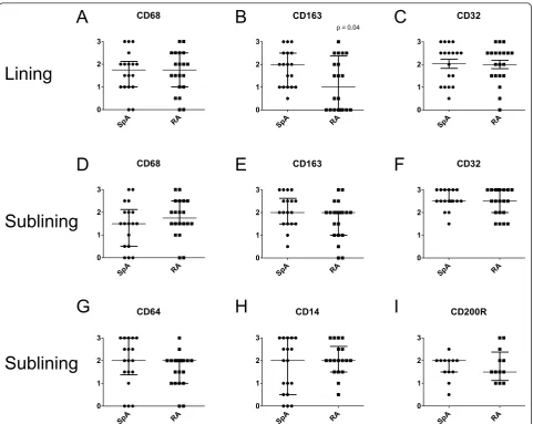
To confirm and extend our previous description of increased CD163 expression in SpA compared with RA synovitis [3,22-25], we performed a detailed histologic analysis of the recently described panel of phenotypic markers for MFIFN-g, MFIL-4, and MFIL-10[21]. As pre-viously shown, the number of CD68+ macrophages in the intimal lining layer and synovial sublining was simi-lar between the two diseases (Figure 1A, D), whereas CD163 expression was higher in the SpA compared with the RA intimal lining layer (P< 0.05) (Figure 1B), but similar in the synovial sublining (Figure 1E). How-ever, the expression of another MFIL-10marker, CD32,
These histologic data confirmed the higher CD163 expression in SpA compared with RA synovitis, but failed to provide additional evidence for a biased M2 polarization in SpA.
The phenotype of peripheral blood monocytes is similar in SpA and RA
To investigate whether the increased expression of CD163 in SpA synovitis is related to preferential MF IL-10 polarization, we set up a series of additional
experi-ments. Because monocytes from peripheral blood of SpA patients display a defective IFN-gsignature [28] and IFN-gis the prototypic M1 polarizing cytokine, we first assessed whether not only intimal lining layer macrophages, but also peripheral blood monocytes have a distinct phenotype in SpA. We quantified the
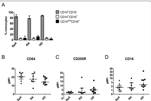
percentage of classic CD14+CD16- monocytes, inter-mediate CD14+CD16+ monocytes, and nonclassic CD14dimCD16+ monocytes [32] in peripheral blood from SpA and RA patients and healthy donors, and did not observe differences in these monocyte subsets between the groups (Figure 2A). We next assessed the expression of macrophage polarization markers on CD14+ monocytes and measured a marked expression of the MFIFN-g marker CD64 and a more modest
expression of the MFIL-4 marker CD200R and the MFIL-10marker CD16 (Figure 2B-D). However, no dif-ferences were found in the expression of these markers between SpA and RA patients and healthy controls. Expression of MFIFN-g marker CD80 and MFIL-10 mar-ker CD163 on monocytes was very low (data not shown). Based on this assessment, no indication exists
A B C
D E F
G H I
Lining
Sublining
Sublining
CD68
SpA RA
0 1 2 3
CD163
SpA RA
0 1 2 3
p = 0.04
CD32
SpA RA
0 1 2 3
CD68
SpA RA
0 1 2 3
CD163
SpA RA
0 1 2 3
CD32
SpA RA
0 1 2 3
CD64
SpA RA
0 1 2 3
CD14
SpA RA
0 1 2 3
CD200R
SpA RA
[image:5.595.58.540.87.470.2]0 1 2 3
Figure 1Immunohistochemical analysis of the expression of polarization markers in spondyloarthritis (SpA) compared with
rheumatoid arthritis (RA) synovial tissue. The expression of CD68, CD163, and CD32 in the intimal lining layer(A through C)and the
of phenotypic differences between monocytes from SpA and RA patients and healthy individuals.
The phenotype ofin vitropolarized monocyte-derived
macrophages is similar in SpA and RA
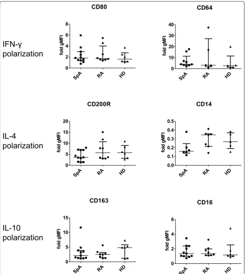
Further to investigate the propensity of monocytes to polarize preferentially toward a specific macrophage subset in SpA, monocytes from SpA and RA patients and healthy donors were polarized in vitro in the pre-sence of IFN-g, IL-4, or IL-10. The expression of specific phenotypic markers was measured with flow cytometry after 4 days of polarization. On spontaneous macro-phage maturation in the absence of polarizing cytokines, no differences were noted in the expression of phenoty-pic markers between SpA and RA patients and healthy donors (data not shown). As previously described in healthy donors [21], IFN-gspecifically upregulated the membrane expression of CD80 and CD64, IL-4 upregu-lated CD200R and downreguupregu-lated CD14, whereas IL-10 upregulated CD163 and CD16. After polarization of patient and healthy donor monocytes, we measured a
similar modulation of these phenotypic markers in all three groups (Figure 3). In conclusion, we found no evi-dence for differences in the polarization potential of monocyte-derived macrophages from SpA and RA patients and healthy individuals.
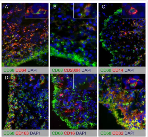
Intimal lining layer macrophages display a MFIL-10-like
phenotype
Because we observed an increased expression of CD163, but no differences in the expression of other polariza-tion markers in SpA compared with RA synovitis, we next performed double-immunofluorescence staining to characterize further the polarization phenotype of syno-vial macrophages. We studied the colocalization of pan-macrophage marker CD68 with MFIFN-g marker CD64 (Figure 4A), MFIL-4markers CD200R and CD14 (Figure 4B, C), and MFIL-10 markers CD163, CD16, and CD32 (Figure 4D-F). Intimal lining layer CD68+ cells highly expressed the MFIL-10markers CD163 and CD32 (Fig-ure 4D, F), whereas the expression of CD64, CD80, CD200R, CD14, and CD16 was almost absent (Figure
A
B
CD64
SpA RA HD
0 10 20 30
gM
FI
CD16
SpA RA HD
0 1 2 3 10 20 30 40
gM
FI
CD200R
SpA RA HD
-5 0 5 10 15 20 25
gM
FI
SpA RA HD
0 20 40 60 80
100 CD14+CD16
-CD14+CD16+ CD14dimCD16+
%
m
onoc
yt
e
s
[image:6.595.58.538.87.408.2]C D
Figure 2Monocyte subsets and expression of phenotypic markers on spondyloarthritis (SpA) and rheumatoid arthritis (RA) patients
and healthy donor monocytes. The percentage of CD14+CD16-, CD14+CD16+, and CD14dimCD16+monocytes from SpA and RA patients and
IFN-
γ
polarization
IL-4
polarization
IL-10
polarization
CD80
SpA RA HD
0 2 4 6 8
fo
ld
g
M
F
I
CD64
SpA RA HD
0 10 20 30 40
fol
d gM
FI
CD200R
SpA RA HD
0 5 10 15 20
fol
d gM
FI
CD163
SpA RA HD
0 5 10 15
fol
d gM
FI
CD16
SpA RA HD
0 2 4 6
fol
d gM
FI
CD14
SpA RA HD
0.0 0.1 0.2 0.3 0.4 0.5
fol
d gM
[image:7.595.57.540.87.627.2]FI
Figure 3Expression of phenotypic markers onin vitropolarized monocyte-derived macrophages from spondyloarthritis (SpA) and
rheumatoid arthritis (RA) patients and healthy individuals. The expression of MFIFN-gmarkers CD80 and CD64, MFIL-4markers CD200R and
CD14, and MFIL-10markers CD163 and CD16 was measured with flow cytometry onin vitropolarized monocyte-derived macrophages. Data are
4A-C, 4E) in both SpA and RA. In contrast, synovial sublining macrophages abundantly expressed both the MFIL-10 markers CD163 and CD32 (Figure 4D, F) and the MFIFN-g marker CD64 (Figure 4A). The positive MFIL-4 marker CD200R showed a low expression and
low colocalization with CD68 (Figure 4B), whereas the negative MFIL-4marker CD14 strongly colocalized with
CD68 (Figure 4C). In agreement with previous reports, CD80 expression was almost absent in synovial tissue (data not shown) [33], whereas CD16 expression was
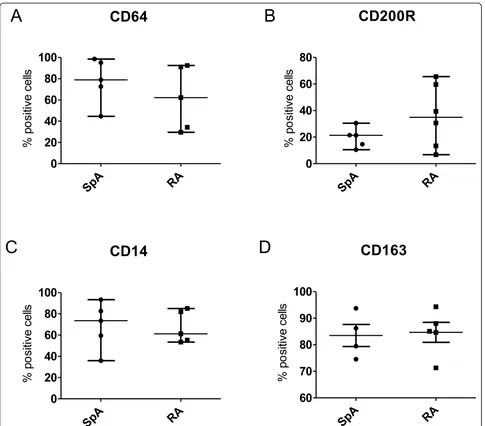
low on CD68+ cells (Figure 4E). Although the number of samples used in these double-immunofluorescence experiments is too small for an accurate statistical analy-sis, quantification of the staining confirmed the lack of marked differences in the expression of these markers between SpA and RA synovial sublining macrophages (Figure 5). These findings suggest an MFIL-10-like
phe-notype of intimal lining layer macrophages in both SpA and RA, whereas synovial sublining macrophages had a more heterogeneous phenotype.
CD68
CD163
DAPI
CD68
CD16
DAPI
CD68
CD32
DAPI
CD68
CD64
DAPI
CD68
CD200R
DAPI
CD68
CD14
DAPI
A B C
[image:8.595.58.541.87.532.2]D E F
Figure 4Double immunofluorescence stainings of CD68 and macrophage polarization markers on synovial tissue from chronic
inflammatory arthritis. Colocalization of CD68 (green) with MFIFN-gmarker CD64(A), MFIL-4markers CD200R(B)and CD14(C), and MFIL-10
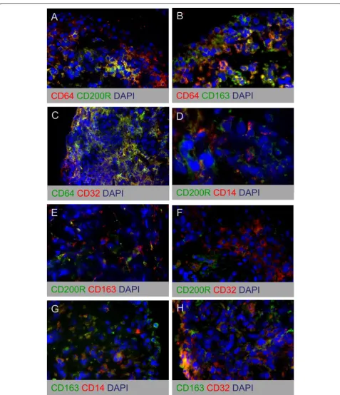
Synovial sublining macrophages co-express MFINF-gand
MFIL-10markers
To characterize further the phenotype of synovial sub-lining macrophages, we studied the colocalization of MFIFN-gmarker CD64 with CD200R, CD163, and CD32 (Figure 6A-C), colocalization of MFIL-4marker CD200R
with CD14, CD163, and CD32 (Figure 6D-F), and colo-calization of MFIL-10 marker CD163 with CD14 and
CD32 (Figure 6G, H). These stainings confirmed that both MFIL-10markers CD163 and CD32 were expressed
on the same cells in SpA and RA synovitis (Figure 6H). However, we also observed a high degree of coexpres-sion between the MFINF-g marker CD64 and the M
FIL-10 markers CD163 and CD32 (Figure 6B, C).
Further-more, the smaller macrophage subset that expressed the MFIL-4 marker CD200R also appeared to coexpress CD64, CD14, CD163, and CD32 (Figure 6D-F). Taken together, these data indicate that synovial sublining macrophages display a mixed MFINF-g/MFIL-10 pheno-type. Within this population, a smaller macrophage sub-set also coexpresses the MFIL-4 marker CD200R.
Discussion
Synovial tissue macrophages play a key role in chronic inflammatory arthritis, but the distribution and the function of different macrophage subsets in this process
A B
C D
CD64
Sp
A
RA
0
20
40
60
80
100
%
p
o
sitive
ce
lls
CD14
SpA
RA
0
20
40
60
80
100
%
p
o
sitive
ce
lls
CD200R
Sp
A
RA
0
20
40
60
80
%
p
o
sitive
ce
lls
CD163
SpA
RA
60
70
80
90
100
%
p
o
sitive
ce
[image:9.595.57.542.87.513.2]lls
CD163
CD32
DAPI
H
CD64
CD163
DAPI
B
CD64
CD32
DAPI
C
CD64
CD200R
DAPI
A
F
CD200R
CD32
DAPI
CD200R
CD163
DAPI
E
CD163
CD14
DAPI
CD200R
CD14
DAPI
D
[image:10.595.58.536.85.640.2]G
Figure 6Double-immunofluorescence stainings of MFIFN-g, MFIL-4, and MFIL-10polarization markers on synovial tissue from chronic
inflammatory arthritis. Colocalization of MFIFN-gmarker CD64 with CD200R(A), CD163(B), and CD32(C), colocalization of MFIL-4marker
CD200R with CD14(D), CD163(E), and CD32(F), and colocalization of MFIL-10marker CD163 with CD14(G)and CD32(H). Figure parts are
remain largely unknown. Based on the increased CD163 expression in SpA synovitis [3,22-25], we proposed the hypothesis of a preferential M2 polarization in SpA, as opposed to an M1 polarization in RA [27]. The present study aimed to assess this hypothesis experimentally by a detailed characterization of macrophages in inflamma-tory arthritis, by using a recently validated panel of phe-notypic markers for in vitro polarized human macrophage subsets [21]. Comparison of macrophage phenotypic marker expression between SpA and RA synovitis confirmed the higher expression of CD163 in the SpA intimal lining layer. Although previous reports showed increased CD163 expression in both the intimal lining layer and synovial sublining, the differences appeared to be larger in the intimal lining layer [3,22].
As CD163 is a prototypical marker for MFIL-10, we
performed three types of experiments to assess whether the increased CD163 expression reflected a preferential M2 polarization in SpA. First, the previously described inverse IFN-gsignature in monocytes from SpA patients [28] suggested that even before entering the synovial compartment, myeloid cells may be skewed to polarize toward M2 rather than M1. Our analysis of peripheral blood monocytes, however, did not reveal phenotypic differences between SpA, RA, and healthy controls. In humans, three monocyte subsets have been identified: classic CD14+CD16-, intermediate CD14+CD16+, and nonclassic CD14dimCD16+ monocytes. Among these subsets, CD14+CD16+ monocytes were described to be recruited in tissues during inflammation and to produce high amounts of TNF [32,34-37]. Although other publi-cations report contrasting findings of either an increased [38-40] or a decreased [41] percentage of CD14+CD16+ monocytes in inflammatory compared with healthy con-ditions, our experiments did not identify differences in these monocyte subsets between SpA and RA patients and healthy donors. Additionally, we did not observe differential expression of other phenotypic markers, such as CD64, CD16, and CD32, whereas the expression of CD80 and CD163 was very low on peripheral mono-cytes, as previously described [42,43].
Second, we assessed whether a potential bias toward M2 may appear during the differentiation of monocytes toward macrophages. In vitropolarization experiments with monocyte-derived macrophages failed, however, to indicate preferential polarization to a distinct subset in SpA compared with RA patients and healthy donors. These data suggest that the CD163+macrophage pheno-type in SpA synovitis is determined by the local inflam-matory milieu rather than by intrinsic myeloid alterations. Previous publications showing CD163 upre-gulation during in vitromacrophage maturation in the presence of SpA synovial fluid [27] and a strong synovial
CD163 downregulation on effective antiinflammatory treatment [12,13] support this observation.
Third, we assessed whether the local upregulation of CD163 on intimal lining layer macrophages in SpA was associated with differential expression of other phenoty-pic polarization markers between SpA and RA. This was not the case, as, for example, both the MFIL-10 marker CD32 and the prototypical MFIFN-g marker CD64 were similarly expressed in both diseases. These data question whether the altered expression of CD163, which is a reliable M2 marker in vitro, necessarily reflects a marked M2 versus M1 polarization in vivo. Supporting the notion that the phenotype of macro-phage subsetsin vivo is more complex than the concep-tualin vitro framework of M1 and M2, detailed double-staining analysis showed that synovial sublining macro-phages display a mixed phenotypic profile with a high colocalization of MFIFN-g and MFIL-10markers. Similar
In contrast with the mixed phenotype of synovial sub-lining macrophages, the intimal sub-lining layer macro-phages clearly displayed an MFIL-10-like phenotype. We
observed the same differences between the phenotype of intimal lining layer and synovial sublining macrophages, not only in SpA and RA but also in gout synovitis (data not shown), suggesting that this macrophage distribu-tion pattern is a global feature of synovial inflammadistribu-tion. Together with the low expression of CD14 in the inti-mal lining layer, this observation may fit a model according to which the intimal lining layer contains mainly mature resident macrophages, whereas the syno-vial sublining is actively infiltrated with immature monocyte-derived macrophages. This model is in agree-ment with publications showing that the number of synovial sublining but not intimal lining layer macro-phages is associated with disease activity in RA and cor-relates with response to therapy [7-9].
Concerning the differences between SpA and RA, this model would predict that the synovial sublining is simi-larly infiltrated by monocyte-derived macrophages with a mixed phenotype in both pathologies. However, the MRP8/14+infiltrating macrophages were shown to accu-mulate highly in the intimal lining layer in RA [3] and secrete a variety of proinflammatory mediators [62]. In contrast, the SpA intimal lining layer appears to consist mainly of resident, MFIL-10-like macrophages, which is
in accordance with the lower levels of M1-derived cyto-kines in SpA compared with RA synovial fluid [27] and with the less-destructive appearance of SpA synovitis.
Based on these findings, modulating the polarization and/or the migration of distinct macrophage subsets to the intimal lining layer would represent interesting ther-apeutic approaches for chronic synovitis.
Abbreviations
CD32: cluster of differentiation 32; CD163: cluster of differentiation 163; FCS: fetal calf serum; gMFI: geometric mean fluorescence intensity; IMDM: Iscove Modified Dulbecco Medium; RA: rheumatoid arthritis; SpA: spondyloarthritis.
Acknowledgements
DB is supported by a VIDI grant from The Netherlands Organization for Scientific Research (NWO) and by a grant from the Dutch Arthritis Foundation (Reumafonds).
Author details
1
Department of Clinical Immunology and Rheumatology, Academic Medical Center/University of Amsterdam, Meibergdreef 9, 1105 AZ Amsterdam, The Netherlands.2Department of Experimental Immunology, University of Amsterdam, Meibergdreef 9, 1105 AZ Amsterdam, The Netherlands.
3
GlaxoSmithKline, Gunnels Wood Road, Stevenage Herts, SG1 2NY, UK.
Authors’contributions
CAA, PPT, and DLPB participated in the design of the study. Acquisition of the data was performed by CAA, TN, and MJH. Data were analyzed and interpreted by CAA and TN. The manuscript was drafted by CAA and was revised by MJH, PPT, and DLPB. All authors read and approved the final manuscript.
Competing interests
The authors declare that they have no competing interests.
Received: 27 February 2012 Revised: 23 March 2012 Accepted: 11 April 2012 Published: 11 April 2012
References
1. Smith MD, Barg E, Weedon H, Papengelis V, Smeets T, Tak PP, Kraan M, Coleman M, Ahern MJ:Microarchitecture and protective mechanisms in synovial tissue from clinically and arthroscopically normal knee joints.
Ann Rheum Dis2003,62:303-307.
2. Tak PP, Smeets TJ, Daha MR, Kluin PM, Meijers KA, Brand R, Meinders AE, Breedveld FC:Analysis of the synovial cell infiltrate in early rheumatoid synovial tissue in relation to local disease activity.Arthritis Rheum1997,
40:217-225.
3. De Rycke L, Baeten D, Foell D, Kruithof E, Veys EM, Roth J, De KF:
Differential expression and response to anti-TNFalpha treatment of infiltrating versus resident tissue macrophage subsets in autoimmune arthritis.J Pathol2005,206:17-27.
4. Tak PP, Bresnihan B:The pathogenesis and prevention of joint damage in rheumatoid arthritis: advances from synovial biopsy and tissue analysis.
Arthritis Rheum2000,43:2619-2633.
5. Baeten D, Kruithof E, De RL, Boots AM, Mielants H, Veys EM, De KF:
Infiltration of the synovial membrane with macrophage subsets and polymorphonuclear cells reflects global disease activity in spondyloarthropathy.Arthritis Res Ther2005,7:R359-R369.
6. Baeten D, Houbiers J, Kruithof E, Vandooren B, Van den Bosch F, Boots AM, Veys EM, Miltenburg AM, De KF:Synovial inflammation does not change in the absence of effective treatment: implications for the use of synovial histopathology as biomarker in early phase clinical trials in rheumatoid arthritis.Ann Rheum Dis2006,65:990-997.
7. Gerlag DM, Haringman JJ, Smeets TJ, Zwinderman AH, Kraan MC, Laud PJ, Morgan S, Nash AF, Tak PP:Effects of oral prednisolone on biomarkers in synovial tissue and clinical improvement in rheumatoid arthritis.Arthritis Rheum2004,50:3783-3791.
8. Haringman JJ, Gerlag DM, Zwinderman AH, Smeets TJ, Kraan MC, Baeten D, McInnes IB, Bresnihan B, Tak PP:Synovial tissue macrophages: a sensitive biomarker for response to treatment in patients with rheumatoid arthritis.Ann Rheum Dis2005,64:834-838.
9. Wijbrandts CA, Vergunst CE, Haringman JJ, Gerlag DM, Smeets TJ, Tak PP:
Absence of changes in the number of synovial sublining macrophages after ineffective treatment for rheumatoid arthritis: implications for use of synovial sublining macrophages as a biomarker.Arthritis Rheum2007,
56:3869-3871.
10. Baeten D, Kruithof E, Van den Bosch F, Demetter P, Van DN, Cuvelier C, De VM, Mielants H, Veys EM, De KF:Immunomodulatory effects of anti-tumor necrosis factor alpha therapy on synovium in spondylarthropathy: histologic findings in eight patients from an open-label pilot study.
Arthritis Rheum2001,44:186-195.
11. Canete JD, Celis R, Hernandez V, Pablos JL, Sanmarti R:Synovial immunopathological changes associated with successful abatacept therapy in a case of severe refractory psoriatic arthritis.Ann Rheum Dis
2010,69:935-936.
12. Kruithof E, De RL, Roth J, Mielants H, Van den Bosch F, De KF, Veys EM, Baeten D:Immunomodulatory effects of etanercept on peripheral joint synovitis in the spondyloarthropathies.Arthritis Rheum2005,
52:3898-3909.
13. Kruithof E, Baeten D, Van den Bosch F, Mielants H, Veys EM, De KF:
Histological evidence that infliximab treatment leads to downregulation of inflammation and tissue remodelling of the synovial membrane in spondyloarthropathy.Ann Rheum Dis2005,64:529-536.
14. van Lent PL, Holthuysen AE, Van RN, Van De Putte LB, van den Berg WB:
Local removal of phagocytic synovial lining cells by clodronate-liposomes decreases cartilage destruction during collagen type II arthritis.Ann Rheum Dis1998,57:408-413.
15. Murray PJ, Wynn TA:Protective and pathogenic functions of macrophage subsets.Nat Rev Immunol2011,11:723-737.
16. Hamilton JA, Tak PP:The dynamics of macrophage lineage populations in inflammatory and autoimmune diseases.Arthritis Rheum2009,
17. Stein M, Keshav S, Harris N, Gordon S:Interleukin 4 potently enhances murine macrophage mannose receptor activity: a marker of alternative immunologic macrophage activation.J Exp Med1992,176:287-292. 18. Gordon S:Alternative activation of macrophages.Nat Rev Immunol2003,
3:23-35.
19. Mantovani A, Sica A, Sozzani S, Allavena P, Vecchi A, Locati M:The chemokine system in diverse forms of macrophage activation and polarization.Trends Immunol2004,25:677-686.
20. Mosser DM, Edwards JP:Exploring the full spectrum of macrophage activation.Nat Rev Immunol2008,8:958-969.
21. Ambarus CA, Krausz S, van EM, Hamann J, Radstake TR, Reedquist KA, Tak PP, Baeten DL:Systematic validation of specific phenotypic markers for in vitro polarized human macrophages.J Immunol Methods2012,
375:196-206.
22. Baeten D, Demetter P, Cuvelier CA, Kruithof E, Van DN, De VM, Veys EM, De KF:Macrophages expressing the scavenger receptor CD163: a link between immune alterations of the gut and synovial inflammation in spondyloarthropathy.J Pathol2002,196:343-350.
23. Baeten D, Moller HJ, Delanghe J, Veys EM, Moestrup SK, De KF:Association of CD163+macrophages and local production of soluble CD163 with decreased lymphocyte activation in spondylarthropathy synovitis.
Arthritis Rheum2004,50:1611-1623.
24. Kruithof E, Baeten D, De RL, Vandooren B, Foell D, Roth J, Canete JD, Boots AM, Veys EM, De KF:Synovial histopathology of psoriatic arthritis, both oligo- and polyarticular, resembles spondyloarthropathy more than it does rheumatoid arthritis.Arthritis Res Ther2005,7:R569-R580. 25. Noordenbos T, Yeremenko N, Gofita I, van de Sande M, Tak PP, Canete JD,
Baeten D:Interleukin-17-positive mast cells contribute to synovial inflammation in spondylarthritis.Arthritis Rheum2012,64:99-109. 26. Buechler C, Ritter M, Orso E, Langmann T, Klucken J, Schmitz G:Regulation
of scavenger receptor CD163 expression in human monocytes and macrophages by pro- and antiinflammatory stimuli.J Leukoc Biol2000,
67:97-103.
27. Vandooren B, Noordenbos T, Ambarus C, Krausz S, Cantaert T, Yeremenko N, Boumans M, Lutter R, Tak PP, Baeten D:Absence of a classically activated macrophage cytokine signature in peripheral spondylarthritis, including psoriatic arthritis.Arthritis Rheum2009,
60:966-975.
28. Smith JA, Barnes MD, Hong D, DeLay ML, Inman RD, Colbert RA:Gene expression analysis of macrophages derived from ankylosing spondylitis patients reveals interferon-gamma dysregulation.Arthritis Rheum2008,
58:1640-1649.
29. van de Sande MG, Gerlag DM, Lodde BM, van Baarsen LG, Alivernini S, Codullo V, Felea I, Vieira-Sousa E, Fearon U, Reece R, Montecucco C, Veale DJ, Pitzalis C, Emery P, Klareskog L, McInnes IB, Tak PP:Evaluating antirheumatic treatments using synovial biopsy: a recommendation for standardisation to be used in clinical trials.Ann Rheum Dis2011,
70:423-427.
30. Dougados M, van der Linden S, Juhlin R, Huitfeldt B, Amor B, Calin A, Cats A, Dijkmans B, Olivieri I, Pasero G:The European Spondylarthropathy Study Group preliminary criteria for the classification of
spondylarthropathy.Arthritis Rheum1991,34:1218-1227.
31. Arnett FC, Edworthy SM, Bloch DA, McShane DJ, Fries JF, Cooper NS, Healey LA, Kaplan SR, Liang MH, Luthra HS:The American Rheumatism Association 1987 revised criteria for the classification of rheumatoid arthritis.Arthritis Rheum1988,31:315-324.
32. Cros J, Cagnard N, Woollard K, Patey N, Zhang SY, Senechal B, Puel A, Biswas SK, Moshous D, Picard C, Jais JP, D’Cruz D, Casanova JL, Trouillet C, Geissmann F:Human CD14dim monocytes patrol and sense nucleic acids and viruses via TLR7 and TLR8 receptors.Immunity2010,
33:375-386.
33. Balsa A, Dixey J, Sansom DM, Maddison PJ, Hall ND:Differential expression of the costimulatory molecules B7.1 (CD80) and B7.2 (CD86) in rheumatoid synovial tissue.Br J Rheumatol1996,35:33-37.
34. Belge KU, Dayyani F, Horelt A, Siedlar M, Frankenberger M, Frankenberger B, Espevik T, Ziegler-Heitbrock L:The proinflammatory CD14+CD16+DR++ monocytes are a major source of TNF.J Immunol2002,168:3536-3542. 35. Geissmann F, Auffray C, Palframan R, Wirrig C, Ciocca A, Campisi L,
Narni-Mancinelli E, Lauvau G:Blood monocytes: distinct subsets, how they relate to dendritic cells, and their possible roles in the regulation of T-cell responses.Immunol Cell Biol2008,86:398-408.
36. Serbina NV, Cherny M, Shi C, Bleau SA, Collins NH, Young JW, Pamer EG:
Distinct responses of human monocyte subsets toAspergillus fumigatus conidia.J Immunol2009,183:2678-2687.
37. Ziegler-Heitbrock L:The CD14+CD16+blood monocytes: their role in infection and inflammation.J Leukoc Biol2007,81:584-592.
38. Blom AB, Radstake TR, Holthuysen AE, Sloetjes AW, Pesman GJ, Sweep FG, van de Loo FA, Joosten LA, Barrera P, van Lent PL, van den Berg WB:
Increased expression of Fcgamma receptors II and III on macrophages of rheumatoid arthritis patients results in higher production of tumor necrosis factor alpha and matrix metalloproteinase.Arthritis Rheum2003,
48:1002-1014.
39. Kawanaka N, Yamamura M, Aita T, Morita Y, Okamoto A, Kawashima M, Iwahashi M, Ueno A, Ohmoto Y, Makino H:CD14+, CD16+blood monocytes and joint inflammation in rheumatoid arthritis.Arthritis Rheum2002,46:2578-2586.
40. Rossol M, Kraus S, Pierer M, Baerwald C, Wagner U:The CD14(bright) CD16 +monocyte subset is expanded in rheumatoid arthritis and promotes Th17 expansion.Arthritis Rheum2012,64(3):671-7.
41. Cairns AP, Crockard AD, Bell AL:The CD14+CD16+monocyte subset in rheumatoid arthritis and systemic lupus erythematosus.Rheumatol Int
2002,21:189-192.
42. Fleischer J, Soeth E, Reiling N, Grage-Griebenow E, Flad HD, Ernst M:
Differential expression and function of CD80 (B7-1) and CD86 (B7-2) on human peripheral blood monocytes.Immunology1996,89:592-598. 43. Sanchez C, Domenech N, Vazquez J, Alonso F, Ezquerra A, Dominguez J:
The porcine 2A10 antigen is homologous to human CD163 and related to macrophage differentiation.J Immunol1999,162:5230-5237. 44. Bourlier V, Zakaroff-Girard A, Miranville A, De BS, Maumus M, Sengenes C,
Galitzky J, Lafontan M, Karpe F, Frayn KN, Bouloumie A:Remodeling phenotype of human subcutaneous adipose tissue macrophages.
Circulation2008,117:806-815.
45. Zeyda M, Farmer D, Todoric J, Aszmann O, Speiser M, Gyori G, Zlabinger GJ, Stulnig TM:Human adipose tissue macrophages are of an anti-inflammatory phenotype but capable of excessive pro-anti-inflammatory mediator production.Int J Obesity (Lond)2007,31:1420-1428. 46. Mantovani A, Germano G, Marchesi F, Locatelli M, Biswas SK:
Cancer-promoting tumor-associated macrophages: new vistas and open questions.Eur J Immunol2011,41:2522-2525.
47. Odegaard JI, Chawla A:Alternative macrophage activation and metabolism.Annu Rev Pathol2011,6:275-297.
48. Wilson HM:Macrophages heterogeneity in atherosclerosis: implications for therapy.J Cell Mol Med2010,14:2055-2065.
49. Gocheva V, Wang HW, Gadea BB, Shree T, Hunter KE, Garfall AL, Berman T, Joyce JA:IL-4 induces cathepsin protease activity in tumor-associated macrophages to promote cancer growth and invasion.Genes Dev2010,
24:241-255.
50. Imtiyaz HZ, Williams EP, Hickey MM, Patel SA, Durham AC, Yuan LJ, Hammond R, Gimotty PA, Keith B, Simon MC:Hypoxia-inducible factor 2alpha regulates macrophage function in mouse models of acute and tumor inflammation.J Clin Invest2010,120:2699-2714.
51. Mantovani A, Sozzani S, Locati M, Allavena P, Sica A:Macrophage polarization: tumor-associated macrophages as a paradigm for polarized M2 mononuclear phagocytes.Trends Immunol2002,23:549-555. 52. Sierra JR, Corso S, Caione L, Cepero V, Conrotto P, Cignetti A, Piacibello W,
Kumanogoh A, Kikutani H, Comoglio PM, Tamagnone L, Giordano S:Tumor angiogenesis and progression are enhanced by Sema4D produced by tumor-associated macrophages.J Exp Med2008,205:1673-1685. 53. Steidl C, Lee T, Shah SP, Farinha P, Han G, Nayar T, Delaney A, Jones SJ,
Iqbal J, Weisenburger DD, Bast MA, Rosenwald A, Muller-Hermelink HK, Rimsza LM, Campo E, Delabie J, Braziel RM, Cook JR, Tubbs RR, Jaffe ES, Lenz G, Connors JM, Staudt LM, Chan WC, Gascoyne RD:Tumor-associated macrophages and survival in classic Hodgkin’s lymphoma.N Engl J Med
2010,362:875-885.
54. Duluc D, Corvaisier M, Blanchard S, Catala L, Descamps P, Gamelin E, Ponsoda S, Delneste Y, Hebbar M, Jeannin P:Interferon-gamma reverses the immunosuppressive and protumoral properties and prevents the generation of human tumor-associated macrophages.Int J Cancer2009,
125:367-373.
56. Odegaard JI, Ricardo-Gonzalez RR, Goforth MH, Morel CR, Subramanian V, Mukundan L, Red EA, Vats D, Brombacher F, Ferrante AW, Chawla A:
Macrophage-specific PPARgamma controls alternative activation and improves insulin resistance.Nature2007,447:1116-1120.
57. Spencer M, Yao-Borengasser A, Unal R, Rasouli N, Gurley CM, Zhu B, Peterson CA, Kern PA:Adipose tissue macrophages in insulin-resistant subjects are associated with collagen VI and fibrosis and demonstrate alternative activation.Am J Physiol Endocrinol Metab2010,299: E1016-E1027.
58. Li AC, Glass CK:The macrophage foam cell as a target for therapeutic intervention.Nat Med2002,8:1235-1242.
59. Pinderski LJ, Fischbein MP, Subbanagounder G, Fishbein MC, Kubo N, Cheroutre H, Curtiss LK, Berliner JA, Boisvert WA:Overexpression of interleukin-10 by activated T lymphocytes inhibits atherosclerosis in LDL receptor-deficient mice by altering lymphocyte and macrophage phenotypes.Circ Res2002,90:1064-1071.
60. Khallou-Laschet J, Varthaman A, Fornasa G, Compain C, Gaston AT, Clement M, Dussiot M, Levillain O, Graff-Dubois S, Nicoletti A, Caligiuri G:
Macrophage plasticity in experimental atherosclerosis.PLoS One2010,5: e8852.
61. Waldo SW, Li Y, Buono C, Zhao B, Billings EM, Chang J, Kruth HS:
Heterogeneity of human macrophages in culture and in atherosclerotic plaques.Am J Pathol2008,172:1112-1126.
62. Bresnihan B, Youssef P:Macrophages in rheumatoid arthritis.InThe Macrophage..2 edition. Edited by: Burke B, Lewis CE. Oxford: Oxford University Press; 2002:231-433.
doi:10.1186/ar3796
Cite this article as:Ambaruset al.:Intimal lining layer macrophages but not synovial sublining macrophages display an IL-10 polarized-like phenotype in chronic synovitis.Arthritis Research & Therapy201214:R74.
Submit your next manuscript to BioMed Central and take full advantage of:
• Convenient online submission
• Thorough peer review
• No space constraints or color figure charges
• Immediate publication on acceptance
• Inclusion in PubMed, CAS, Scopus and Google Scholar
• Research which is freely available for redistribution