Estrogen Receptor Alpha 36 Gene Knockdown Promote the
Expression of NF-
κ
B in PC12 Cells
*
Ping Zou1, Chao Qu2, Yihui Xu2,3, Hongyan Li2, Dannv Han2, Dan Shi2, Wei Zou2,4#
1Teachers College, Dalian University, Dalian, China 2College of Life Science, Liaoning Normal University, Dalian, China
3School of Life Science, Shandong University, Jinan, China
4The Research Center of Developmental and Educational Psychology, Liaoning Normal University, Dalian, China
Email: #weizou60@hotmail.com
Received May 14, 2013; revised June 14, 2013; accepted July 14,2013
Copyright © 2013 Ping Zou et al. This is an open access article distributed under the Creative Commons Attribution License, which permits unrestricted use, distribution, and reproduction in any medium, provided the original work is properly cited.
ABSTRACT
The nuclear transcription factors κB (NF-κB) is widely existed in various kinds of cell types in the nervous system and plays an important role in neuron apoptosis and neurodegenerative diseases. Estrogen receptor alpha 36 (ER-α36) is a novel variant of ERα (as known ER-α66) which can transduce both estrogen- and antiestrogen-dependent activation of MAPK signal pathway and stimulate cell growth.Here, we aimed to detect the effect of ER-α36 gene silencing on the expression of NF-κB in normal cultured PC12 cells and to provide an experimental foundation for understanding the function of ER-α36 in nerve cells. PC12 cells with ER-α36 expression knocked down by the shRNA method. Then Western blot and immunocytochemical staining were performed to detect the expression and translocation of NF-κB after transfection. The results showed that NF-κB expression was significantly higher comparing with the control group after transfection (P < 0.01). Also, NF-κB subunit entered nuclear after transfection; Immunofluorescence staining and immunocytochemical staining of PC12 cells demonstrated that ER-α36 was expressed mainly on the plasma membrane and on the cell nucleus membrane. These data indicate that ER-α36 gene silencing can increase the expression of NF-κB and promote its nuclear translocation in PC12 cells.
Keywords:
NF-
κB; Estrogen Receptor Alpha 36; PC12 Cells1. Introduction
Nuclear factor kappa B (NF-κB), as a dimeric transcrip- tion factor, is widely existing in neurons of central ner- vous system. Activated (NF-κB) controls the expression of genes that regulate a broad range of biological proc- esses through canonical and non-canonical pathways, such as synaptic plasticity, cell injury, and the adjustment of the immune and inflammatory response factors ex- pressions, such as cell adhesion molecules and cytokines. In the central nervous system, NF-κB controls inflam- matory reactions and the apoptotic cell death following nerve injury [1], which plays a regulating role in the course of inflammation and immunoreaction during neu- ron apoptosis and neurodegenerative diseases and the change of NF-κB expression caused the neuron death and astrocyte activation. It is also reported that the activation
of NF-κB and CREB is involved in the protection of chromaffin cells and the sympathoadrenal PC12 cells (an established model for the study of neuronal cell apoptosis and survival) against serum deprivation-induced apop- tosis by the neuroactive steroids dehydroepiandrosterone (DHEA), its sulfate ester DHEAS and allopregnanolone (Allo) [2]. However, NF-κB is essential for neurosurvival as well. NF-κB activation is a part of recovery process that may protect neurons against oxidative-stresses or brain ischemia-induced apoptosis and neurodegeneration [3].
Recent studies show that estrogen receptor-α mediates the brain anti-inflammatory activity of estradiol. It has also been reported that estradiol-induced enhancement of object memory consolidation involves hippocampal ex- tracellular signal regulated kinase activation and mem-brane-bound estrogen receptors [4]. ER-α36, as a newly discovered estrogen receptor subtype, lacks both transac-tivation domains and functions as a dominant-negative effector of transactivation activities of the full-length *This work was supported by the Chinese National Natural Science
ER-α66 and ERβ [5]. ER-α36 primarily localizes to the cytoplasm and plasma membrane, it can transduce both estrogen-and antiestrogen-dependent activation of MAPK signal pathway, stimulating proliferation of breast cancer cells [6].
The interaction between ER-α66 and NF-κB has been studied in many kinds of cell types. ER can affect the NF-κB transcript activity by several aspects. It has been reported that the ER protects against debilitating effects of the inflammatory response by inhibiting the NF-κB in the MCF-7 breast cancer cell line. Tamoxifen treatment in ER-positive breast cancer up regulate NF-κB gene [7]. And the activity of NF-κB relies on the expression of ER in MCF-7 and HER2 cell line: the more ER expressed, the less NF-κB there was [8]. The activity of the NFκB signaling cascade is associated with mammary carcino-genesis, especially tumors with an aggressive and ER- negative phenotype [9]. ER-α36 expression is regulated differently from ER-α66, consistent with the findings that ER-α36 is expressed in specimens from ER-negative breast cancer patients and established ER-negative breast cancer cells that lack ER-α66 expression [10,11]. Al- though the function of ER-α36 has been studied in cancer, to our knowledge, it has not yet been known in the nerv- ous system. As ER-α36 may play important roles in these progresses, it is of great importance to detect the function of ER-α36 and its interaction with NF-κB in neuron apoptosis and neurodegenerative diseases.
2. Results
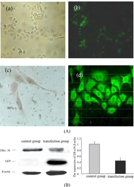
2.1. The Transfection of ER-α36 shRNA down Regulate the Expression of ER-α36 in PC12 Cells (Figure 1)
The transfection efficiency of ER-α36 shRNA plasmid was detected by observing the fluorescence 48 hours after transfection and it was nearly 87%. ER-α36 expres- sion was also detected by Western blot and the expres- sion level of ER-α36 protein was suppressed by up to 25% which shows that there was high interference effi- ciency.
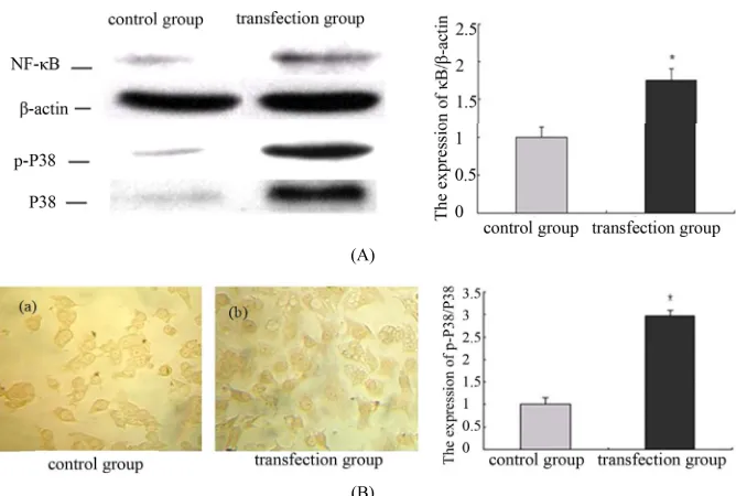
2.2. The Down Regulation of ER-α36 Promote the Expressions of NF-κB and Related Proteins in PC12 Cells (Figure 2)
NF-κB is widely existed in various kinds of cell types in nervous system. After transfection in PC12 cells for 48 hours, NF-κB was activated and had translocated from plasma to the nucleus. Compared with the control group, the expression level of NF-κB increased significantly after transfection. These date indicate that ER-α36 gene- silencing can increase the expression of NF-κB and pro- mote its nuclear translocation in PC12 cells. The expres-
(a) (b)
(c)
(d)
(A)
[image:2.595.308.538.83.404.2](B)
Figure 1. The transfection of ER-α36 shRNA down regulate the expression of ER-α36 in PC12 cells. (A) Control group (a) ×100). GFP protein expressions in the PC12 cells after transfection for 48 hours (b) ×100) immunocytochemical staining detection ER-α36 location in PC12 (c) ×400). Im-munofluorescence staining detection ER-α36 location in PC12 (d) ×400). (B) Western blot detection on ER-α36 ex-pression after transfection for 48 hours in PC12 cells. Den-sitometric analysis of ER-α36/β-actin. The results are pre-sented as means ± SEMs of three independent experiments.
sion of p-P38 and P38 were also detected by Western blot and there is a markerable increase after transfection.
3. Discussion
(A)
[image:3.595.133.467.81.306.2](B)
Figure 2. The down regulation of ER-α36 promote the expressions of NF-κB and related proteins in PC12 cells. (A) The ex-pressions of NF-κB, p-P38 and P38 were detected by western blot after transfection for 48 hours in PC12 cells. Densitometric analysis of NF-κB/β-actin; Densitometric analysis of p-P38/P38. The results are presented as means ± SEMs of three inde-pendent experiments (*significantly different from the control group); (B) Immunocytochemical staining showed that NF-κB
subunit expression and location in PC12 cells after transfection for 48 hours.
NF-κB is a transcription factor that regulates the ex- pression of a large number of genes that are critical for the regulation of cellular process, such as inflammatory responses, apoptosis, and cell proliferation [12]. NF-κB pathways may promote central nervous system (CNS) cell survival through the inhibition of caspase-1 and -3 activity [13], and NF-κB as key mediators of the neuro-protective against inflammatory stress in nigral dopa-minergic (DA) neurons [14].
In recent studies, NF-κB transrepression of steroid hormone receptors has been found [15]. 17 beta-estradiol (E2β) treatment produced strong protective effects by reducing infarct volume, neuronal apoptosis, and in-flammatory responses caused by NF-κB activation through estrogen receptors in transient cerebral ischemia model [16]. Several groups have reported the direct interaction between ERα and NF-κB in the nucleus of living cells [17] and a reciprocal transcription inhibition between agonist-bound ERα and activated NF-κB [18]. Previous work demonstrated that the NF-κB transcription factor promotes survival and chemoresistance in human breast cancer [19]. The NF-κB signaling pathways have been implicated as mediators of breast cancer drug resistance. The literature reported that ER-α36 existed in breast cancer patient samples and found that its expression lev-els were high in ER-negative tumors and low in ER- positive tumors [20,21], in addition, ER-α36 mediates nongenomic antiestrogen signaling in ER-negative breast cancer cells such as activation of the MAPK/ERK sig- naling in these cells [22,23], is involved in the resistance
of breast cancer to endocrine therapy, for example, ta- moxifen [24]. It is also been demonstrated that the nu- clear translocation and activation of NF-κB was signifi- cantly blocked by p38 MAP kinases inhibitor SB 203580 [25]. In our study, the expression of NF-κB in transfec-tion group was increased together with the increase of p-P38 expression after ER-α36 gene was silenced 48 hours later. These results demonstrate that p38 MAP kinase might be upstream of NF-κB which plays an im- portant role in nervous system.
In summary, we reported the function of ER-α36 might be associated with NF-κB transcription factor and showed that ER-α36 gene silencing promoted the expres- sion of NF-κB in ER-negative PC12 cells. It’s suggesting that non-genomic estrogen signaling mediated by ER-
α36 contributes to development and progression of PC12 cells that express NF-κB. This is the first report to our knowledgethat demonstrates that ER-α36 gene silencing promote the expression of NF-κB in PC12 cells, but which kind of cross-talk is involves in this process re-mains to be determined.
4. Materials and Methods
4.1. Chemicals and Antibodies
University, CaliforniaPlaza, USA).
4.2. Cell Culture and Transfections
PC12 cells were grown in RPMI medium 1640 (Sigma) supplemented with 5% FBS and 10% HS at 37˚C under a humidified 5% CO2 atmosphere. For transient transfec- tion experiments, 6 × 10⁵ cells per well were seeded in 6- well plates in 2ml of RPMI medium 1640 without antibi-otics. Transient transfections were performed with lipid- LipofectamineTM 2000 for 48 hours. Each experiment was performed on triplicate samples and repeated at least three times.
4.3. Western Blot Analysis
The cells were collected in ice-cold PBS, and then the cells extracts were prepared in RIPA buffer with pro- teinase inhibitor cocktail from Sigma (St. Louis, MO). Cell lysates were boiled with gel-loading buffer for 5 min at 100˚C, resolved on 10% SDS-PAGE, transferred to PVDF membrane, probed with appropriate antibodies and visualized with enhanced chemiluminescence (ECL) detection reagents (Amersham Pharmacia Biotech, Pis- cataway, NJ).
4.4. Immunocytochemistry
Cells were fixed in 4% (w/v) paraformaldehydein PBS (pH 7.4) followed by Permeabilization in 0.2% (v/v) Tri- ton X-100 in PBS (PBST). Background staining was minimalized by incubating these ctionsins 50% (v/v) ethanol, 0.9 (v/v) hydrogen peroxide in PBS for 30 min to block endogenous peroxidaseactivity. 0.9% followed by 4% (w/v) bovine serum albumin in PBS for 1 h at room temperature. 1:200 dilution of the anti-NF-κB an- tibody was added to the slides and incubated for 1 hour at 37˚C. After removing unbound antibody with PBST washes, immuno reactivity was detected with a bioti- nylated secondary antibody followed by an avidin horse- radish peroxidase complex. Immunoreactivity was visu- alized using a diaminobenzidine staining kit for 30 min.
4.5. Immunofluorescent
Cells were plated onto laminin-coated coverslips whereas were plated onto poly-l-lysine-coated coverslips. Cells grown on cover glasses were washed twice with PBS and then fixed with 4% ice cold paraformaldehyde for 15 min. The cover slips were washed three times with 0.1% Tri-ton X-100 in PBS and blocked with 5% bovine serum albumin (BSA) (Roche) in phosphate buffered saline solution for 1 h, and after that incubated with anti-ER-α36 antibodies (rabbit polyclonal, 1:100, Millipore). After washing (three times for 10 min in PBS), cells were in-cubated with Rhodamine-conjugated affinipure goat anti-
rabbit antibodies (1:300, Zhongshan Goldenbridge Bio- technology, China). Nuclei were stained using DAPI (0.2 μg/mL, Sigma). Fluorescence was imaged with a Bio- Rad MRC 600 confocal imaging system. Images were acquired using a Leica TCS SP2 MultiPhoton confocal microscope.
4.6. Statistical Analysis
Statistical analyses were performed using SPSS statisti- cal software. Treatment effects were analyzed using one- way analysis of variance. Significance was set at P < 0.05.
REFERENCES
[1] F. Cavttaneo, G. Guerra and R. Ammendola, “Expression and Signaling of Formyl-Peptide Receptors in the Brain,” Neurochemical Research, Vol. 35, No. 12, 2010, pp. 2018- 2026. doi:10.1007/s11064-010-0301-5
[2] I. Charalampopoulos, V. I. Alexaki, C. Tsatsanis, V. Mi- nas, E. Dermitzaki, I. Lasaridis, L. Vardouli, C. Stourna- ras, A. N. Margioris, E. Castanas and A. Gravanis, “Neu- rosteroids as Endogenous Inhibitors of Neuronal Cell Ap- optosis in Aging,” Annals of the New York Academy of Sciences, Vol. 1088, 2006, pp. 139-152.
doi:10.1196/annals.1366.003
[3] C. H. Nijboer, C. J. Heijnen, F. Groenendaal, M. J. May, F. van Bel and A. Kavelaars, “Strong Neuroprotection by Inhibition of NF-KappaB after Neonatal Hypoxia-Ische- mia Involves Apoptotic Mechanisms but Is Independent of Cytokines,” Stroke, Vol. 39, No. 7, 2008, pp. 2129- 2137. doi:10.1161/STROKEAHA.107.504175
[4] S. M. Fernandez, M. C. Lewis, A. S. Pechenino, L. L. Harburger, P. T. Orr, J. E. Gresack, G. E. Schafe and K. M. Frick, “Estradiol-Induced Enhancement of Object Memory Consolidation Involves Hippocampal Extracel- lular Signal-Regulated Kinase Activation and Membrane- Bound Estrogen Receptors,” The Journal of Neuroscience, Vol. 28, No. 35, 2008, pp. 8660-8667.
doi:10.1523/JNEUROSCI.1968-08.2008
[5] Z. Wang, X. Zhang, P. Shen, B. W. Loggie, Y. Chang and T. F. Deuel, “Identification, Cloning, and Expression of Human Estrogen Receptor-Alpha36, a Novel Variant of Human Estrogen Receptor-Alpha66,” Biochemical and Biophysical Research Communications, Vol. 336, No. 4, 2005, pp. 1023-1027. doi:10.1016/j.bbrc.2005.08.226 [6] L. M. Lee, J. Cao, H. Deng, P. Chen, Z. Gatalica and Z. Y.
Wang, “ER-Alpha36, a Novel Variant of ER-Alpha, Is Expressed in ER-Positive and -Negative Human Breast Carcinomas,” Anticancer Research, Vol. 28, No. 1B, 2008, pp. 479-483.
[7] C. Hicks, R. Kumar, A. Pannuti and L. Miele, “Integra- tive Analysis of Response to Tamoxifen Treatment in ER-Positive Breast Cancer Using GWAs Information and Transcription Profiling,” Breast Cancer, Vol. 6, 2012, pp. 47-66.
Sharma and G. L. Greene, “CBP Is a Dosage-Dependent Regulator of Nuclear Factor—KappaB Suppression by the Estrogen Receptor,” Molecular Endocrinology, Vol. 22, No. 2, 2008, pp. 263-272. doi:10.1210/me.2007-0324 [9] J. W. Antoon, M. D. White, E. M. Slaughter, J. L. Driver,
H. S. Khalili, S. Elliott, C. D. Smith, M. E. Burow and B.S. Beckman, “Targeting NFκB Mediated Breast Cancer Chemoresistance through Selective Inhibition of Sphin- gosine Kinase-2,” Cancer Biology & Therapy, Vol. 11, No. 7, 2011, pp. 678-689. doi:10.4161/cbt.11.7.14903 [10] Y. Zhou, S. Eppenberger-Castori, C. Marx, C. Yau, G. K.
Scott, U. Eppenberger and C. C. Benz, “Activation of Nu- clear Faetor-KappaB (NFkappaB) Identifies a High-Risk Subset of Hormone-Dependent Breast Cancers,” The In- ternational Journal of Biochemistry & Cell Biology, Vol. 37, No. 5, 2005, pp. l130-l144.
doi:10.1016/j.biocel.2004.09.006
[11] L. Shi, B. Dong, Z. Li, Y. Lu, T. Ouyang, J. Li, T. Wang, Z. Fan, T. Fan, B. Lin, Z. Wang and Y. Xie, “Expression of ER-(α)36, a Novel Variant of Estrogen Receptor-(α), and Resistance to Tamoxifen Treatment in Breast Can- cer,” Journal of Clinical Oncology, Vol. 27, No. 21, 2009, pp. 3423-3429. doi:10.1200/JCO.2008.17.2254
[12] G. Zhang and S. Ghosh, “Toll-Like Receptor-Mediated NFκB Activation: A Phylogenetically Conserved Para- digm in Innate Immunity,” Journal of Clinical Investiga- tion, Vol. 107, No. 1, 2001, pp. 13-19.
doi:10.1172/JCI11837
[13] A. L. Bernardino, T. A. Myers, X. Alvarez, A. Hasegawa and M. T. Philipp, “Toll-Like Receptors: Insights into Their Possible Role in the Pathogenesis of Lyme Neuro- borreliosis,” Infection and Immunity, Vol. 76, No. 10, 2008, pp. 4385-4395. doi:10.1128/IAI.00394-08
[14] J. K. Lee, J. Chung, K. M. Druey and M. G. Tansey, “RGS10 Exerts a Neuroprotective Role through the PKA/ c-AMP Response- Element (CREB) Pathway in Dopami- nergic Neuron-Like Cells,” Journal of Neurochemistry, Vol. 122, No. 2, 2012, pp. 333-343.
[15] L. I. McKay and J. A. Cidlowski, “Molecular Control of Immune/Inflammatory Responses: Interactions between Nuclear Factor-Kappa B and Steroid Receptor-Signaling Pathways,” Endocrine Reviews, Vol. 20, No. 4, 1999, pp. 435-459. doi:10.1210/er.20.4.435
[16] N. Maulik, M. Sato, B. D. Price and D. K. Das, “An Es- sential Role of NFkappaB in Tyrosine Kinase Signaling of p38 MAP Kinase Regulation of Myocardial Adaptation to Ischemia,” FEBS Letters, Vol. 429, No. 3, 1998, pp. 365-369. doi:10.1016/S0014-5793(98)00632-2
[17] I. Sarnico, A. Lanzillotta, M. Benarese, M. Alghisi, C. Baiguera, L. Battistin, P. Spano and M. Pizzi, “NF-Kap- paB Dimers in the Regulation of Neuronal Survival,” In-ternational Review of Neurobiology, Vol. 85, 2009, pp. 351-362. doi:10.1016/S0074-7742(09)85024-1
[18] C. H. Nijboer, C. J Heijnen, F. Groenendaal, M. J. May, F. Bel and A. Kavelaars, “Strong Neuroprotection by Inhibi- tion of NF-KappaB after Neonatal Hypoxia-Ischemia In- volves Apoptotic Mechanisms but Is Independent of Cy- tokines,” Stroke, Vol. 39, No. 7, 2008, pp. 2129-2137. doi:10.1161/STROKEAHA.107.504175
[19] C. B. Weldon, A. P. Parker, D. Patten, S. Elliott, Y. Tang, D. E. Frigo, C. M. Dugan, E. L. Coakley, N. N. Butler, J. L. Clayton, J. Alam, T. J. Curiel, B. S. Beckman, B. M. Jaffe and M. E. Burow, “Sensitization of Apoptotically- Resistant Breast Carcinoma Cells to TNF and TRAIL by Inhibition of p38 Mitogen-Activated Protein Kinase Sig- naling,” International Journal of Oncology, Vol. 24, No. 6, 2004, pp. 1473-1480.
[20] J. Zhang, G. Li, Z. Li, X. Yu, Y. Zheng, K. Jin, H. Wang, Y. Gong, X. Sun, X. Teng, J. Cao and L. Teng, “Estro- gen-Independent Effects of ER-a36 in ER-Negative Breast Cancer,” Steroids, Vol. 77, No. 6, 2012, pp. 666-673. doi:10.1016/j.steroids.2012.02.013
[21] X. T. Zhang, L. Ding, L. G. Kang and Z. Y. Wang, “In- volvement of ER-α36, Src, EGFR and STAT5 in the Bi- phasic Estrogen Signaling of ER-Negative Breast Cancer Cells,” Oncology Reports, Vol. 27, No. 6, 2012, pp. 2057- 2065.
[22] X. Zhang, L. Ding, L. Kang and Z. Y. Wang, “Estrogen Receptor-Alpha 36 Mediates Mitogenic Antiestrogen Signaling in ER-Negative Breast Cancer Cells,” PLOS One, Vol. 7, No. 1, 2012, pp. e30174-30186.
[23] L. Kang, Y. Guo, X. Zhang, J. Meng and Z. Y. Wang, “A Positive Cross-Regulation of HER2 and ER-α36 Controls ALDH1 Positive Breast Cancer Cells,” Molecular Biol-ogy, Vol. 127, No. 3-5, 2011, pp. 262-268.
[24] J. Rao, X. Jiang, Y. Wang and B. Chen, “Advances in the Understanding of the Structure and Function of ER-α36, a Novel Variant of Human Estrogen Receptor-Alpha,” Mo- lecular Biology, Vol. 127, No. 3-5, 2011, pp. 231-237. [25] M. E. Quaedackers, C. E. van den Brink, P. T. van der