ISSN Online: 2161-4512 ISSN Print: 2161-4105
In Vitro Effects of the Phytocomplex
TrichoTech
TM
on Human Fibroblasts:
Proliferative Potential and Effects on
Gene Expression of FGF-7 and FGF-10
Fernando Amaral
1, Maira Jardim
1, Valeria Maria de Souza Antunes
1,2,
Luis Felipe Gomes Michelin
3, Bárbara Anaís Rodrigues dos Santos
2,
Christiano Marcelo Vaz Barbosa
4, Daniel Gonsales Spindola
1,2,5,
Claudia Bincoletto
5, Carlos Rocha Oliveira
1,2,5*1Instituto de Osmologia e Óleos Essenciais, Minas Gerais, Brazil
2Grupo de Fitocomplexos e Sinalização Celular, Escola de Ciência da Saúde, Universidade Anhembi Morumbi, São Paulo, Brazil 3Centro Interdisciplinar de Investigação Bioquímica, Universidade de Mogi das Cruzes, São Paulo, Brazil
4Departamento de Análises clínicas e Toxicológicas, Faculdade de Ciências Farmacêuticas, Universidade de São Paulo, São Paulo,
Brazil
5Departamento de Farmacologia, Universidade Federal de São Paulo, São Paulo, Brazil
Abstract
The human hair follicle, a mini-organ formed with neuroectodermal-meso- dermal interaction, is a complex structure, in the active steady state (anagen) the dermal papilla can be considered as a ball of extracellular matrix, sur-rounding specialized fibroblasts. The cross-talk of dermal papilla with neigh-bouring matrix cells results in the maintenance of hair fibre production. This study aimed to investigate the proliferative potential of the compound Tri-chotechTM, a phytocomplex obtained from a mixture of essential oils, on
cul-tured human fibroblasts and its ability to modulate the gene expression of FGF-7 and FGF-10. TrichotechTM was shown to enhance fibroblasts
prolifera-tion in concentraprolifera-tions of 0.5% to 2.0%, and also increase the percentage of cells in the S/G2/M phases of the cell cycle. TrichotechTM at both 1.0% and
2.0% induced a statistically significant effect on wound healing assay com-pared to the untreated control. We examined the interaction between cell sur-vival (PI3K/Akt) and mitogenic (Ras/MAPK) signal transduction pathways after TrichotechTM treatment (1.0% and 2.0%) on the fibroblast cell line.
Tri-chotechTM caused phosphorylation of ERK1/2, as well as greater
phosphoryla-tion of MEK in comparison with both the untreated control and ERK1/2. PI3K and AKT, however, were not shown to be significantly more phospho-rylated following TrichotechTM exposure. To verify the relative expression of How to cite this paper: Amaral, F., Jardim,
M., de Souza Antunes, V.M., Michelin, L.F.G., dos Santos, B.A.R., Barbosa, C.M.V., Spin-dola, D.G., Bincoletto, C. and Oliveira, C.R. (2017) In Vitro Effects of the Phytocomplex TrichoTechTM on Human Fibroblasts: Pro-liferative Potential and Effects on Gene Expression of FGF-7 and FGF-10. Journal of Cosmetics, Dermatological Sciences and Applications, 7, 1-13.
https://doi.org/10.4236/jcdsa.2017.71001
Received: December 14, 2016 Accepted: January 16, 2017 Published: January 19, 2017
Copyright © 2017 by authors and Scientific Research Publishing Inc. This work is licensed under the Creative Commons Attribution International License (CC BY 4.0).
http://creativecommons.org/licenses/by/4.0/
mRNA for FGF-7 and FGF-10 genes, a real-time polymerase chain reaction (qPCR) protocol was used. Results show the increase in mRNA expression by fibroblasts after treatment with TrichotechTM. In both concentrations tested,
TrichotechTM was found to increase the expression of FGF-7 and FGF-10.
Si-rius red staining allows for rapid assessment of collagen content, it showed a significant increase in collagen content in treated fibroblasts. Further investi-gation concerning TrichotechTM could be helpful towards the development of
new bioactive phytocomplexes for dermatological and trichological use.
Keywords
TrichotechTM, Fibroblasts, Proliferation, FGF-7, FGF-10
1. Introduction
The human hair follicle, a mini-organ formed with neuroectodermal-mesodermal interaction [1], is a complex structure consisting of an outer root sheath, an in-ner root sheath, the hair shaft, the bulge and the sebaceous gland [2].
The follicle undergoes successive steps of fibre production, regression and rest, which in humans last for an average of 3 years, 3 weeks and a few months, respectively. An additional phase involving the active release of the club fibre has also been described, and is thought to be independent from the rest of the hair cycle [3], while bearing no direct consequence on fibre production initiation [4].
Human hair follicle dynamics are regulated through a bi-stable equilibrium state, including an active steady state (the anagen stage) and a resting steady state (the telogen stage); the transition between these two steady states involves either a degradation phase (the catagen phase) or a neo-morphogenesis phase (the neogen phase). It is now believed that mesenchymal and epithelial oscilla-tors control the stochastic autonomous switching between these two steady states
[5].
In the active steady state (anagen), the dermal papilla can be considered as a ball of extracellular matrix, surrounding specialized fibroblasts. The cross-talk of dermal papilla with neighbouring matrix cells results in the maintenance of hair fibre production [6].
The dermal papilla maintains bulge stem cells and secondary hair germ cells quiescent during telogen through production of bone morphogenetic protein 4 (BMP4) and fibroblast growth factor 18 (FGF-18). Cell proliferation during anagen is triggered via production of BMP inhibitors (e.g. Sosrdc1 and Bmbi), as well as secretion of FGF-7 and FGF-10 [7][8]. Thus, a combination of factors secreted by dermal papilla fibroblasts generates a signaling environment that dictates whether hair follicles will remain dormant or enter the anagen stage.
upre-gulation of β-catenin and Shh signaling.
The use of certain phytocompounds as stimulants of hair growth has been considered an effective secondary measure for the treatment of hair loss, espe-cially when common first-line treatments such as minoxidil application or fi-nasteride administration yield poor results or cause adverse reactions. Many plant extracts and fractions thereof have been shown to elicit hair growth in mice [10] [11] [12], thus the prospecting of plant extracts as a source of hair growth-promoting compounds is a promising strategy.
The present study aimed to investigate the proliferative potential of the com-pound TrichotechTM, a phytocomplex obtained from a mixture of essential oils,
on cultured human fibroblasts and its ability to modulate the gene expression of FGF-7 and FGF-10, as well as to propose further applications on hair growth.
2. Materials and Methods
2.1. Chemicals
Propidium iodide, Direct Red 80 (Sirius Red) and 3-(4,5-Dimethylthiazol-2-yl)- 2,5-diphenyltetrazolium Bromide (MTT) were purchased from Sigma Chemical Co. (St. Louis, MO, USA). The AnnexinV/FITC Apoptosis Detection Kit was obtained from BD Pharmigen (CA, USA). Iscove’s Modified Dulbecco’s Medium (IMDM) and all cell culture reagents were purchased from Life Technologies (Thermo Fisher Scientific, USA). MEK, ERK, PI3K and AKT primary antibodies and Alexa Fluor 488-conjugate monoclonal antibodies were acquired from Santa Cruz Biotechnology, Inc. (Santa Cruz, CA, USA).
2.2. Cell Culture
CCD-1072Sk (ATCC® CRL2088TM) fibroblasts were cultured in ISCOVE’S
me-dium with 10% fetal bovine serum, 0.292 g/l L-glutamine, 1.0 g/l D-glucose, 2.2 g/l NaHCO3, 10.000 UI penicillin, and 0.060 g/l streptomycin. Cells were kept in
25 cm2 flasks (1 × 10⁵ cells/ml) in a humidified incubator at 37˚C with an
at-mosphere of 5% CO2 for a maximum of 30 population doublings. In all
experi-ments, the fibroblast cultures were subjected to cell viability assays using Trypan blue dye, and readings were performed in a hemocytometric chamber under a light microscope. All experiments described were performed when cell viability was equal or above 95%.
2.3. MTT Reduction Cell Viability Assay
cells/well and treated with different concentrations of TrichotechTM (0.5, 1.0, 1.5
and 2.0%) for 24 hours. Next, 10 μl of a 5 mg/ml MTT solution (Sigma-Aldrich) were added to each well. After 4 hours the samples was reincubated with 100 µl of Sodium dodecyl sulfate (SDS) solution [10%] for 12 hours, and then optical density was measured in a FlexStation® 3 multimode Benchtop Reader (Molecu-lar Devices, CA, USA) at 540 nm.
2.4. Propidium Iodide (PI) Incorporation Assay
Propidium iodide incorporation assays were performed using flow cytometry to assess the cellular fraction in the S/G2/M phase of the cell cycle (i.e. proliferating cells). To summarize, cells were seeded in 24-well plates at an initial density of 2 × 10⁵ cells/well, to which was added a hypotonic fluorochrome solution (HFS—
0.1% w/v sodium citrate, 0.5% w/v Triton X-100 and 50 µg/ml propidium iodide). After an incubation period of 4 h at 4˚C and shielded from light, the cells and supernatant were collected and analyzed. A FACS can flow cytometer and the CellQuest software were employed, and the data obtained were analyzed with WinMDI 2.8, considering 20,000 events per analysis for each assay.
2.5. Wound Healing
For this assay, fibroblasts were seeded in 6-well microplates and cultured as de-scribed above until observation of a confluent monolayer. The cell monolayers were carefully “scratched” with a sterile pipette tip, and washed with saline and PBS to remove loose cells and debris. Next, the cells were incubated at 37˚C with culture medium without fetal bovine serum (nutrient deprivation) and with 0.2% low molecular weight HA. Reference points near the “wound” were de-marcated to ensure the same area of image acquisition. Images were obtained at different times using a digital camera attached to the microscope, and the per-centage of wound closure was calculated using IMAGEJ (NIH, USA).
2.6. Measurement of MEK/ERK and PI3K/AKT Signaling Activity
in Fibroblasts
Protein phosphorylation is a dynamic process controlled by the enzymatic activ-ities of kinases and phosphatases. In order to inhibit these processes rapidly, fix-ation was done by adding BD FACSTM Lysing Solution. Following fixation, the
cyto-meter.
2.7. Real-Time PCR (qPCR)
Total RNA extracted from fibroblast samples was converted to cDNA using a SuperScript® III RT kit (Invitrogen, Carlsbad, CA). A qPCR analysis was per-formed in 10 μL reactions with the SYBR GREEN PCR Master Mix and analyzed on a StepOnePlusTM Real-Time PCR instrument (Invitrogen, Carlsbad, CA).
Rela-tive standard curves were generated by serial dilutions and all samples were run in triplicates. Primers used are: FGF-7 forward (5’-ATCAGGACAGTGGCAGT TGGA-3’); FGF-7 reverse (5’-AACATTTCCCCTCCGTTGTGT-3’) and FGF-10 forward (5’-CACATTGTGCCTCAGCCTTTC-3’); FGF-10 reverse (5’-AGGTGA TTGTAGCTCCGCACA-3’). The PCR reaction was performed under the fol-lowing conditions: 50˚C (2 min), 95˚C (10 min), and 40 cycles of 95˚C (15 s) and 55˚C (1 min). GAPDH was used as a control gene.
2.8. Sirius Red Collagen Quantification
After cells were cultured, the medium was removed and the wells were washed three times with 0.1 M PBS. Next, 100 μl of Bouin’s solution (picric acid 0.9%, formaldehyde 9.0% and glacial acetic acid 5.0%) were added for fixation for 1 h. Samples were washed with PBS, then the Sirius Red dye was added. After 1 h, the maximum possible amount of dye was removed, followed by washing with 150 μl of a 0.01 M hydrochloric acid solution for 30 seconds to remove the dye that did not bind to collagen. Next, the dye was removed from cell layers by the addi-tion of 0.1 M NaOH for 30 min. 100 μl aliquots of the soluaddi-tion contained in the wells were transferred to a new plate. Absorbance was measured with an Elx- 800-UV (Bio-Tek Instruments, USA) microplate reader at 570 nm.
2.9. Statistical Analysis
Results were given as mean ± SEM (standard error of the mean). The results ob-tained were statistically analyzed using a one-way analysis of variance (ANOVA), followed by Tukey’s test a posteriori. Semi-quantifications were analyzed using Student’s t-test. P-values < 0.05 were considered significantly different. Analyses were performed using GraphPad Prism version 5.0 (GraphPad Software Inc., CA, USA).
3. Results and Discussion
3.1. MTT to Formazan Reduction Assay
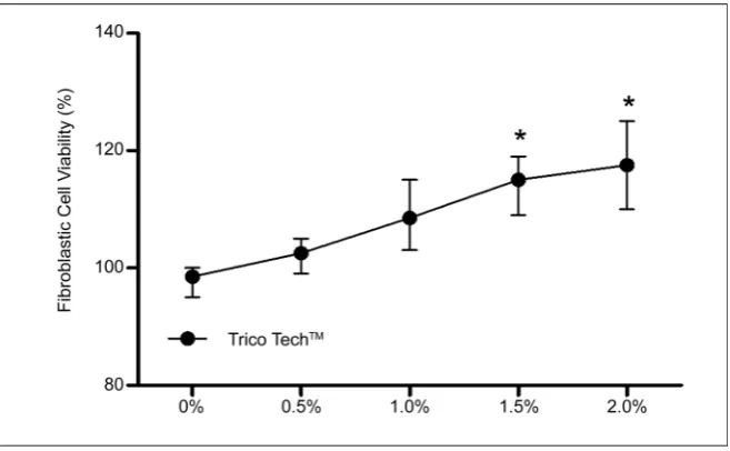
MTT exposure [14]. TrichotechTM was shown to enhance fibroblasts
prolifera-tion (Figure 1). This effect of TrichotechTM in concentrations of 0.5% to 2.0%
was visible by MTT assay after 24 hours of treatment (P < 0.05). TrichotechTM at
concentrations of 1.5% and 2.0% significantly enhanced the proliferation of fi-broblasts compared to the untreated group. Notice a dose-dependent increase in fibroblast proliferation.
3.2. Propidium Iodide (PI) Incorporation Assay
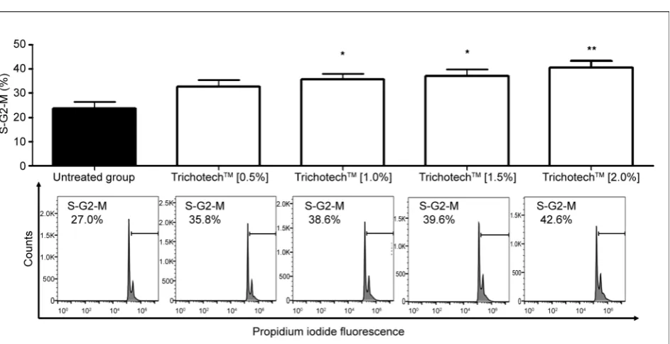
Progression through the cell cycle is one of the most fundamental features of cells and can be measured by staining cells with propidium iodide (PI). The level of PI fluorescence in a cell is, directly proportional to the DNA content of that cell, the quantification of which indicates the percentage of cells in each phase of the cell cycle in a sample [15]. As seen in Figure 2, the percentage of cells in the S/G2/M phases of the cell cycle increased in a dose-dependent fashion upon TrichotechTM treatment. This shows that TrichotechTM acts in accordance with
several lines of evidence which support a molecular mechanism in the response to stimulation by natural compounds, increasing G2/M phase in fibroblasts [16] [17].
3.3. Wound Healing
[image:6.595.210.539.480.683.2]Cell migration and proliferation coupled with controlled cell cycle are beneficial for the repair of sagged and wrinkled skin, dermal, and gastrointestinal wound healing. The in vitro scratch assay is a well-developed method to measure cell migration and its steps involve creating a “scratch” in a cell monolayer, captur-ing the images at the beginncaptur-ing and at regular intervals durcaptur-ing cell migration to close the scratch, and comparing the images to quantify the migration rate of the
Figure 1. Results of cell proliferation from MTT reduction assay after 24-hour exposure to different concentrations of TricotechTM. Before starting the tests, cells were deprived of
Figure 2. (a) Percentage of cells in S/G2/M phase obtained after 24-hour exposure of CCD-1072Sk cells to different concentra-tions of TricotechTM. Before starting the tests, cells were deprived of fetal bovine serum. (*) P < 0.05—significant in relation to
control. ANOVA, Tukey. GraphPad Prism v5.0. (b) Histogram representing statistically significant concentration (10%). FlowJo v10.0.
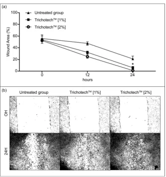
cells [18]. We evaluated TrichotechTM wound healing stimulating activity on
fi-broblast cells using the scratch assay. Scratches were made on confluent fibrob-last monolayers, which were then exposed to TrichotechTM for 24 h at two
con-centrations (1.0% and 2.0%). TrichotechTM at both 1.0% and 2.0% induced a
sta-tistically significant effect on wound closure compared to the untreated control
(Figure 3(b)). In Figure 3(a) and Figure 3(b), respectively, we can see images
obtained at different times and the graphic representation of the distance be-tween the edges of the scratch.
3.4. Measurement of MEK/ERK and PI3K/AKT Signaling Activity
in Fibroblasts
An understanding of the mechanisms that regulate the cell migration and proli-feration of dermal fibroblast cells by a natural compound could be beneficial in devising novel therapies to regulate fibrosis and wound contraction to ultimately improve the wound healing process [19]. The most highly studied intracellular signaling cascades in the context of cancer are the mitogen activated protein ki-nase (MAPK) and phosphoinositide 3-kiki-nase (PI3K)/AKT pathways [20]. We examined the interaction between cell survival (PI3K/Akt) and mitogenic (Ras/ MAPK) signal transduction pathways after TrichotechTM treatment on the
fi-broblast cell line. The cells were stimulated with 1.0% and 2.0% TrichotechTM.
The treatment with TrichotechTM caused phosphorylation of ERK1/2, as well as
Figure 3. (a) Photographic representation of in vitro samples subjected to a simulated wound and exposed for 24 hours to different concentrations of TrichotechTM (1% and
2%). (b) Representative graphic of the percentage of wounded area at 0 h and 24 h after the same treatment. Before starting the tests, cells were deprived of fetal bovine serum. (*) P < 0.05—significant in relation to group 0 h treated with TrichotechTM, Student’s t-test,
GraphPad Prism v5.0.
were obtained [21], whereby the proliferative effects of camphor were shown to be mediated by the PI3K/AKT/mTOR and MAP kinase pathways—the key sig-naling pathways involved in the control of cell proliferation. In this same study, camphor-induced phosphorylation of ERK, but not PI3K and AKT, was also re-ported. Taken together, this evidence indicates that TrichotechTM induced
fi-broblast proliferation possibly through upregulation of MAP kinase signaling pathways.
3.5. Real-Time PCR (qPCR)
Figure 4. Graphic and corresponding representative histogram of MEK (a), ERK (b), PI3K (c) and AKT (d) phosphorylated pro-teins signaling after exposure to TrichotechTM [1%] and [2%] for 1 h. Results are expressed by the MFI (median fluorescence
in-tensity) and compared with the untreated group (CTL). (*) P < 0.05—significant in relation to CTL group. ANOVA, Tukey, GraphPad Prism v5.0.
catagen or telogen [23]. FGF-10 is found in the dermal papilla fibroblasts and its receptor FGFR2IIIb is found in the neighboring outer root sheath of the kerati-nocytes [24], suggesting that FGF-10 is a mesenchymally derived stimulator of hair follicle cells, which contribute to the hair-promoting activity. To verify the relative expression of mRNA for fibroblast growth factor-7 (FGF-7) and fibrob-last growth factor-10 (FGF-10) genes, a real-time polymerase chain reaction (qPCR) protocol was used. Figure 5 shows the increase in mRNA expression by fibrob-lasts after treatment with TrichotechTM. In both concentrations tested,
Tricho-techTM was found to increase the expression of FGF-7 and FGF-10 by several
fold, thereby constituting a dermal papilla signal instructing hair germ cells to proliferate and initiate a new hair cycle.
3.6. Sirius Red Collagen Quantification
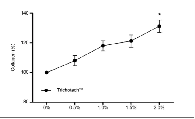
Sirius red staining of collagen has been used for many years. The present colo-rimetric plate assay allows for rapid assessment of collagen content [25]. The si-rius red assay showed a significant increase in collagen content in treated fi-broblasts (Figure 6). The magnitude of the increase in collagen between control and treated samples was markedly increased following treatment with 2.0% TrichotechTM. Previous reports have demonstrated similar increases in collagen
Figure 5. Relative expression levels of mRNA for FGF-7 and FGF-10 in human fibrob-lasts assessed by quantitative RT-PCR. Bars represent the range of relative expression. *P < 0.05, **P < 0.01, and ***P < 0.0001 compared to internal control GAPDH. Graphs were plotted with GraphPad Prism v5.0.
Figure 6. Total collagen content in fibroblasts as measured by incorporation of Sirius Red dye after 24-hour exposure to different concentrations of TrichotechTM. Before starting
the tests, cells were deprived of fetal bovine serum. The exposure did not cause a signifi-cant increase in relation to the control group (untreated cells). (*) P < 0.05—signifisignifi-cant in relation to control. Student’s t-test. GraphPad Prism v5.0.
4. Conclusion
Fibroblasts are found in the dermal papilla of hair follicles, and as such are heav-ily implicated in hair growth regulation. Taken together, our data show a stimu-lating effect of TrichotechTM on cultured fibroblasts. Indeed, a pronounced
in-crease in cell growth was observed after exposure to TrichotechTM at
concentra-tions of 0.5% - 2.0%. Our results suggest that TrichotechTM induced fibroblast
proliferation by activating ERK signaling pathways. In addition, FGF-7 and FGF-10 mRNA levels were shown to be increased compared with untreated controls. Further investigation concerning TrichotechTM could be helpful towards the
[image:10.595.210.539.288.489.2]Acknowledgements
This work was supported by the Fagron Group-São Paulo/SP-Brazil.
References
[1] Rishikaysh, P., Dev, K., Diaz, D., Qureshi, W.M., Filip, S. and Mokry. J. (2014) Sig-naling Involved in Hair Follicle Morphogenesis and Development. International Journal of Molecular Sciences, 15, 1647-1670. https://doi.org/10.3390/ijms15011647 [2] Schmid, D., Belser, E. and Zülli, F. (2013) The FGF7 and Noggin Genes Are Key
Targets to Treat Hair Loss. SOFW-Journal, 139, 9.
[3] Halloy, J., Bernard, B.A., Loussouarn, G. and Goldbeter, A. (2002) The Follicular Automaton Model: Effect of Stochasticity and of Synchronization of Hair Cycles.
Journal of Theoretical Biology, 214, 469-479. https://doi.org/10.1006/jtbi.2001.2474 [4] Higgins, C.A., Westgate, G.E. and Jahoda, C.A. (2009) From Telogen to Exogen:
Mechanisms Underlying Formation and Subsequent Loss of the Hair Club Fiber.
Journal of Investigative Dermatology, 129, 2100-2108. https://doi.org/10.1038/jid.2009.66
[5] Bernard, B.A. (2012) The Human Hair Follicle, a Bistable Organ? Experimental Dermatology, 21, 401-403. https://doi.org/10.1111/j.1600-0625.2012.01457.x [6] Bassino, E., Gasparri, F., Giannini, V. and Munaron, L. (2015) Paracrine Crosstalk
between Human Hair Follicle Dermal Papilla Cells and Microvascular Endothelial Cells. Experimental Dermatology, 24, 388-390. https://doi.org/10.1111/exd.12670 [7] Solanas, G. and Benitah, S.A. (2013) Regenerating the Skin: A Task for the
Hetero-geneous Stem Cell Pool and Surrounding Niche. Nature Reviews Molecular Cell Bi-ology, 14, 737-748. https://doi.org/10.1038/nrm3675
[8] Geyfman, M., Plikus, M.V., Treffeisen, E., Andersen, B. and Paus, R. (2014) Resting No More: Re-Defining Telogen, the Maintenance Stage of the Hair Growth Cycle.
Biological Reviews, 90, 1179-1196.
[9] Lin, W., Xiang, L., Shi, H., Zhang, J., Jiang, L., Cai, P., Lin, Z., Lin, B., Huang, Y., Zhang, H., Fu, X., Guo, D., Li, X., Wang, X. and Xiao, J. (2015) Fibroblast Growth Factors Stimulate Hair Growth through β-Catenin and Shh Expression in C57BL/6 Mice. BioMed Research International, 2015, 730139.
[10] Gottumukkala, V.R., Annamalai, T. and Mukhopadhyay, T. (2011) Phytochemical Investigation and Hair Growth Studies on the Rhizomes of Nardostachys jatamansi
DC. Pharmacognosy Magazine, 7, 146-150. https://doi.org/10.4103/0973-1296.80674
[11] Saumendu, D.R., Raj, K.P., Suvakanta, D., Jashabir, C. and Biswajit, D. (2014) Hair Growth Stimulating Effect and Phytochemical Evalutation of Hydro-Alcoholic Ex-tract of Grycyrrhiza glabra. Global Journal of Research on Medicinal Plants & Indi-genous Medicine, 3, 40-47.
[12] Li, Y., Han, M., Lin, P., He, Y., Yu, J. and Zhao, R. (2015) Hair Growth Promotion Activity and Its Mechanism of Polygonum multiflorum. Evidence-Based Comple-mentary and Alternative Medicine, 2015, 517901.
[13] Mosmann, T. (1993) Rapid Colorimetric Assay for Cellular Growth and Survival: Application to Proliferation and Cytotoxicity Assays. Journal of Immunological Me-thods, 65, 55-63. https://doi.org/10.1016/0022-1759(83)90303-4
[15] Mea Crowley, L.C., Chojnowski, G. and Waterhouse, N.J. (2016) Measuring the DNA Content of Cells in Apoptosis and at Different Cell-Cycle Stages by Propi-dium Iodide Staining and Flow Cytometry. Cold Spring Harbor Protocols.
[16] Savio, M., Coppa, T., Bianchi, L., Vannini, V., Maga, G., Forti, L., Cazzalini, O., Lazzè, M.C., Perucca, P., Prosperi, E. and Stivala, L.A. (2009) The Resveratrol Ana-logue 4,4’-Dihydroxy-Trans-Stilbene Inhibits Cell Proliferation with Higher Effi-ciency but Different Mechanism from Resveratrol. International Journal of Bioche-mistry and Cell Biology, 41, 2493-2502. https://doi.org/10.1016/j.biocel.2009.08.005 [17] Madhyastha, H., Madhyastha, R., Nakajima, Y., Omura, S. and Maruyama, M. (2012)
Regulation of Growth Factors-Associated Cell Migration by C-Phycocyanin Scaffold in Dermal Wound Healing. Clinical and Experimental Pharmacology and Physiol-ogy, 39, 13-19. https://doi.org/10.1111/j.1440-1681.2011.05627.x
[18] Liang, C.C., Park, A.Y. and Guan, J.L. (2007) In Vitro Scratch Assay: A Convenient and Inexpensive Method for Analysis of Cell Migration in Vitro. Nature Protocols, 2, 329-333. https://doi.org/10.1038/nprot.2007.30
[19] Harishkumar, M., Masatoshi, Y., Hiroshi, S., Tsuyomu, I. and Masugi, M. (2013) Revealing the Mechanism of in Vitro Wound Healing Properties of Citrus tamurana
Extract. BioMed Research International, 2013, Article ID: 963457. http://dx.doi.org/10.1155/2013/963457
[20] Paraiso, K.H.T., van der Kooi, K., Messina, J.L. and Smalley, K.S.M. (2010) Mea-surement of Constitutive MAPK and PI3K/AKT Signaling Activity in Human Can-cer Cell Lines. Methods in Enzymology, 484, 549-567.
https://doi.org/10.1016/B978-0-12-381298-8.00027-7
[21] Tran, T.A., Ho, M.T., Song, Y.W., Cho, M. and Cho, S.K. (2015) Camphor Induces Proliferative and Antisenescence Activities in Human Primary Dermal Fibroblasts and Inhibits UV-Induced Wrinkle Formation in Mouse Skin. Phytotherapy Re-search, 29, 1917-1925. https://doi.org/10.1002/ptr.5484
[22] Lino, M., Ehama, R., Nakazawa, Y., Iwabuchi, T., Ogo, M., Tajima, M. and Arase, S. (2007) Adenosine Stimulates Fibroblast Growth Factor-7 Gene Expression via Adeno-sine A2b Receptor Signaling in Dermal Papilla Cells. Journal of Investigative Der-matology, 127, 1318-1325. https://doi.org/10.1038/sj.jid.5700728
[23] Rosenquist, T.A. and Martin, G.R. (1996) Fibroblast Growth Factor Signalling in the Hair Growth Cycle: Expression of the Fibroblast Growth Factor Receptor and Li-gand Genes in the Murine Hair Follicle. Developmental Dynamics, 205, 379-386. https://doi.org/10.1002/(SICI)1097-0177(199604)205:4<379::AID-AJA2>3.0.CO;2-F [24] Saksena, S., Priyamvada, S., Kumar, A., Akhtar, M., Soni, V., Anbazhagan, A.N.,
Alakkam, A., Alrefai, W.A., Dudeja, P.K. and Gill, R.K. (2013) Keratinocyte Growth Factor-2 Stimulates P-Glycoprotein Expression and Function in Intestinal Epithelial Cells. The American Journal of Physiology—Gastrointestinal and Liver Physiology, 304, G615-G622. https://doi.org/10.1152/ajpgi.00445.2012
[25] Kliment, C.R., Englert, J.M., Crum, L.P. and Oury, T.D. (2011) A Novel Method for Accurate Collagen and Biochemical Assessment of Pulmonary Tissue Utilizing One Animal. International Journal of Clinical and Experimental Pathology, 4, 349-355. [26] Jung, E., Lee, J., Baek, J., et al. (2007) Effect of Camellia japonica Oil on Human
Type I Procollagen Production and Skin Barrier Function. Journal of Ethnophar-macology, 112, 127-131. https://doi.org/10.1016/j.jep.2007.02.012
[27] Lee, J., Jung, E., Lee, J., et al. (2007) Panax ginseng Induces Human Type I Collagen Synthesis through Activation of Smad Signaling. Journal of Ethnopharmacology, 109, 29-34. https://doi.org/10.1016/j.jep.2006.06.008
Cin-namon Extract Promotes Type I Collagen Biosynthesis via Activation of IGF-I Sig-naling in Human Dermal Fibroblasts. Journal of Agricultural and Food Chemistry, 60, 1193-1200. https://doi.org/10.1021/jf2043357
Submit or recommend next manuscript to SCIRP and we will provide best service for you:
Accepting pre-submission inquiries through Email, Facebook, LinkedIn, Twitter, etc. A wide selection of journals (inclusive of 9 subjects, more than 200 journals) Providing 24-hour high-quality service
User-friendly online submission system Fair and swift peer-review system
Efficient typesetting and proofreading procedure
Display of the result of downloads and visits, as well as the number of cited articles Maximum dissemination of your research work
Submit your manuscript at: http://papersubmission.scirp.org/
![Figure 4. Graphic and corresponding representative histogram of MEK (a), ERK (b), PI3K (c) and AKT (d) phosphorylated pro-teins signaling after exposure to TrichotechTM [1%] and [2%] for 1 h](https://thumb-us.123doks.com/thumbv2/123dok_us/7787172.724140/9.595.61.538.61.304/graphic-corresponding-representative-histogram-phosphorylated-signaling-exposure-trichotechtm.webp)