Automated Human Bone Age Assessment using Image
Processing Methods - Survey
Rajitha Bakthula
Suneeta Agarwal
CSED CSEDMNNIT Allahabad, Uttar Pradesh, India. MNNIT Allahabad, Uttar Pradesh, India.
ABSTRACT
Computers have been widely used in the field of medical research over the past few decades. One of the emerging researches in medical imaging is to estimate age of the live or dead human being. Bone Age Assessment (BAA) is directly proportional to Skeletal (Bone) growth assessment. The study for Age Estimation (AE) has been categorized into two: one from live human where the AE is analysed through radiograph images such as X-ray, MRI, CT and DICOM and another from dead human body remains, where the AE can be performed manually (Forensic Study). AE is purely based on the measuring the length and shape of various bones, so radiographic images are must for live human. These radiographic medical images must be well processed for better assessment using various Image Processing Methods. Therefore, this paper present’s an overview of various image processing methods applied for automated bone age estimation of live human and discuss some major challenges in storing the medical image datasets.
Keywords
Bone Age Assessment, Image Processing, Automated Bone Growth Assessment, Medical Diagnosis, Statistical Analysis, Computer PACS, Fuzzy Logic, Neural Network
1. INTRODUCTION
In Forensic research of Anatomy, the study of skeleton remains is an on-going research over the years. In this research first main characteristic is, to estimate the gender form skeleton remains, second age estimation, third diseases identification, fourth to understand cause of death and finally the understanding of life style (culture), health conditions and climatic changes over the decades. This study helps to compare the historical changes in human history. Amongst all the above, gender and age determinations are the most prominent features to be determined. But in general when we get skeleton remains or half decomposed body due to natural disasters (earth calamities, bomb blasts and tsunamis) it's hard to identify the gender and age in these scenarios. So, different scientists have determined gender and age using various parts of the body individually. Research in forensic is driven into different areas of interest, one is for dead skeleton bones study, half decomposed human bodies (in crime investigation) or directly for the live human (for skeleton growth and medical diagnosis).
1.1 Manual Approaches for Bone Age
Assessment:
"Dead human do tell stories" about its sex, age, height and diseases they had at the time of death. Bone age estimation is a clinical method for identifying the skeleton maturation of
the human as per the age. Initially forensic scientists have proposed tooth eruption [1][2][3][4] as the bone age assessment method. Though it was reliable and robust for age assessment, but what if the skeleton is found only in parts not as a whole? Because of this alternative solutions of age estimation have been investigated. In the long run many researchers proposed different methods for age estimation using different parts of the bones such as skull[5][6], foot-bones [7][8], knees [9][10], spinal cord[11][12], pelvis [13][14], rib [15], femur [16][17], Carpals and Epiphyses of the Ulna and Radius and the left hand skeleton structure [18][19][20]. The main characteristic of bone age assessment is fusion of bones.
1.1.1 Problems in Manual Approaches:
Forensic study on bone analysis mainly focuses on measuring the bone lengths, angles and shape variations.
These measurements may vary from scientist to scientist and from observation to observation.
The manual measurements may lead to errors.
Time consuming processes.
All calculations are specific laboratory tool dependent for measuring the values.
These methods can be applied on dead human bones only.
In case of age related bone surgeries or bone replacements the measurements must be accurate. The surgery may fail due to over estimation or under estimation of bone length or bone angle and hence leads to disability or deformity.
1.2 Why Age Assessment for Live human:
1.2.1 In Social Development:
A. Aggrawal and A. Busuttil, from Maulana Azad Medical College, New Delhi, India stated many applications where in medico-legal AE tests from doctors are required for Indian Penal Court (IPC) judgements [23]. According to IPC, children under 5 years must be in the custody of mother(as per section 6(a)), crime by the children of age below 7 years are considered as negligible since they are treated as incapable of doing the crime(under section 82). Similarly for the children age between 7 to 12 years, the judgement depends on judge (as per section 83). Sexual relationship with females below 16 years with or without their consent is considered to be rape (as per section 375). As per Article 24, child labour is restricted. There are many other age dependent sections available [24][25] in the IPC. All the above stated reasons are focusing the importance of age estimation of live human.
1.2.2 In Medical Diagnosis
Age estimation or skeleton growth play an important role in medical diagnosis [26][27]. As it’s known that the bones have common property of fusion i.e. bones grow or diffuse in length and width for certain range of adult period (from zero age to teen age). In growing stage some of the bones grow and make the joints stronger. But, in contrast they start diffusing after the age 40. According to Centre for Disease Control and Prevention (CDC) many diseases start at the age of 40 years [28]. Some of them are Osteoporosis, Arthritis, Osteoarthritis, and Rheumatoid Arthritis etc. [29]. Osteoporosis is a disease where a bone weakens to a stage, such that it can break easily. Arthritis attacks the joints at any part of the body and makes the bone swelling. CDC report [28] states that there are around 19 million people in the world with these diseases. Osteoarthritis is a disease, because of disappearing of cartilage in the joints hence the bones rub each other and causes pain and also may break. Rheumatoid Arthritis’s is also a painful disease due to stiffness of bones. So, for all the above mentioned diseases, age assessment and bone growth stage play a major role for diagnosis. Bone growth and age assessment don't limit to diseases identification other applications are as follows
Skeletal Maturity.
Management of Bone Dysplasia.
Predicting the Height.
Monitoring growth disorders.
Bone Fracture Treatment.
1.3 A Brief on Radiographic Images:
Computational Forensics (CF) is an emerging area in the research domain. Forensic problems investigated manually for exact results are time consuming. So, CF is an automated approach for faster results. This includes computer based analysis for solving the forensic problems. Therefore computer forensic is also treated as digital forensic or forensic information technology. Digital forensic relies on X-ray image analysis, CT/MRI scan image analysis and ultra sound analysis. The first two are the digitized images of bones through laser beam and the later one is continuous video frames such as Digital Imaging and Communications in Medicine (DICOM) [30] analysis. The radiologists are responsible to visualize the internal structure of human body using electronic devices. These images are further studied by the specialized doctor for medical diagnosis. Therefore digitized image must be well enhanced. This requires Image Processing (IP) methods like image enhancement methods in spatial domain or in frequency domain followed by restoration, morphological processing, wavelet transforms,
color image processing, image compression and image segmentation as per requirement [31].
The Bones can be visualized through X-ray, MRI (Magnetic Resonance Image), CT (Computed Tomography) and DICOM images. The MRI and Ultrasound produce DICOM images. These are shown in the Figure.1, Figure.2 and Figure.3 respectively. From these images it's clear that image processing techniques such as pre-processing, background removal, enhancement, segmentation etc. must be used for extracting the features correctly specially in X-ray images due to its low intensity values. Estimating the gender, age, diseases etc. are tedious tasks unless they are clear enough.
[image:2.595.318.537.208.334.2](a) (b)
Figure. 1: X-ray of Hand and Pelvic of Human [68]
The DICOM images are sufficiently clear due to advanced software and hardware available. But further color based enhancements methods can be applied for even better results, especially in surgeries.
2. AUTOMATED SYSTEMS IN BONE
AGE ASSESSMENT
Bone age, Bone growth, Bone Gender and Bone Disease assessments all are assessed using the radiographic images. These images are prone to noise, low intensity, low contrast etc. According to Dinesh M S [32] every visual imaging system for radiograph analysis must have pre-processing, segmentation and measurement stages. These three stages can be further categorised into five stages as (i) Image Enhancement (such as background removal, Noise filtering,
[image:2.595.318.523.519.734.2]Figure 3. Ultrasound of baby [70]
Histogram equalization and etc.) (ii) Rotation of bone pixels to proper angle (if required) (iii) Segmentation (such as dynamic thresholding, edge detection, etc.), (iv) Measurement analysis and (v) Decision making.
Every x-ray image of human bone has three sets of pixels bone pixels, soft-tissue pixels and background pixels. The background and soft-tissue pixels are not needed for analysis so image enhancement in spatial or frequency domain is required for removing them. Further noise has to be removed using specialized filters. The positions of bones vary from image to image so rotation of bony pixels is also required. Thereafter identification of Region of Interest (ROI) for measurement is achieved through Segmentation techniques. From these ROI's the lengths and angles are to be measured automatically through specialized algorithms. Finally the Decision making is performed based on specific application. These five stages are performed in different ways on different bone images.
2.1
Left Hand Image Analysis for BAA
(Standard Methods):
2.1.1 Greulich and Pyle Method (GP):
Left hand image is widely used for age assessment or skeleton assessment. Since the right hand is used too much to work it effects on the growth of bones. So the Left hand is preferred for analysis. The hand bone development stages were given in as an Atlas by W. W. Greulich and S. I. Pyle [33] in 1959 from Stanford University Press, Stanford, California, USA. The atlas has been published by the name "Atlas of Skeletal Development of the Hand and Wrist". These scientists started the research in the year 1929 at the Western Reserve University School of Medicine in Ohio. Large number of children of different age groups was enrolled for this study. Different Radiographs of left shoulder, elbow, hand, hip and knee were taken. These Radiographs were taken at the interval of three months up to five years and thereafter taken on yearly basis. The data collection process was carried out from 1931 to 1942 years comprising around 1000 children radiographs as a source. Finally in the year 1950, research Atlas has been published. For Male and Female different Atlas has been proposed since they mature at different rates. These atlases are the base model for analysing age related changes in the human bone architectural structure. This method is popularly referred as GP method by most of the authors.
2.1.2 Tanner and Whitehouse Method (TW):
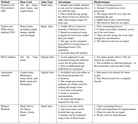
Another atlas has been proposed by Tanner and Whitehouse (TW: TW1, TW2, TW3)[34][35] in the year 1962. Here the study was focused on the age estimation but relies on the bone standard maturity. The TW method used bone joints locationas ROIs for bone maturity (shown in Figure.4, Figure.5 and Figure.6). Each ROI is further divided into three parts: epiphysis, diaphysis and metaphysis. Out of these three, the epiphysis ossifies from the age zero to teen age and later gets combined with diaphysis. So the age assessment of the TW and GP methods is only up to 19 years. Epiphysis has 9 stages in total starting with A (as no epiphysis bone) to B, C, D ...I as shown in Figure.6. Further authors have developed alternative methods for skeleton growth assessment system. They are stated as below
TW2 (20 bones): This is a modified method published in the year 1975 of initial TW1 method. It uses 20 ROIs for bone analysis including first, middle, fifth fingers and the carpal bones.
RUS (Radius, Ulna and Short bones): This is same as TW2 method but excludes the carpal bones.
CARPAL: This study is using the carpal bones alone. These bones ossify till the age of 9 years. So the age assessment using this method is limited to 9 years.
GP and TW2 methods are the base models with initial Atlas and look-up-tables for human age assessment. In the radiograph image epiphysis-ROI or carpal-ROI index stage must be identified and manually checked with the hand atlas. So, many researchers tried to develop fully automated systems for verifying and validating (digitally) their results using these two methods. A new atlas has been developed by combining children of both sexes of different countries [36][37].
2.1.3 FELS Method:
After a long gap, an alternative method FELS (Fels Longitudinal Study) was proposed by A.F. Roche et al. [38] in the year 1989. This is a computerized system using the scores/grades for each bone as an input for evaluating the age. Unlike the GP and TW methods here the grade distribution is evaluated for within the same age group as well. More than 130 points are chosen for each and every bone for analysis. This method also predicts the error correction for assessment. It is 0.3 to 0.6 years for boys from 1 month to 2 years and 0.2 to 0.3 years for girls from 1 to 14 years of age. For age > 2 it was 0.3 for boys. The errors are more for younger and older children (due to bones fusion stages). The authors have assumed that the input images are clear images without any distortion. The FELS method is complicated with huge set of points. It is not available as a software/package for analysis/verifying but, one can work on for implementing this code.
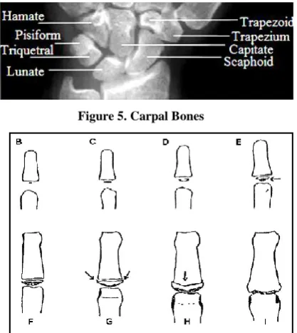
[image:3.595.346.503.571.732.2]Figure 5. Carpal Bones
Figure 6. Epiphysis region of ossification [71]
2.2 Automated Systems Design using X-ray
Image of Hand
2.2.1 HANDX Software System:
David J Michael and Alan C Nelson [39] developed the first automated system called HANDX for BAA in the year 1989. System is modulated into three stages pre-processing, segmentation and measurement. A Model-Based Histogram modification algorithm is used to enhance the image. According to this algorithm histogram is constructed first and then it is segmented into three classes as background, soft-tissues and bone pixels using a Gaussian distribution function. Then an adaptive contour process is used to outline the bone shape by overlaying the segmented bone image on the binary image of input image. This overlaying process is repeated for each and every bone of the hand image. Finally measurements are calculated by finding major and minor axes for each bone. These axis values are compared manually with the atlas of GP and TW2 for BAA. This has driven the research to computer assisted software development design. The proposed pre-processing algorithm might not be successful for all cases, since most of the soft-tissue pixels and bone pixels may overlap due to which the background subtraction may not be accurate. Moreover the noise at the shape edges will affect the segmentation process.
2.2.2 Computer PACS System:
In the year 1991 Eva Pietka, Michael F. McNitt-Gray and co-authors [40] developed a computer assisted system using phalangeal bones. Research was carried out to integrate with system to their Computer PACS lab (Picture Archiving and Communication systems) [41]. They maintained a separate database in the lab. Image pixels were categorised as uniform/non-uniform image pixels for background removal. Then boundaries were extracted by finding the edges from four sides of the image and drawing a line between the contour points. Further the edges are included or excluded using an error-correction method. Once the image edges are found it is rotated if required. Phalangeal ROIs are found using the sobel gradient on the image both on x and y directions. Finally the distal (d), proximal (p) and middle (m)
distances are measured and a ratio m/d and p/m are calculated for analysis. These results are compared with length table of phalangeal [42] for BAA. According to the authors algorithm was successful in 94% of cases. Here the noise in the image is not estimated and results are not compared with GP or TW2 methods. Image enhancement procedure still needs advancements. Eva Pietka published another research paper with some improvements in segmentation procedure in the year 2002[43].
2.2.3 Point Distribution Model
A.T. AI-Taani from Yarmouk University, Irbid, Jordan [44] have classified images according to age using point distribution model comprising of 120 images. For each bone a set of boundary points are chosen manually using mouse cursor by joining the existing points dynamically. These values are aligned for extracting the bone shape and finally classified to a model age group using fitting function (The least distance from the models). This experiment had 70.5% success for distal bone and 73.7% for epiphysis bone. The main drawback of the model is manual selection of points for edge identification. This problem was solved by new methods like active shape models in further research.
2.2.4 CASAS System:
Some of the diseases for human may also depend on the skeleton growth disorders. H. Frish, S Reidl and T. Waldhor from Austria [45] had evaluated age for both diseased and normal people by comparing with the manual GP and TW2 (RUS) methods using with Computer Aided Skeletal Age Scores (CASAS) system in the year 1996. Research included the children with Turner's syndrome, familial short stature and growth hormone deficiency under the age group of 2.4 years to 18.9 years. Separate investigators were assigned for each of the methods. A total of 100 images were analysed including 30 from Turner’s syndrome, 34 from familial short stature and 36 from growth hormone deficiency. Results showed that TW2 and CASAS with almost same age estimations. Due to disorders in bone shapes manual interruption (9% to 12.3 %.) was needed and thereafter the test was repeated in some cases for accuracy (due to differences in the measurements of short fingers). The proposed CASAS had a 21% of refusal cases at max and with a +1.1 years difference in the absolute average between TW2 and CASAS methods. These results state a wide scope for further research in designing the CASAS system with better image clarity and statistical analysis.
2.2.5 Active Shape/Active Appearance Models:
In the same year 1999 Sasan Mahmoodi et al. [47] considered 57 images including males and females and used binary threshold method and concave convex detection methods to remove the soft tissue pixels and to enhance the finger bone boundaries. Later an active shape model is used to detect the edges of the bony pixels by super imposing the trained image on the processing image. For this they used a fixed number of points distributed over the boundary to extract the shape of bone. These points are further used to find the width variations between the proximal tip and phalanx, epiphysis and metaphysis bones. Principal component method is also used for analysing. Finally, among these values the best confident growth values are chosen as the result and age is estimated based on them. The author claims that the success rate as 82% for males and 84% for females. This approach gives better results but takes a very high processing time for detecting the deformable shape analysis and also needs a huge set of training and test samples. As a whole the process is complicated.Automated system using both the epiphysis and carpal bone regions is proposed by D. Giordano and et.al [58] in the year 2009. These regions are separated using wedge functions and pre-processed them (background removal using threshold, orientation of the image through centroid calculation). For epiphysis region analysis they used Gaussian differentiation filter (DoG) for ossification bone measurement. Carpal region analysis is performed by applying DoG filter and matching with active contour method. These results are compared with TW and GP methods for the age group of 1 to 10 years. Authors claim that the average success rate was 87%. Further their analysis is to be extended for 11 to 19 years children.
2.2.5.1 BoneXpert Software System:
BoneXpert method is automated software developed by Hans Henrik Thodberg et.al [57] in the year 2008. This is very well known image processing software for skeleton maturity available over web. This software works is in three stages 1) Constructing bone borders 2) Estimate the age and 3) Compare results with GP and TW methods. For border estimation active shape driven appearance models have been used and age is estimated by bone health index calculation after applying principal component analysis and by calculating the mean, variance and standard deviation. Around 1559 images were considered of the age group of 2 to 8 years. This system is available over web as a commercial product from the year 2009. The software predicts age only between 2.5 to 17 years for boys and 2 to 15 years for girls. Their bone age accuracy was 0.72 years for GP method and 0.65 years for TW method. It is a licensed product and for each x-ray image they charge around 10€.
2.2.6 Dynamic Threshold Method:
Another computer based bone age assessment was proposed by Ewa Pieta et. al. [48] in the year 2000. In this, images were rotated for specified direction if required and the background pixels are removed using a dynamic threshold method. In this method a fixed sliding window has been used to locate the background pixels and remove them by calculating mean, variance and etc. Noise has been restored using morphological filters. Author estimated age using only the middle finger oscillation bones. So, this ROI is identified by using step wedge function and cropping from the entire image. Resulted image is smoothened by median filtering. The age is estimated using the ratios among the diameter of epiphysis, diameter of metaphysis and distance between metacarpal and epiphysis. These ratios are compared with all the ratios of dataset images
with respect to age and also categorised to different age groups. Here the background pixels are not removed from the image and this may cause some dominance in edge evaluation (gradient image). Middle finger alone may not lead to accurate results. Moreover the results are not compared with the defined age charts. M. Niemeijer et. al [49] also developed an automated system using only the centred phalanx of the middle finger having a success rate of 73.3% using active shape models.
2.2.7 Semi-Automated System:
A Semi-automated approach for skeleton maturity was proposed by Ana Maria Marques Da Silva et. al.[50] using image processing methods in the year 2001. For each finger bone image a point is chosen inside and thereafter contours are detected using multi-scale imaging system [51]. Later the middle finger is isolated from the image and respective profile curves are plotted for each image. Edges of epiphysis and cartilage thickness are identified using these profile curves only. Finally based on the ossification of epiphysis and cartilage thickness gap, measurements are evaluated by measuring the distances among peaks of profile curves (total proximal, metacarpal and phalanx bone). Age is estimated using these distances only. This approach is developed with manual intervention for analysis.
2.2.8 Fuzzy Logic Based System:
Age assessment based on carpal bones alone was given by Aifeng Zhang and et. al. in the year 2007[52]. Background is removed by thresholding. Carpal ROI is segmented using the horizontal line crossing the middle two metacarpal bones, vertical lines crossing the corner carpal bones and the line crossing the bottom forearm bones. Then the image is smoothed by using the filter proposed by Malik and Perona [53]. This method works on diffusion i.e. removing the noise by preserving the border information. Thereafter the edge is found using canny edge function. Then the image is refined to get only the carpal bones by excluding the unwanted bony pixels (morphological operators are used for this). Centroid is estimated for capitate bone. ROI is divided into five sectors with respect to the centroid for checking the presence of bones and their size and shape. Finally a fuzzy logic classifier is used for age assessment by giving these results as inputs. According to the authors the success rate was near to 100% for age>=2 and 80% for age<2 years. Authors had included radiographs of 0-7 years of males and 0-5 years for females. After the age above 7 years for males and 5 years for females the some of the carpal bones may overlap so, these age groups are excluded from research.
2.2.9 Neural Network Based System:
2.2.10 Score Index Method:
Metin Yildiz and et. al [59] developed a web site (using C# language and .NET environment) for age calculation in the year 2010. Only wrist bones were used for age assessment. So, the children less than 4 years have been excluded from the survey. The web site asks the user to load the image and thereafter prompts to choose the stages for each wrist bone. Based on these stages a score index gets created for each stage and finally bone age is estimated using the formula Bone age= 1+index/10; The author found that the observer variations vary in TW and GP from -0.77 to -0.97 and -0.45 to 0.37 with 95% confidence. The average computational time for TW2 method is 2.4min and for GP is 0.8min while the manual approach was taking 9min. In this the user is expected to have prior knowledge to give the input for various stages, which is the key factor for index calculation. This may lead to errors in measurements and age predication.
2.2.11 System Based on Histogram Technique:
Histogram based automated system was developed by Marjan Mansourvar et. al [60] in 2012, initially user is provided with an option to feed an image over web link and then a histogram is generated using the ImageMagik software. Thereafter the content based image retrieval system is used to find the most relevant images from the database by assigning the ranks to the similar images (ranks are assigned using similarity score). Age is estimated using the ratio between highest ranked age from the retrieved images to the total number of ranked images. The system showed a huge variation for age less than 10 years and on an average the error rate was -0.170625 years. The system also fails in most of the cases since an image histogram will be same for multiple images. Hough transform is used for the age assessment by Markus Brunk et. al [61] where the only the middle finger is used for analysis with a success rate of 95%.Recently in 2013 Shao-Yan Zhang and et. al [62] have given a research article on the automated bone age assessment on Chinese population. It was purely based on the BoneXpert software system. They developed a bone age scale table named as BX-China05 by comparing the age chart given by GP. The table describes the correction value needed to estimate the age for Chinese children age charts for boys and girls separately in the age group of 2 to 17 years. A BX-China05 atlas has excluded age < 2 since it's the limitation of BoneXpert software.
2.3 Limitations of Hand x-ray Analysis:
The main limitation of hand x-ray analysis is the age group. This can be used to estimate only the age up to 18 or 19 years.
The results vary for male and female.
The atlases developed will vary with regions. So, different atlases have to be generated for various countries and regions.
Age can't be estimated if the images have loss of data such as less no. of fingers, joint fingers, cracked bones etc.
2.4 Automated Systems using Tooth
Radiographs:
In 1944 the first atlas was developed by Schour et. al [63] based on eruption of teeth. Later another atlas was designed by Moorres which was further modified by Anderson
including all the tooth stages. Other than atlas there is also a score method available which assigns scores to each individual tooth and takes the average for age estimation [64]. Queen Mary University of London designed an atlas (UMAL-Atlas)[65] for the age group between 7 months to 23 years by collecting the data from England, UK and London. They used Moorres method for age estimation and made atlas available over web for education. The automated systems are not much developed using this bone.
2.5 Automated Systems using other human
bone radiograph images:
Automated bone age assessments using radiographs of other parts of the bones are not well studied in the research till date. Age assessment is mostly the manual approach. The hand-held age atlases might not be available for other parts of the bones. There is a huge scope in the research for age assessment using the knee image for age group above 19 years. Skull image analysis can be used for age group zero to adult. So, one can think of automating these approaches.
3. STORAGE CHALLENGES FOR
IMAGES DATA SETS:
Medical diagnoses by the researchers or scientists need a huge set of images for processing and analysing the desired tasks. The medical researchers of different countries need data exchange and outsourcing the image data over national and international health care research institutes. So, these images must be stored and managed through well-defined database models and tools. The current researchers are facing following challenges for storing and managing the medical data.
Data Collection: The image data will be in different formats such as X-ray, MRI, CT and DICOM etc. (png, jpeg, tif and etc.). Integrating into one common format is not an easy task.
Data Storing: The image data can't be stored in the available models (which are for text data). A new software or indexing method must be applied for this task.
Data Sharing: Providing the security for patient's information while sharing the data over multiple recipients is the biggest challenge in medical image analysis. So, image compression and image crypt-analysis techniques must be used in this stage.
Data Managing and Management: Every health care organization has a limited storage space and memory capacities (in contrast the image data grows vary rapidly). So, storing and managing the data at one place for medical diagnosis is very tedious task.
4. CONCLUSION AND FUTURE
RESEARCH SCOPE:
This paper was focused on the current research challenges for age assessment of human using radiographs of different bones. This paper is a survey paper of current approaches applied for age assessment using the image processing techniques. Various implementation approaches or algorithms were discussed which were used by many researchers for background pixels removal from x-ray images, bony pixels enhancement, edge detection or boundary extraction for bone shape, segmentation techniques for extracting the region of interest and finally decision making for age assessment (A summary of all methods are given in Table 1.
This paper covered both manual and automated age assessment methods available till date. This ranges from skull, hand, pelvic and tooth etc. The automated system design is still an on-going research using these bones radiographic images. There are many automated approaches for the hand x-ray analysis due to available atlases. For other bone radiographs the automated age assessment is not up to the mark.
Many researchers have used the pre-processing, enhancement, segmentation techniques, fuzzy logic, neural network, active shape models and clustering methods for automated bone age assessment but still some of the unwanted pixels like background and soft tissue pixels still remain in the image during measurement calculations (for statistical analysis). This might cause miss-classification. This leads to future scope of research for using the morphological image processing, color image processing, frequency and spatial domain enhancement methods etc. for better age assessment and bone length measurement analysis.
Storing images is also a major issue in this research. This focuses to the research direction in which better image compression techniques, image encryption and decryption techniques may be applied for storing, safe-sharing and managing the medial data over web for accurate age analysis.
[image:7.595.51.535.325.760.2]
Table 1: Summary of Techniques for Bone Age Assessment
Method Bone Used Atlas Type Advantages Disadvantages
Greulich and Pyle method (GP)
All the finger joint bones and carpal bones.
Hand Atlas 1. Simple and reliable method to use, just by comparing the x-ray with the atlas.
2. Once found the age group the observer have to check for older and younger stages for exact age.
1. Time consuming process. 2. Around 22 joints have to be processed.
3. The image must be very clear for estimating the age.
4. Might lead to miss classification 5. The observer must be an expert. Tanner and
White-house method (TW)
Bones joints including carpal, thump, middle and last finger.
Hand Atlas 1. Unlike GP it is based on maturity of skeleton bones. 2. Based on numerical score assigned for each bone (rather than just shape).
3. The age can be estimated using RUS or Carpal bones or Phalangeal bones (20) separately.
4. Accurate than GP method.
1. Time consuming process.
2. Complex procedure (maturity scores are 8 for each bone).
3. Here also the image has to be clear enough for classification.
4. The observer must be an expert.
FELS method All the bone joints
Digital Atlas 1. A new approach to age assessment using the maturity scores for all joints bones. 2. Ease to understand and implement as a program.
1. Too many points (above 130) are chosen for each bone.
2. Not available as software/package: so the researchers can't test and verify.
Automated methods
Carpal alone, Phalangeal bones alone, and Both carpal and Phalangeal
Digital Atlas 1. Available over web either as free/licensed product for evaluation.
2. The image processing methods are widely used for making the image clear. 3. Less time consuming. 4. Leads to accurate measurements.
5. No prior knowledge is required for the user.
1. Still need to be refined for better results.
2. The observer must be a computer literate.
Manual Methods
Skull, Pelvis, rib, Spine, Knees, femur, Hand
Hand Atlas 1. Easy to use and verify. 2. Age assessment can be beyond teen age group. 3. Gender can be estimated using some of these bones.
1. Time consuming Process
2. Lab tools dependent for measurement. 3. Observer must be an expert
5. REFERENCES
[1] Liversidge HM, Dean MC, Molleson TI, Increasing human tooth length between birth and 5.4 years, Am J Phys Anthropol 1993; 90: 307–13.
[2] Hugo F. V. Cardoso, A Test of the Differential Accuracy of the Maxillary Versus the Mandibular Dentition in Age Estimations of Immature Skeletal Remains Based on Developing Tooth Length, J Forensic Sci, March 2007, Vol. 52, No. 2.
[3] Hong-Seop Kho and Young-Ku Kim, Jong-Il Yun, Jeong-Yun Lee, Jin-Woo Chung, Age Estimation of Korean Adults by Occlusal Tooth Wear, J Forensic Sci, May 2007, Vol. 52, No. 3.
[4] Roberto Cameriere, Giuseppe Brogi, Luigi Ferrante, Dora Mirtella, Claudia Vultaggio, Mariano Cingolani and Gino Fornaciari, Reliability in Age Determination by Pulp/Tooth Ratio in Upper Canines in Skeletal Remains, J Forensic Sci, July 2006, Vol. 51, No. 4
[5] Dr. Rajesh Kumar Verma, Dr Mukesh K Goyal, Dr Shiv. Kochar, Age Assessment from Radiological Cranial Suture closure in Fourth to Seventh decades (A Jaipur Based Study), J Indian Acad Forensic Med, 32(2), ISSN 0971-0973.
[6] Vyas P.C. and Sischer, H. Age estimation by closure of suture of skull in individuals of 25-45 yrs of age of Jaipur area. Dissertation for M.D. (Forensic Medicine) University of Rajasthan (1996).
[7] Derya Atamturk and Izzet Duyar, Age-Related Factors in the Relationship Between Foot Measurements and Living Stature and Body Weight, J Forensic Sci, November 2008, Vol. 53, No. 6
[8] Lucina Hackman, Catriona M. Davies, and Sue Black, Age Estimation Using Foot Radiographs from a Modern Scottish Population, J Forensic Sci, January 2013, Vol. 58, No. S1.
[9] Pyle SI, Hoerr NL, A radiographic standard of reference for the growing knee, 1st edn. Springfield, IL: Charles C. Thomas, 1969.
[10] Lucina Hackman and Sue Black, Age Estimation from Radiographic Images of the Knee, J Forensic Sci, May 2013, Vol. 58, No. 3.
[11] Atif Khan, Daciana Iliescu, Evor Hines, Charles Hutchinson, Robert Sneath, Neural Network Based Spinal Age Estimation Using Lumbar Spine Magnetic Resonance Images (MRI), 2013 4th International Conference on Intelligent Systems, Modelling and Simulation.
[12] Martell M, Fescina RH, Martínez E, Bolívar N, Estimation of gestational age by the length of the dorsal spine, J Perinat Med. 1997;25(2):168-72.
[13] A. Schmitt, Age-at-Death assessment using the OsPubis and the Auricular Surface of the llium: a Test on an Identified Asian Sample, Int. J. of Osteoarchaeology:14:1-6(2004).
[14] Erin H. Kimmerle, Lyle W. Konigsberg, Richard L. Jantz and Jose Pablo Baraybar, Analysis of Age-at-Death Estimation Through the Use of Pubic Symphyseal Data, J Forensic Sci, May 2008, Vol. 53, No. 3
[15] Iscan MY, Loth SR, Wright RK, Age estimation from the rib by phase analysis: white males, J Forensic Sci 1984;29:1094–104.
[16] George J. R. Maat, Ann Maes, M. Job Aarent and Nico J. D. Nagelkerke, Histological Age Prediction from the Femur in a Contemporary Dutch Sample The decrease of nonremodeled bone in the anterior cortex, J Forensic Sci, March 2006, Vol. 51, No. 2.
[17] Seung-Ho Han, Sang-Hyun Kim, Yong-Woo Ahn, Gi-Yeong Huh, Dai-Soon Kwak, Dae-Kyoon Park, U-Young Lee and Yi-Suk Kim, Microscopic Age Estimation from the Anterior Cortex of the Femur in Korean Adults, J Forensic Sci, May 2009, Vol. 54, No. 3
[18] Roberto Cameriere, Stefano De Luca, Roberto Biagi, Mariano Cingolani, Giampietro Farronato and Luigi Ferrante, Accuracy of Three Age Estimation Methods in Children by Measurements of Developing Teeth and Carpals and Epiphyses of the Ulna and Radius, J Forensic Sci, 2012, doi: 10.1111/j.1556-4029.2012.02120, Available online at: onlinelibrary.wiley.co
[19] Sven Schmidt, Inna Nitz, Ronald Schulz and Andreas Schmeling, Applicability of the skeletal age determination method of Tanner and Whitehouse for forensic age diagnostics, Int J Legal Med (2008) 122:309–314
[20] M. Vignolo, S. Milani, E. DiBattista, A. Naselli, M. Mostert, G. Aicardi, Modified Greulich-Pyle, Tanner-Whitehouse and Roche-Wainer-Thissen (knee) methods for skeletal age assessment in a group of Italian children and adolescents, Eur J Pediatr (1990) 149 : 314-317
[21] C.Cattaneo, D.De Anhelis, M.Ruspa, D.Gibelli, R.Cameriere, M. Grandi, How old am I? Age estimation in living adults: a case report. J Forensic Odontostomatol. 2008 Dec 1;26(2):39-43.
[22] Rajendra Kumar Sharma, Demography and Population Problems, Atlantic (2007).
[23] A. Aggrawal and A. Busuttil, Age estimation in the living, The Police Surgeon (Journal of The Association of Police Surgeons). No. 38, Jan 1991, Pp 33-36.
[24] Anil Aggrawal, Estimation of age in the living: in matters civil and criminal, J Anat. 2009 May 11
[25] http://www1.umn.edu/humanrts/research/Philippines/rule_ on_children.html
[26] Sundeep Khosla, B. Lawrence Riggs, Pathophysiology of Age-Related Bone Loss and Osteoporosis, Endocrinol Metab Clin N Am, 34 (2005) 1015–1030
[27] http://www.nlm.nih.gov/medlineplus/ency/article/004015.h tm
[28] John Wiley & Sons, Inc., Hoboken, NJ, USA, ASBMR (2008) Chapter 20, Age-Related Bone Loss, in Primer on the Metabolic Bone Diseases and Disorders of Mineral Metabolism, doi: 10.1002/9780470623992.ch20.
[29] B. Lawrence Riggs, The Burden of Disease: Bone Health, Osteoporosis, and Related Bone Diseases, Report of the Surgeon General's Workshop on Osteoporosis and Bone Health: December 12 – 13, 2002, Washington, DC.
[30] http://medical.nema.org/Dicom/about-DICOM.html
[32] Dinesh M.S, Bhanuprakash, Dr. Ashok Rao, Vision System for bone measurement from digital hand radiographs, Proceedings RC-IEEE-EMBS & 14th BMESl, 1995
[33] W. W. Greulich and S. I. Pyle, Radiographic Atlas of Skeletal Development of Hand Wrist, 2 ed. Palo Alto, CA: Stanford Univ. Press, 19
[34] J. M. Tanner, R. H. Whitehouse, W. A. Marshall, M. J. R. Healy, and H. Goldstein, Assessment of Skeletal Maturity and Prediction of Adult Height (TW2 Method), New York, NY: Academic, 1975.
[35] Tanner JM, Whitehouse RH, Cameron N, Marshall WA, Healy MJR, Goldstein NH. Assessment of skeletal maturity and prediction of adult height (TW3 method), 3rd ed. London: W.B Saunders; 2001.
[36] Gertych A, Zhang A, Sayre J, Pospiech-Kurkowska S, Huang KH, Bone age assessment of children using digital hand atlas, Computer Medical Imaging Graph 31:322-331, 2007.
[37] University of South California, Image Processing and Informatics Lab: Digital Hand Atlas Database System, Available online at: htto://ipilab.org/BAAweb/
[38] WM. Cameron Chumlea, Alex F. Rouche, David Thissen, The FELS method of assessing the skeletal maturity of the hand-wrist, American. J. of Human Biology, 1:175-183, 1989
[39] David J. Michael, Alan C. Nelson, HANDX: A Model-Based System for Automatic Segmentation of Bones from Digital Hand Radiographs, IEEE Transactions on Medical Imaging, vol 8, No.1, 1989
[40] Ewa Pietka, Michael F. McNitt-Gray, M. L. Kuo, and H. K. Huang, Computer assisted phalangeal analysis in skeletal Age Assessment, IEEE Transactions on Medical Imaging, vol. 10, No.4, December 1991.
[41] R. T. Taira, N. J . Mankovich, M. I. Boechat, H. Kangarloo, and H. K. Huang, Design and implementation of a picture archiving and communication system for pediatric radiology, Amer. J . Roentgenol., vol. 250, pp. 1117-1121, 1988.
[42] S. M. Gam, K. P. Hertzog, A. K. Poznanski, and J. M. Nagy, Metacarpophalangeal length in the evaluation of skeletal malformation, Radiology, vol. 105, pp. 375-381, 1972.
[43] Ewa Pietka, Sylwia Pospiech-Kurkowska, Arkadiusz Gertych, Fei Cao, Integration of computer assisted bone age assessment with clinical PACS, Computerized Medical Imaging and Graphics , 27 (2003) 217–228
[44] A.T. Al-Taani, Classification of Hand Bones for Bone age Assessment, ICECS-1996
[45] H. Frisch, S. Riedl, T. Waldhor, Computer aided estimation of skeletal age and comparition with bone age evaluations by the Greulich-Pyle and tanner-whitehouse, Pediatr Radiol (1996) 26:226-231
[46] F. Caoa, H.K. Huanga, E. Pietkab, V. Gilsanzc, Digital Hand Atlas and web-based bone age assessment system design and implementation, Computerized Medical Imaging and Graphics 24 (2000) 297-307
[47] Sasan Mahmoodi, Bayan S. Sharif, E. Graeme Chester, John P. Owen, and Richard Lee, Skeletal Growth Estimation Using Radiographic Image Processing and Analysis, IEEE Transactions on Information Technology in Bio-Medicine, vol. 4, no. 4, December 2000.
[48] Ewa Pietka, Arkadiusz Gertych, Sylwia Pospiech, Fei Cao, H. K. Huang and V. Gilsanz, Computer-Assisted Bone Age Assessment: Image Preprocessing and Epiphyseal/Metaphyseal ROI Extraction, IEEE Transactions on Medical Imaging, vol 20, No.8, 2001.
[49] Meindert Niemeijer, B. Van. Ginneken, Casper A. Maas, Frederik J. A. Beek, Max A. Viergever, Assessing the skeletal age from hand radiograph automating the Tanner-Whitehouse method, Proc. SPIE 5032.
[50] Ana Maria Marques Da Silva, Silvia Delgado Olabarriaga, Carlos Augusto Dietrich and Carlos Andre Aita Schmitz, On Determining a Signature for Skeletal Maturity, Proceedings of XIV Brazilian Symposium on Computer Graphics and Image Processing, 2001.
[51] M.K.Schneider, P.W. Fieguth, W.C. Karl and A.S. Willsky, Multiscale methods for the segmentation and reconstruction of signals and images, IEEE Trans. Image Process. vol. 9, no. 3, 2000, pp: 456-468.
[52] Aifeng Zhang, Arkadiusz Gertych and Brent J. Liu, Automatic bone age assessment for young children from newborn to 7-year-old using carpal bones, Comput Med Imaging Graphics, 2007;31(4-5):299-310.
[53] Perona P, Malik J, Scale-space and edge detection using anisotropic diffusion, PAMI 1990:12(7).
[54] Jian Liu, Jing Qi, Zhao Liu, Qin Ning, Xiaoping Luo, Automatic bone age assessment based on intelligent algorithms and comparison with TW3 method, Comput Med Imaging and Graphics 32 (2008) 678–684
[55] Krit Somakanta, Nipon Theera Umpon, Sansanee Auephanwiriyakul, Bone age assessment in young children using automatic carpal bone feature extraction and support vector regression, J. Digit Imaging, 2011, 24:1044-1058.
[56] Gilsanz V, Ratib O, Hand Bone age: A Digital Atlas of Skeletal maturity, 2005.
[57] Hans Henrik Thodberg, Sven Kreiborg, Anders Juul, and Karen Damgaard Pedersen, The BoneXpert Method for Automated Determination of Skeletal Maturity, IEEE Transactions on Medical Imaging, vol 28, No.1, 2009.
[58] D. Giordano, C. Spampinato, G. Scarciofalo and R. Leonardi, Automated skeletal bone age assessment by integrating EMROI and CROI processing, IEEE International Workshop on Medical Measurements and Applications, 2009, pp:141-145.
[59] Metin Yildiz, Albert Guveni, Esra Guven, Didar Talat and Mahmut Haktan, Implementation and Statistical Evaluation of a Web-Based Software for Bone Age Assessment, J Med Syst, Published Online, 2nd February 2010.
[61] Markus Brunk, Heike Ruppertshofen, Sarah Schmidt, peter Beyerlein, Hauke Schramm, Bone age classification using discriminative generalized Hough transform, Springer, Bildverarbeitung für die Medizin 2011, pp. 284-288.
[62] Zhang SY, Liu G, Ma CG, Han YS, Shen XZ, Xu RL, Thodberg HH, Automated Determination of Bone Age in a Modern Chinese Population, ISRN Radiology, Volume 2013, Article ID 874570.
[63] Willems G. A review of the most commonly used dental age estimation techniques. J Forensic Odontostomatol. 2001;19(1):9–17.
[64] Malik P, Saha R, Agarwal A. Applicability of Demirjian’s method of age assessment in a North Indian female population, Eur J Paediatr Dent. 2012;13(2):133–135.
[65] https://atlas.dentistry.qmul.ac.uk/
[66] http://www.bridgeheadsoftware.com/products/healthcare_d ata_management_hdm_platform/
[67] http://www.firehost.com/secure-cloud/enterprise/healthcare
[68] http://ganymed.imib.rwth-aachen.de/irma/datasets_en.php
[69] http://www.magnet.fsu.edu/education/tutorials/magnetacad emy/mri/fullarticle.html.
[70] K. Kirk Shung, Diagnostic Ultrasound Imaging and Blood Flow Measurements, CRC Press, 19-Sep-2005.
![Figure. 1: X-ray of Hand and Pelvic of Human [68]](https://thumb-us.123doks.com/thumbv2/123dok_us/8031341.768876/2.595.318.523.519.734/figure-x-ray-hand-pelvic-human.webp)