R E S E A R C H A R T I C L E
Open Access
High grade isthmic spondylolisthesis; can
reduction always re-align the unbalanced
pelvis?
Konstantinos Martikos
*, Tiziana Greggi and Cesare Faldini
Abstract
Background:Recently, various studies have reported the importance of distinguishing between balanced and
unbalanced SL, sustaining the importance of SL reduction in unbalanced cases. In this study we present our experience in the treatment of isthmic spondylolisthesis in young patients, observing the correlation between SL reduction and sagittal correlation between spine and pelvis.
Methods:This is a retrospective study of a series of patients treated surgically for isthmic spondylolisthesis. Inclusion criteria were L5-S1 isthmic spondylolisthesis of III° or IV°, pediatric age, clinical and radiographic follow up of at least 1 year. Radiographic evaluation included the following elements: grade and percentage of listhesis (%L), lumbar lordosis (LL), lumbar-sacral angle (LSA), pelvic incidence (PI), sacral slope (SS), pelvic tilt (PT)
distinguishing between“balanced”and“unbalanced”patients. Radiographic values were confronted by using Student’s t- test, obtaining a statistically significant difference for values inferior to 0,05.
Results:Based on inclusion criteria, 28 patients were selected for our retrospective analysis, 19 female and 9 male. Mean age at surgery was 15,6 years. Mean follow up was 3 years and 3 months (min. 1 year–max 6 years and 7 months). Spondylolisthesis reduction was statistically significant both in balanced and in unbalanced patients, but pelvic incidence values did not improve significantly. We observed fewer mechanical complications in patients treated with interbody support.
Conclusion:In our study, differences between pre-op and post-op spinopelvic alignment values were not
statistically significant, even though spondylolisthesis reduction was statistically significant in all cases. Our study could be considered an initial attempt to correlate spinopelvic changes to spondylolisthesis reduction in a progressive manner, and possibly in the future, generate threshold values of reduction for ideal spinopelvic alignment in every different patient.
Keywords:Isthmic spondylolisthesis, Pelvic balance, Interbody fusion, Meyerding classification
Background
Isthmic spondylolisthesis (SL) represents a sagittal de-formity of the spine, thus influencing both sagittal align-ment of the pelvis both from the clinical and radiographic point of view. Therefore, the objective of surgical treat-ment is not only the stabilization of the instable segtreat-ment but also reduction of the slipped vertebra over S1. Re-cently, various studies have reported the importance of distinguishing between balanced and unbalanced SL, sus-taining the importance of SL reduction in unbalanced
cases. More precisely, various studies in the recent years sustain that reducing a high-grade spondylolisthesis may balance un unbalanced pelvis, without focusing on results in cases of incomplete spondylolisthesis reduction.
In this study we present our experience in the treat-ment of isthmic spondylolisthesis in young patients, observing the correlation between SL reduction and sa-gittal pelvic alignment in terms of pelvic tilt, sacral slope and lumbar-sacral angle, with a critical view on pelvic balance especially in cases of satisfactory but not complete spondylolisthesis reduction.
© The Author(s). 2019Open AccessThis article is distributed under the terms of the Creative Commons Attribution 4.0 International License (http://creativecommons.org/licenses/by/4.0/), which permits unrestricted use, distribution, and reproduction in any medium, provided you give appropriate credit to the original author(s) and the source, provide a link to the Creative Commons license, and indicate if changes were made. The Creative Commons Public Domain Dedication waiver (http://creativecommons.org/publicdomain/zero/1.0/) applies to the data made available in this article, unless otherwise stated. * Correspondence:[email protected]
Methods
This is a retrospective study of a series of patients treated surgically for isthmic spondylolisthesis in a single spinal deformities division between 2003 and 2012. In-clusion criteria were the following: patients affected by L5-S1 isthmic spondylolisthesis of III o IV grade accord-ing to Meyerdaccord-ing, age between 10 and 18 years, posterior pedicle screw instrumentation, clinical and radiographic follow up of at least 1 year.
Radiographic evaluation included the following elements: grade and percentage of listhesis (%L) according the Mayerding scale, lumbar lordosis (LL, angle between L1 and S1 upper endplates), lumbar-sacral angle (LSA, angle be-tween L5 lower endplate and S1 upper endplate), pelvic inci-dence (PI, angle between the line perpendicular to the sacral plate at its midpoint and the line connecting this point to the femoral heads axis), sacral slope (SS, angle between S1 upper endplate and the horizontal plane), pelvic tilt (PT, angle between a vertical line and the line joining the middle of the sacral plate and the axis of the femoral heads).
Our series was analyzed both in its entirety as well as divided in two groups of“balanced”and“unbalanced” pa-tients. The concepts of balanced and unbalanced spondylo-listhesis were based on previous reports of Hresko at al [1]., that distinguish high SS-low PT as balanced ones and low SS-high PT as unbalanced ones, with the latter ones presenting retroverted pelvis and vertical sacrum.
All radiographic measurements were performed upon pre-operatory, post-operatory and last follow up x-rays by the author of the study. Radiographic values were confronted by using Student’s t- test, obtaining a statisti-cally significant difference for values inferior to 0,05.
Based on inclusion criteria, 28 patients were selected for our retrospective analysis, 19 female (67.9%) and 9 male (32.1%) (Table 1). Mean age at surgery was 15,6
years (min. 10 - max 18 years). All patients had spondy-lolisthesis superior to 50%, being therefore classified as grade III or IV according to Meyerding. Mean slippage percentage was 81,04% (min 57% - max 100%). Mean follow up was 3 years and 3 months (min. 1 year – max 6 years and 7 months).
Baseline surgical treatment was posterolateral instru-mented fusion in all patients. Posterior arch of L5 was removed in all cases prior to reduction maneuvers for an optimal radicular release. Spondylolisthesis was reduced to some point, although not totally in all patients. In 19 cases, instrumentation was limited to L5, in 9 patients it was extended to L4. In 15 patients interbody support was associated to posterior instrumentation:
– Posterolateral lumbar interbody fusion (PLIF) in 4 cases (cages in peek material in 2 cases, titanium in 2 cases).
– Transforaminal lumbar interbody fusion (TLIF) in 6 cases (cages in peek material in 4 cases, titanium in 2 cases).
– Posterior L5-S1 transdiscal screw in 1 case.
– Anterior fusion in 4 cases, performed at mean 4,2 months after initial posterior fusion (min 1–max 8 months). Of these patients, two were treated with a single L5-S1 screw, one was treated with an anterior L5-S1 transdiscal screw and one was treated with autogenous interbody bone graft.
Our mean pre-operatory spinal-pelvic values are con-sistent with previously reported values in high grade isthmic spondylolisthesis, mainly characterized by high PI and PT [2–4]:
PI: 72.78° (normal value 48.4°). PT: 28° (normal value 7.2)°. SS: 43.89° (normal value 41.2°). LL: 57.81° (normal value 48.5°).
Results
[image:2.595.57.291.536.731.2]Spinal and pelvic radiographic measurements of the en-tire series are reported in Table 2. Considering the whole series (without distinguishing between balanced and unbalanced patients), we found a statistically signifi-cant improvement in %L (p= 0,0000017) and LSA (p= 0, 003) between pre-operative and post-operative values. No statistically significant differences were found regard-ing LL, SS, PT and PI between pre and post-operative values. Although post-operative values of SS and PT showed a trend towards normal population values when compared to preoperative values (SS increased and PT declined) the difference was not statistically significant. Values remained stable at final FU, with no statistically significant differences between post-op and final FU. Table 1Mean demographic values of patient population
Age 15.61years (10–18)
Sex 19 F(67,9%),9 M(32,1%)
Grade III° SL 12(42,8%)
Grade IV° SL 16(57,2%)
Posterior instrumentation with PLIF 4(14,3%)
Posterior instrumentation with TLIF 6(21,4%)
Only anterior stabilization 1(3,5%)
Posterior and anterior stabilization 4(14,3%)
-Anterior L5-S1 screw 2(7,1%)
-ALIF 1(3,5%)
-Autologous bone only 1(3,5%)
Sonly posterior pedicle screw instrumentation 15(53,5%)
L5-S1 fusion 19
The series was afterwards split in two groups, balanced and unbalanced patients, based on pre-operatory pelvic values as defined in the Hresko et al. paper [1]: balanced patients group with high SS and low PT (11 patients) and unbalanced patients group with low SS and high PT (17 patients). Results were similar to the entire series evalu-ation. Neither group showed statistically significant differ-ences in pelvic parameters between pre-op ad post-op values. Although there was a statistically significant listh-esis reduction as well as LSA improvement, pelvic param-eters (SS, PT, LL) did not seem to show a statistically significant improvement in neither balanced or unbal-anced group of patients (Tables3and4). Lastly, our case series was divided into two groups based on the presence or not of interbody support at first surgical procedure (PLIF, TLIF, ALIF, transdiscal screw) and sagittal spinal-pelvic parameters were calculated before and after surgery (Tables5and6). Both posterior + interbody instrumented cases as well as those with only posterior instrumentation showed a statistically significant %L reduction: 78% mean pre-op vs 23,5% mean post-op, p= 0,0001 in posterior + interbody instrumented cases 83,2% pre-op vs 56% post-op,p= 0,00001 in posterior only instrumented cases with-out interbody support.
At final FU we observed that the interbody support group maintained listhesis reduction while the posterior only instrumented patients presented a loss of listhesis reduction when compared to immediate post-operative results. This difference between the two groups was sta-tistically significant (p = 0,0001). %L: 23.5% post-op vs 29.5% at final FU in the interbody support patients and 56.65% post-op vs 75.31% at final FU in patients without interbody support.
Patients treated with interbody support showed statisti-cally better LSA improvement when compared to patients treated without interbody support: LSA improvement of 25% in patients treated with interbody support (p< 0.0001) vs 8.1% (p= 0.17) in patients treated without inter-body support. LSA: 4.7° post-op vs 0.6° at final FU in interbody support patients and−13.9° post-op vs −19.5° at final FU in patients without interbody fusion. The remaining sagittal spinal-pelvic parameters (LL, SS, PT and PI) remained statistically similar, both within each group before and after surgical treatment, and between the two groups when compared to each other.
Mechanical complications with loss of correction were observed in 4 patients (14.2%). Revision surgery was per-formed in each case, with the objective to obtain satisfac-tory listhesis stabilization, not necessarily reduction. All mechanical complications occurred in patients treated without interbody support at primal surgery. Therefore, mechanical complications incidence in patients treated without interbody support at primal surgery was 24%. No mechanical complications were observed in patients treated with interbody support. Difference in mechanical complica-tions was therefore statistically significant between these two groups of patients (p< 0,00001).
[image:3.595.57.538.99.196.2]We observed 2 cases of bilateral sacral screw breakage (7.1%) that occurred after a mean 4,5 months (2 and 7 months respectively). Both patients were initially treated with posterior instrumented arthrodesis L4-S1 without any interbody support. In both cases revision surgery was performed by removing the broken screws and re-placing them with greater diameter ones. In one case, anterior instrumentation with two L5- S1 screws was performed.
Table 2Mean pre-op, post-op and final FU values, in all patients
X-Ray index Pre-op Post-op FU
Listhesis percentage 81%(57–141) 43,6%(0–149) 58,8%(5–154)
Lumbar-sacral angle (LSA) −19,5°(−47 to + 17) −6,6°(−58 to + 15) −12,3°(−47 to + 15)
Lumbar lordosis (LL) 57,8°(29–72) 56,4°(40–72) 55,1°(38–71)
Sacral slope (SS) 43,9° (20–59) 45,9° (18–57) 43,9° (19–60)
Pelvic tilt (PT) 28,2° (17–41) 25,6° (11–45) 28,8° (14–49)
Pelvic incidence (PI) 72,8° (49–88) 72,1° (51–88) 73,1° (41–89)
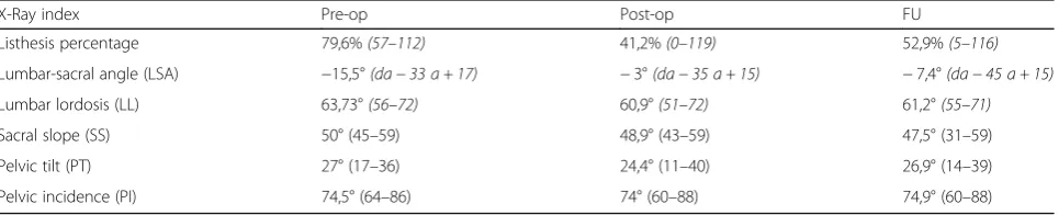
Table 3Mean pre-op, postop and FU values of“balanced”group patients
X-Ray index Pre-op Post-op FU
Listhesis percentage 79,6%(57–112) 41,2%(0–119) 52,9%(5–116)
Lumbar-sacral angle (LSA) −15,5°(da−33 a + 17) −3°(da−35 a + 15) −7,4°(da−45 a + 15)
Lumbar lordosis (LL) 63,73°(56–72) 60,9°(51–72) 61,2°(55–71)
Sacral slope (SS) 50° (45–59) 48,9° (43–59) 47,5° (31–59)
Pelvic tilt (PT) 27° (17–36) 24,4° (11–40) 26,9° (14–39)
[image:3.595.58.541.634.732.2]L5 screw pull out was observed in 2 patients (7.1%), unilateral in 1 case and bilateral in the other, after a mean 4,5 months from initial surgery (3 and 6 months respectively). Both cases underwent revision surgery by substituting mobilized screws with greater diameter ones. In one patient instrumentation was was extended to L4 and anterior stabilization with AxiaLIF was per-formed. In one patient treated with TLIF cages, subsid-ence was observed (3.5%) 1 month after surgery, without symptoms and without listhesis progression.
No infections were registered.
Two patients experienced neurologic symptoms imme-diately after surgery. Both patients had unilateral L5 nerve root incomplete motor deficit that recovered com-pletely after a mean two months period.
Discussion
The limits of our study are its retrospective design, the limited number of patients and the diversity in surgical technique applied through a 10-year period by spine sur-geons of our Institute. On the other hand, we should consider that high grade isthmic spondylolisthesis is a relatively rare disease and surgical techniques have dras-tically changed over time.
Patients treated with interbody support showed statis-tically better LSA improvement.
when compared to patients treated without interbody support. At final follow up, patients treated with inter-body support showed a statistically significant better sta-bility both in terms of LSA and %L.
Mechanical complications occurred in 4 patients all of whom were treated with posterior only fusion without interbody support, confirming therefore the utility of interbody support. All mechanical complications were
treated with revision surgery, obtaining segmental stabil-ity at final FU in all patients.
We did not have any major or permanent neurologic deficits or infections.
The most important outcome of this study is repre-sented by considerations upon pelvic balance. Our study confirms literature data [5], regarding elevated spinalpel-vic radiographic parameters in pediatric patients affected by high grade isthmic spondylolisthesis. Our mean pre-operatory spinal-pelvic values are consistent with previ-ously reported values in high grade isthmic spondylo-listhesis, mainly characterized by high PI and PT.
Our study data differs from those of other studies when we take under consideration spinopelvic radiographic changes that occur after spondylolisthesis surgical treat-ment. Recent papers regarding surgical treatment of high grade isthmic spondylolisthesis, sustain that listhesis reduction in patients with unbalanced pelvis can restore sagittal pelvic alignment and therefore adapt spinopelvic radiographic parameters towards normal values [1, 6, 7]. In our study, we did observe spinopelvic alignment improvement but differences between pre-op and post-op values were not statistically significant. Even when we considered only “unbalanced” patients according do the Hresco et al. nomogram [1], we did not observe statistically significant differences in spinopelvic alignment parameters between preop and postop, although spondylolisthesis re-duction was statistically significant, in terms of both %L and LSA. Even when we divided patients according to the pres-ence of interbody support, neither group of patients showed a statistically significant difference between pre and post-op.
[image:4.595.57.541.99.196.2]The 2009 paper from Hresko et al. [8] sustains similar results to our data, sustaining that“in unbalanced spon-dylolisthesis, pelvic sagittal balance improved in 75% of Table 4Mean pre-op, postop and FU values of“unbalanced”group patients
X-Ray index Pre-op Post-op FU
Listhesis percentage 81%(63–141) 45,6%(4–149) 62,5%(19–154)
Lumbar-sacral angle (LSA) −22°(−58 to + 15) −9,1°(−58 to + 15) −15,4°(−47 to + 11)
Lumbar lordosis (LL) 54,53°(68–29) 53°(40–63) 50,7°(38–65)
Sacral slope (SS) 40° (20–58) 44° (19–53) 41,6° (19–60)
Pelvic tilt (PT) 29,4° (17–41) 24,1° (13–45) 30,8° (15–49)
Pelvic incidence (PI) 71,9° (49–88) 71,6° (51–87) 72,7° (41–89)
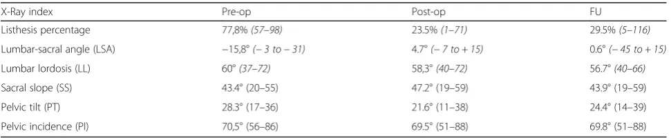
Table 5Mean pre-op, post-op and final FU values in patients treated with interbody support
X-Ray index Pre-op Post-op FU
Listhesis percentage 77,8%(57–98) 23.5%(1–71) 29.5%(5–116)
Lumbar-sacral angle (LSA) −15,8°(−3 to−31) 4.7°(−7 to + 15) 0.6°(−45 to + 15)
Lumbar lordosis (LL) 60°(37–72) 58,3°(40–72) 56.7°(40–66)
Sacral slope (SS) 43.4° (20–55) 47.2° (19–59) 43.9° (19–59)
Pelvic tilt (PT) 28.3° (17–36) 21.6° (11–38) 24.4° (14–39)
[image:4.595.56.542.632.732.2]patients with reduction, but did not correlate to the amount of reduction of spondylolisthesis”.
Conclusions
According to our data, spondylolisthesis reduction may not provide ideal pelvic alignment, in other words incom-plete spondylolisthesis reduction in an initially unbalanced pelvis may still provide unbalanced pelvis after surgery. Presenting a case series of continuous patients affected by high grade isthmic spondylolisthesis that did not have a total spondylolisthesis reduction in each and every case represents a realistic approach to this pathology, in con-trast to other recent studies reporting total listhesis reduc-tion in all patients correlated to ideal pelvic sagittal restoration in all patients. This study may therefore con-tribute to explore the vast amount of information that extends between a high grade listhesis and its perfect re-duction. Our study could be considered an initial attempt to correlate spinopelvic changes to spondylolisthesis re-duction in a progressive manner, and possibly in the fu-ture, generate threshold values of reduction for ideal spinopelvic alignment in every different patient.
Abbreviations
FU:Follow up; L: Listhesis (or slippage); LL: Lumbar lordosis; LSA: Lumbar-sacral angle; PI: Pelvic incidence; PLIF: Posterior lumbar interbody fusion; Post-op: Post-operatory; Pre-op: Pre-operatory; PT: Pelvic tilt; SS: Sacral slope; TLIF: Transforaminal lumbar interbody fusion
Acknowledgements Not applicable.
Authors’contributions
KM, TG, CF participated in the conception and design of the study. They contributed to analysis and interpretation of the data and drafting of the manuscript. They approved the final version of the manuscript to be submitted, and agreed to be accountable for all aspects of the work.
Funding
The authors state that this work has not received financial support.
Availability of data and materials
The datasets used and/or analyzed during the current study are available from the corresponding author on reasonable request.
Ethics approval and consent to participate
The Rizzoli Orthopaedic Institute Ethics Comitee was consulted prior to undertaking this study. Written informed consent for participating in this study was obtained from all patients and patient’s parents.
Consent for publication
This manuscript has the consent of the patient and patient’s parent for the use of her data. The authors obtained the written consent of the patient and patient’s parent for the publication of the data and images that appear in the article.
Competing interests
The authors declare that they have no competing interests.
Received: 9 January 2019 Accepted: 9 October 2019
References
1. Hresko MT, Labelle H, Roussouly P, Berthonnaud E. Classification of high grade spondylolisthesis based on pelvic version and spine balance: possible rationale for reduction. Spine (Phila Pa 1976). 2007;32(20):2208–13. 2. Labelle H, Roussouly P, Berthonnaud E, et al. Spondylolisthesis, pelvic
incidence and spinopelvic balance: a correlation study. Spine (Phila Pa 1976). 2004;29(18):2049–54.
3. Curylo L, Edwards C, DeWald RW. Radographic marlers in spondyloptosis: implications for spondylolisthesis progression. Spine (Phila Pa 1976). 2002;27: 2021–5.
4. Hanson DS, Bridwell KH, Rhee J, et al. Correlation of pelvic incidence with low and high grade isthmic spondylolisthesis. Spine. 2002;27:2026–9. 5. Mac-Thiong JM, Labelle H, Berthonnaud E, et al. Sagittal alignment of the
spine and pelvis during growth. Spine. 2004;29:1642–7.
6. Mac-Thing JM, Wang Z, de Guise JA, Labelle H. Postural model of sagittal spino-pelvic alignment and its relevance for lumbosacral developmental spondylolisthesis. Spine (Phila Pa 1976). 2008;33(21):2316–25.
7. Labelle H, Roussouly P, Chopin D, Berthonnaud E, Hresko T. Spino-pelvic alignment after surgical correction for developmental spondylolisthesis. Eur Spine J. 2008;17:1170–6.
8. Hresko MT, Hirschfeld R, Buerk AA, Zurakowski D. The effect of reduction and instrumentation of spondylolisthesis on spinopelvic sagittal alignment. J Pediatr Orthop. 2009 Mar;29(2):157–62.
Publisher’s Note
[image:5.595.56.548.100.196.2]Springer Nature remains neutral with regard to jurisdictional claims in published maps and institutional affiliations.
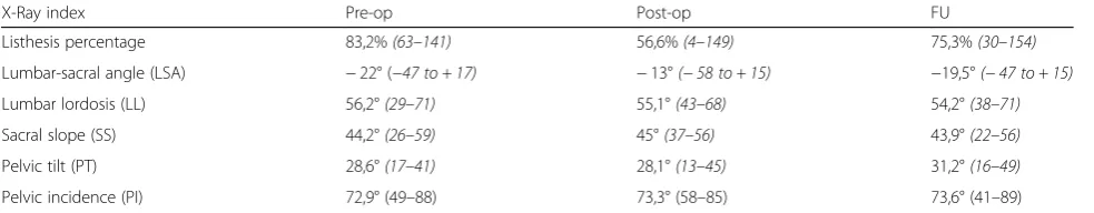
Table 6Mean pre-op, post-op and final FU values in patients treated without interbody support
X-Ray index Pre-op Post-op FU
Listhesis percentage 83,2%(63–141) 56,6%(4–149) 75,3%(30–154)
Lumbar-sacral angle (LSA) −22° (−47 to + 17) −13°(−58 to + 15) −19,5°(−47 to + 15)
Lumbar lordosis (LL) 56,2°(29–71) 55,1°(43–68) 54,2°(38–71)
Sacral slope (SS) 44,2°(26–59) 45°(37–56) 43,9°(22–56)
Pelvic tilt (PT) 28,6°(17–41) 28,1°(13–45) 31,2°(16–49)