0095-1137/95/$04.0010
Copyrightq1995, American Society for Microbiology
Molecular Identification of Bacteria by Fluorescence-Based
PCR–Single-Strand Conformation Polymorphism
Analysis of the 16S rRNA Gene
MYRA N. WIDJOJOATMODJO,1,2* AD C. FLUIT,1,2ANDJAN VERHOEF1
Eijkman Winkler Institute for Medical and Clinical Microbiology, University Hospital Utrecht,1 and U-gene Research B.V.,2Utrecht, The Netherlands
Received 30 January 1995/Returned for modification 7 April 1995/Accepted 6 July 1995
PCR–single-strand conformation polymorphism (PCR-SSCP) analysis is a rapid and convenient technique for the detection of mutations and allelic variants. We have adapted this technique for the identification of bacteria by PCR with fluorescein-labeled primers chosen from the conserved regions of the 16S rRNA gene flanking a variable region. The PCR product was denatured, separated on a nondenaturing gel, and detected by an automated DNA sequencer. The mobility of the single-stranded DNA is sequence dependent and allows the identification of a broad panel of bacteria. A single nucleotide difference in the amplified region was sufficient to obtain different PCR-SSCP patterns. The simultaneous amplification of multiple polymorphic regions by multiplex PCR with subsequent multiplex SSCP increased the discriminatory power of PCR-SSCP. A broad range of gram-negative and gram-positive bacteria were tested by PCR-SSCP, including, e.g.,
Esch-erichia coli, Enterobacter spp., Klebsiella spp., Haemophilus spp., Neisseria spp., Staphylococcus spp, Streptococcus
spp., Enterococcus spp., and Bacillus spp. In total, a panel of 178 strains of bacteria representing 51 species in 21 genera was examined. Although a limited number of strains from each species were tested, the strains tested gave species-specific patterns, with only one exception: Shigella species were indistinguishable from E. coli. PCR is a sensitive technique; as few as 10 CFU of E. coli was sufficient to produce PCR-SSCP patterns suitable for identification. The whole fluorescence PCR-SSCP procedure takes approximately 8 h for the detection and identification of low numbers of bacteria. Fluorescence PCR-SSCP seems to be a promising method for the differentiation of a broad range of pathogens found in usually sterile clinical sites, such as blood and cere-brospinal fluid.
The use of PCR as a diagnostic tool for the detection of pathogens has been expanding during the past few years. Most PCR detection systems described are based on the use of PCR with genus- or species-specific primers (20). These specific detection systems are not suitable for the detection of patho-gens in usually sterile clinical sites, such as blood and cere-brospinal fluid, since a broad spectrum of pathogens can be expected. A more universal method for the detection of micro-organisms is based on the use of broad-range conserved prim-ers for PCR (7, 23). Although this format is able to detect a pathogen in a usually sterile site, the identity of the microor-ganism remains unknown. Probe hybridization requires an ex-tensive panel of specific probes (12), and sequencing (3, 5) is not a suitable tool for routine diagnostics. Amplification of the 16S/23S rRNA gene spacer region by using conserved primers generated multiple PCR products with size distribution pro-files that might be used for the differentiation of bacterial species (16).
Recently, we have presented a new, general approach for directly determining the identities of bacteria that is based on the principle of single-strand conformation polymorphism (SSCP) electrophoresis (19) of PCR-amplified 16S rRNA gene fragments (22). PCR of bacterial cell lysates was performed with conserved primers flanking a variable region. The am-plified PCR product was denatured to two single-stranded DNAs (ssDNAs) and subjected to nondenaturing
polyacryl-amide gel electrophoresis. SSCP patterns were detected by silver staining the nucleic acids (1). The mobility of the ssDNA is sequence dependent and could be used to identify the un-known bacteria. The feasibility of the technique was demon-strated for a broad panel of gram-negative and gram-positive bacteria. We tested over 100 strains of bacteria representing 15 genera and 40 species and obtained species- or genus-specific patterns.
However, in conventional SSCP, the examination of patterns is visual and correction for lane-to-lane and gel-to-gel variation is not possible. Also, silver staining of nucleic acids has a poor reproducibility. PCR-SSCP with fluorescence-labeled primers and analysis on an automated DNA sequencer could over-come these disadvantages (11, 17). The separated strands are detected during electrophoresis as laser-excited fluorescence at the bottom of the gel, and differences in the mobilities of the ssDNAs are detected as differences in retention times. The use of an automated sequencer permits strict control of the gel temperature, which is an important variable when PCR-SSCP is performed (13). Furthermore, the sequencer is coupled to a computer, which enables direct data management by compu-ter.
In this report, we describe the use of fluorescence PCR-SSCP, using conserved 16S rRNA gene primers, for the detec-tion and identificadetec-tion of bacteria. We focused in this study on the identification of a broad range of bacterial pathogens, including bacterial species causing bacteremia. The general features of fluorescence PCR-SSCP are described, and the application of (multiplex) fluorescence PCR-SSCP for the dis-crimination of 178 bacterial strains is shown.
* Corresponding author. Mailing address: Eijkman Winkler Institute for Medical and Clinical Microbiology, University Hospital Utrecht, Room G04.614, P.O. Box 85500, 3508 GA Utrecht, The Netherlands.
2601
on May 15, 2020 by guest
http://jcm.asm.org/
MATERIALS AND METHODS
Bacterial strains.The bacterial strains used were either clinical isolates
col-lected from the University Hospital Utrecht (Utrecht, The Netherlands), clinical isolates from the National Institute for Public Health and Environmental Pro-tection (RIVM, Bilthoven, The Netherlands), or isolates obtained from the American Type Culture Collection. After observation of colony morphology and Gram staining, the clinical isolates were identified either by the automated Vitek system (bioMe´rieux Vitek Inc., Hazelwood, Mo.) or by the Enterotube II system (Roche Diagnostic Systems, Nutley, N.J.) in combination with the API Staph or API 20S system (bioMe´rieux Vitek Inc.). For both systems, supplementary well-established biochemical tests were performed (14).
Bacterial strains were cultured overnight at 378C on blood agar plates, scraped from the plates, and lysed in 10% Chelex 100 (Bio-Rad, Richmond, Calif.)– 0.03% sodium dodecyl sulfate–1% Tween 20–1% Nonidet P-40 for 5 min at 958C. After centrifugation for 10 s, 5ml of the supernatant was directly used for PCR amplification.
The number of CFU was determined by plating on blood agar and culturing for 16 h at 378C.
DNA amplification.The (multiplex) PCR mixture (50ml) contained 50 mM
Tris-HCl (pH 9.0), 50 mM KCl, 7 mM MgCl2, 2 mg of bovine serum albumin per
ml, 16 mM (NH4)2SO4, 100mM each deoxynucleoside triphosphate (dNTP), 0.4
mM each primer, and 0.1 U of Super-Taq polymerase (HT Biotechnology, Cam-bridge, England). The target DNA sequence was the 16S rRNA gene, and conserved sequences were selected for oligonucleotide primers (Table 1). The primers were either obtained commercially (Pharmacia, Roosendaal, The Neth-erlands) or synthesized on a Pharmacia Gene Assembler Plus (Pharmacia LKB, Upsala, Sweden) for primers labeled with a 59-fluorescein phosphoramidite, which was characterized by an extended spacer arm (Fluorescein-ON; Clonetech, Palo Alto, Calif.).
The PCR was performed for 25 cycles of 1 min at 948C, 10 s at 728C, and 1 min at 558C on a DNA thermocycler (Perkin-Elmer Cetus, Norwalk, Conn.). After amplification, 5ml of the amplified product was run on a 1.5% agarose gel in 0.53TBE. DNA bands were detected by ethidium bromide staining and visu-alized by UV light photography. Other thermostable DNA polymerases used were Tth (Boehringer, Mannheim, Germany) Ampli-Taq (Perkin-Elmer Cetus), Ampli-Taq Stoffel fragment (Perkin-Elmer Cetus), and Pfu (Stratagene, La Jolla, Calif.) with buffers as supplied by the manufacturers.
For determining the sensitivity of PCR-SSCP, Ampli-Taq Stoffel fragment was used in the buffer supplied by the manufacturer with the addition of 10 mM MgCl2. The whole PCR mixture, without the target DNA, was incubated with
DNase I (Boehringer) for 15 min at room temperature and then subjected to DNase I inactivation by an incubation of 10 min at 958C. Five microliters of cell lysate was then added to the PCR mixture, and PCR was subsequently performed for 35 cycles.
PCR product polishing.After PCR amplification, the mixture was precipitated
with ethanol, resuspended in distilled water, and incubated for 1 h in the appro-priate DNA polymerase buffer with 100mM dNTPs and 5 U of DNA polymerase. Both the Pfu DNA polymerase (Stratagene) and T4 DNA polymerase (Boehr-inger) enzymes possess 39-to-59proofreading exonuclease activity, according to the instructions of the manufacturers. Incubation was at 728C for Pfu DNA polymerase or at 378C for T4 DNA polymerase. The reaction was stopped by the addition of sequencing sample buffer (5 mM EDTA and 0.05% blue dextran in formamide), and the products were subsequently analyzed on an SSCP gel.
SSCP electrophoresis on an automated sequencer.SSCP electrophoresis was
performed on a Pharmacia A.L.F. DNA sequencer. After thermal cycling, 0.5ml of the PCR mixture was added to 10ml of sequencing sample buffer and heated for 3 min at 958C. The denatured DNA was then placed directly on ice for 5 min before being loaded onto the gel. The SSCP gel composition was 0.53MDE gel (J. T. Baker, Phillipsburg, N.J.), 10% glycerol, and 0.63Tris-borate-EDTA. Electrophoresis was done at 308C with 30 W for 360 min for a gel of 20 by 34 by
0.05 cm. Electrophoresis was evaluated with the Fragment Manager Software (Pharmacia LKB).
Manual SSCP electrophoresis.SSCP electrophoresis was performed at room
temperature with an overnight run on a Bio-Rad Protean II electrophoresis apparatus on a gel of 16 by 20 cm with 0.75-mm spacers at 5 W. The gel matrix and running buffer were the same as those for the automated sequencer. Elec-trophoresis and detection of the nucleic acids by silver staining were as previously described (22).
Internal control for automated SSCP electrophoresis.Two internal controls
were added to each SSCP sample lane, with the toxin B gene of Clostridium
difficile as the target sequence. PCR was performed with a toxigenic C. difficile
strain (ATCC 9689) with PCR conditions as described above. A multiplex PCR was performed with one fluorescein-labeled primer (59-GTC AGA GAA TAC TGT AGT CG, positions 508 to 527) (2) and two unlabeled primers, generating either a 150-bp fragment (fragment I) (primer 59-CTT TAG CTC TAA TAC TTC TG, positions 638 to 657) or a 257-bp fragment (fragment II) (primer 59-CTA TTT ACA TCT TTC CAT TG, positions 755 to 764). For each lane, 0.5 ml of the PCR product was added to the sequencing sample buffer as an internal control. The peak positions of the curves from the shorter ssDNA fragment I and larger fragment II were set arbitrarily at 100 and 200, respectively, in the Frag-ment Manager Software. All retention times were correlated to those for the internal standards.
RESULTS
Fluorescence PCR-SSCP.PCR amplification with universal
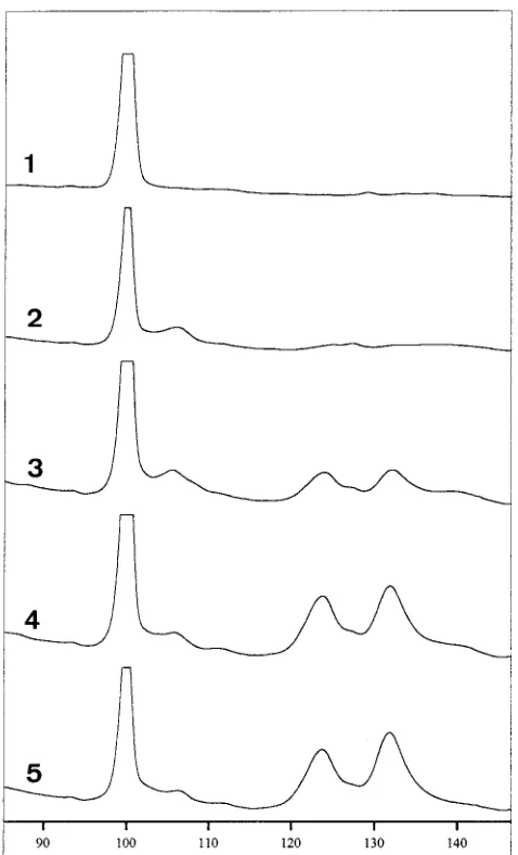
bacterial primers is hampered by low-level DNA contamina-tions in the PCR reagent mixture, especially in Taq DNA polymerase (21). Even the low-DNA Taq polymerase prepa-ration still contains low levels of DNA contaminants (18). A PCR of 25 cycles did not show contaminating DNA in the controls without added DNA by agarose gel electrophoresis or SSCP analysis. When a greater number of cycles was used, PCR components had to be treated to reduce the amounts of contaminating DNA. Therefore, we performed a short DNase I treatment for the whole PCR mixture, excluding the target DNA, followed by heat inactivation of the DNase (22). This protocol required a high magnesium concentration (7 to 10 mM) (4) and was feasible only in combination with Super-Taq and Ampli-Taq Stoffel fragment DNA polymerases. With this pre-PCR sterilization protocol included, the sensitivity of the fluorescein PCR-SSCP was investigated. Serial dilutions of Escherichia coli cells were made and subjected to a 35-cycle PCR with 59-fluorescein-labeled primers (P11P and P13P), and 5ml of PCR product was applied to an SSCP gel on an auto-mated sequencer. The resulting electropherogram (Fig. 1) is normalized by using the internal-standard fragments to correct for the lane-dependent mobility. The mobility profile resulted in two major bands representing the two ssDNAs amplified by PCR. As few as 10 CFU of E. coli resulted in SSCP patterns suitable for identification.
The mobilities of the ssDNAs on a manual SSCP electro-phoresis gel were altered when 59-fluorescein-labeled primers instead of nonlabeled primers, which were used in a previous study (22), were used for PCR amplification (data not shown). In addition, we studied the effect of the spacer length between the primer and the fluorescein label. FLU1 labeled primers carry the normal spacer arm, whereas FLU2 labeled primers carry an extended spacer arm. A small difference in retention times was observed for the two fluorescein-labeled primers P11P and P13P; e.g., the E. coli relative retention times for FLU1 primers were 135.1 and 122.0, while FLU2 primers showed retention times of 135.6 and 122.5 (results are the means of three independent experiments). Comparison of both types of fluorescein-labeled primers in PCR-SSCP on an au-tomated sequencer for a larger panel of bacteria showed sim-ilar resolutions for both types of primers. Further research was performed with the commercially available fluorescein primers with a normal spacer arm (FLU1).
[image:2.612.57.299.92.201.2]Automated PCR-SSCP enables strict temperature control of TABLE 1. Primers located in the 16S rRNA gene
used for PCR-SSCP
Oligonucleotide (sequence) Positiona Fragment size (bp)
P11P (59-GAG GAA GGT GGG GAT GAC GT) 1173–1192 217 P13P (59-AGG CCC GGG AAC GTA TTC AC) 1370–1389 ER10 (59-GGC GGA CGG GTG AGT AA) 103–119 255 ER11 (59-GAC TGC TGC CTC CCG TAG) or
ER11G (59-G GAC TGC TGC CTC CCG TAG)
341–357
ER14 (59-GCT AAC TCC GTG CCA GCA) 506–523 305 ER15 (59-GCG TGG ACT ACC AGG GTA TC) 791–810
a
Based on the E. coli sequence as a reference (GenBank accession no. JO1695).
on May 15, 2020 by guest
http://jcm.asm.org/
electrophoresis, an important variable in PCR-SSCP, and in each lane internal controls can be added to increase reproduc-ibility. To investigate the reproducibility of the automated SSCP, the day-to-day variation and lane-to-lane variation for E. coli SSCP patterns were investigated. When 20 different E. coli strains were tested on the same gel, automated PCR-SSCP proved to be quite accurate, with a lane-to-lane variation of less than 0.15%. The day-to-day variation for 5 consecutive days was less than 0.5%. The same numbers of bands were reproducibly observed. In some cases multiple SSCP bands from one ssDNA were consistently observed, reflecting the fact that an ssDNA possesses multiple conformations during mi-gration on a nondenaturing gel.
Effect of different thermostable DNA polymerases on SSCP
patterns. PCR amplification by different thermostable DNA
polymerases might influence SSCP patterns because of differ-ent enzymatic characteristics of the polymerases. Therefore, the PCR-SSCP patterns obtained for the Taq, Tth, and Pfu DNA polymerases with primer set P11P-P13P were investi-gated. Strikingly, a minor difference between Taq and Pfu
DNA polymerases was observed for both E. coli and Staphy-lococcus epidermidis cell lysates. One explanation might be the terminal deoxynucleotide transferase activity of Taq DNA polymerase, which is mostly limited to the extension of a single nucleotide, predominantly a single A nucleotide, whereas Pfu DNA polymerase possesses 39-to-59exonuclease proofreading activity. This hypothesis was tested by polishing the PCR frag-ments with Pfu and T4 DNA polymerase; both enzymes pos-sess 39-to-59exonuclease proofreading activity in the presence of dNTPs (10). Treatment of Taq DNA polymerase-amplified PCR products with either T4 DNA polymerase or Pfu DNA polymerase resulted in SSCP patterns similar to those obtained with Pfu DNA polymerase (Table 2). Thus, a single overhang-ing 39nucleotide has a significant effect on the retention times of the ssDNAs.
PCR-SSCP for bacterial identification. PCR products
ob-tained from 119 bacterial strains belonging to 27 species in 9 genera were tested with the primer set P11P-P13P. Twenty different patterns were observed; in most cases these were species-specific patterns, and in some cases they were genus-specific patterns. For example, species-genus-specific patterns were observed for Enterobacter and Klebsiella spp., and genus-spe-cific patterns were observed for Proteus spp. The closely re-lated E. coli, Shigella, and Salmonella species all gave identical PCR-SSCP patterns. PCR-SSCP with primer set P11P-P13P gave marginal differences in retention times for some species, which hampered the identification of species.
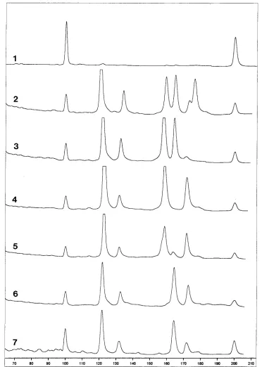
To increase the resolution of PCR-SSCP, an additional con-served primer set, ER10-ER11, was developed for use in a multiplex fluorescence PCR-SSCP. For example, the P11P-P13P amplification products with Citrobacter species showed a minor difference between the patterns of C. freundii and the conserved patterns of C. diversus and C. amalonaticus, whereas the second primer set, ER10-ER11, resulted in three distinct species-specific patterns (Fig. 2). Also, PCR-SSCP with these two primer sets enabled the distinct discrimination of all List-eria species (species-specific patterns for L. innocua, L. mono-cytogenes, L. ivanovii, L. murray, L. welshimeri, and L. seeligeri were observed), whereas the use of either primer set alone did not result in species-specific patterns. Fluorescence-based PCR-SSCP was capable of reproducibly detecting a 2-nucle-otide difference between L. innocua and L. ivanovii in the amplified region of P11P-P13P (9). For some species the flu-orescence signal of the ssDNA extended from the fluorescein-labeled ER11 primer was very weak. This reduction of signal was overcome by the insertion of an extra G base at the 59end of the primer, as described by Makino et al. (17). Also, this single addition of a base altered the retention times slightly because of the presence of an extra base.
When we first tested the applicability of primer set
[image:3.612.60.299.70.464.2]ER10-FIG. 1. Sensitivity of fluorescence PCR-SSCP with P11P-P13P. A serial di-lution of E. coli cells ranging from 0 to 100 CFU was tested with a 35-cycle PCR. Curves 1 to 5, 0, 3, 10, 30, and 100 CFU of E. coli cells, respectively. Retention times are correlated to those for the internal controls, which were assigned values of 100 and 200.
TABLE 2. Effect of post-PCR modification on SSCP patterns of E. coli with primer set P11P-P13P
DNA polymerase used in PCR
DNA polymerase used for post-PCR treatment
Relative retention timesa
Taq None 121.98, 135.66
Pfu 120.94, 133.12
T4 120.95, 133.52
Pfu None 120.63, 133.53
Pfu 120.80, 133.28
T4 120.82, 133.43
a
Retention times are correlated to those for the two internal controls, which were assigned values of 100 and 200. Results are means of two independent assays.
on May 15, 2020 by guest
http://jcm.asm.org/
ER11 with a broad panel of microorganisms, we also tested three strains of E. coli, four species of Shigella, and four strains of Salmonella. For these strains we obtained distinct genus-specific patterns. However, when we tested 30 E. coli strains, although there was a conserved pattern for the P11P-P13P region, polymorphism was observed within the ER10-ER11 region, yielding five different patterns. Remarkably, one of these patterns was similar to the genus-conserved pattern of Shigella spp. Polymorphism in the ER10-ER11 region was also
[image:4.612.121.493.70.592.2]observed for Proteus vulgaris. Thus, this region is less conserved for some species than the P11P-P13P region. This polymor-phism makes identification based on SSCP patterns more dif-ficult; therefore, we developed another primer set located in the 16S rRNA sequence, ER14-ER15, which proved to be more conserved than ER10-ER11. ER14-ER15 was capable of discriminating E. coli and Shigella species from Salmonella species and of distinguishing P. vulgaris from Proteus mirabilis while showing conserved patterns.
FIG. 2. Multiplex fluorescence PCR-SSCP for Citrobacter species with P11P-P13P and ER10-ER11. Curves: 1, internal control; 2, E. coli reference; 3, C.
amalonaticus; 4 and 5, C. freundii; 6 and 7, C. diversus.
on May 15, 2020 by guest
http://jcm.asm.org/
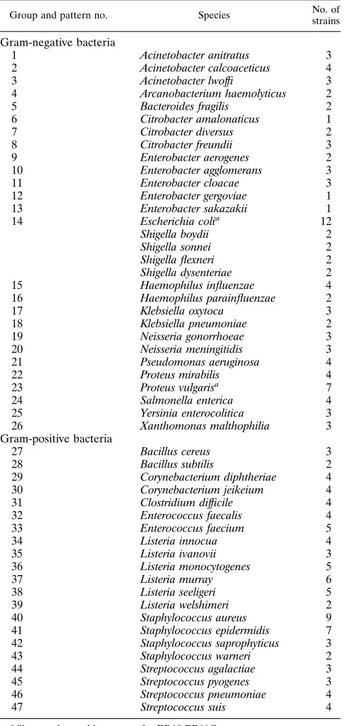
PCR-SSCP with a multiplex PCR based on three regions of the 16S rRNA was tested on 178 bacterial isolates (Table 3). This panel included a large number of gram-negative and gram-positive bacteria. The universal primers used for PCR were highly conserved, and amplification was observed for all species tested. Besides the species mentioned above, species-specific SSCP patterns were observed for the following gram-negative genera: Acinetobacter (A. anitratus, A. calcoaceticus, and A. lwoffi), Enterobacter (E. aerogenes, E. agglomerans, E.
cloacae, E. gergoviae, and E. sakazakii), Haemophilus (H. influ-enzae and H. parainfluinflu-enzae), Klebsiella (K. oxytoca and K. pneumoniae), and Neisseria (N. gonorrhoeae and N. meningiti-dis). Species-specific PCR-SSCP patterns were observed for the gram-positive genera Bacillus (B. cereus and B. subtilis), Corynebacterium (C. diphtheriae and C. jeikeium), Enterococcus (E. faecalis and E. faecium), Staphylococcus (S. aureus, S. epi-dermidis, S. saprophyticus, and S. welshimeri), and Streptococcus (S. agalactiae, S. pyogenes, S. pneumoniae, and S. suis). Ar-canobacterium haemolyticus, Bacteroides fragilis, Salmonella enterica, Pseudomonas aeruginosa, Xanthobacter malthophilia, Yersinia enterocolitica, and C. difficile each gave conserved pat-terns different from those observed for the other species. The 178 tested strains belonging to 51 species resulted in 47 differ-ent PCR-SSCP patterns, although a limited number of strains per species were tested.
DISCUSSION
The currently used molecular biological approach for the detection and identification of a broad spectrum of microor-ganisms involves the use of PCR with conserved 16S rRNA primers followed by sequencing or hybridization with a large panel of probes (5, 12). Recently, we have developed a method based on PCR-SSCP analysis for directly determining the iden-tity of a bacterium (19, 22). PCR was performed with con-served primers located in the 16S rRNA gene sequence flank-ing a variable region. The amplified products contained specific sequences which were utilized for identification by SSCP. The initial PCR-SSCP protocol (22) has been modified by labeling the PCR product with fluorescein-labeled primers instead of silver staining the nucleic acids in the SSCP gel. A major advantage of fluorescein PCR-SSCP over silver staining is that the system is coupled to a computer, which enables data management by computer. The fluorescein label changed the mobility of the ssDNA slightly, but the observed patterns of fluorescence PCR-SSCP are in accordance with the results obtained by manual PCR-SSCP with silver staining. The addi-tion of internal standards to each SSCP lane made the proce-dure highly reproducible, with an interassay variation of less than 0.15% and an intra-assay variation of less than 0.5%. This reproducibility is most likely sufficient for the discrimination of different species.
The conserved primers chosen for PCR amplification were highly conserved as shown by the amplification of DNAs from the panel of 178 bacterial strains from phylogenetically diver-gent bacteria. However, the use of these universal primers is hampered by low-level contamination of PCR reagents with DNA. For the detection of low numbers of pathogens, a pre-treatment of these reagents is necessary. We successfully per-formed a DNase treatment of the whole PCR mixture (omit-ting the target DNA) followed by heat inactivation of this enzyme. The PCR-SSCP sensitivity obtained with this pretreat-ment was the detection of as few as 10 CFU of E. coli.
[image:5.612.58.300.93.603.2]Manually performed SSCP detects ssDNA bands after elec-trophoresis, which makes the running time crucial for the dis-crimination of closely related species, such as L. innocua and L. ivanovii, which differ by only 2 nucleotides in the noncon-served region of P11P-P13P (9). The obnoncon-served differences were marginal and could not always be visualized. Automated fluo-rescence SSCP, which detects fluofluo-rescence at the bottom of the gel during electrophoresis, was able to detect these small dif-ferences reproducibly. Also, fluorescence PCR-SSCP with the same primer set could discriminate C. freundii from C. diversus and C. amalonaticus, whereas silver-stained PCR-SSCP prod-ucts showed a genus-conserved pattern (22) Automated SSCP TABLE 3. Multiplex fluorescence PCR-SCCP with primer sets
P11P-P13P, ER10-ER11G, and ER14-ER15
Group and pattern no. Species No. of
strains
Gram-negative bacteria
1 Acinetobacter anitratus 3
2 Acinetobacter calcoaceticus 4
3 Acinetobacter lwoffi 3
4 Arcanobacterium haemolyticus 2
5 Bacteroides fragilis 2
6 Citrobacter amalonaticus 1
7 Citrobacter diversus 2
8 Citrobacter freundii 3
9 Enterobacter aerogenes 2
10 Enterobacter agglomerans 3
11 Enterobacter cloacae 3
12 Enterobacter gergoviae 1
13 Enterobacter sakazakii 1
14 Escherichia colia 12
Shigella boydii 2
Shigella sonnei 2
Shigella flexneri 2
Shigella dysenteriae 2
15 Haemophilus influenzae 4
16 Haemophilus parainfluenzae 2
17 Klebsiella oxytoca 3
18 Klebsiella pneumoniae 2
19 Neisseria gonorrhoeae 3
20 Neisseria meningitidis 3
21 Pseudomonas aeruginosa 4
22 Proteus mirabilis 4
23 Proteus vulgarisa 7
24 Salmonella enterica 4
25 Yersinia enterocolitica 3
26 Xanthomonas malthophilia 3
Gram-positive bacteria
27 Bacillus cereus 3
28 Bacillus subtilis 2
29 Corynebacterium diphtheriae 4
30 Corynebacterium jeikeium 4
31 Clostridium difficile 4
32 Enterococcus faecalis 4
33 Enterococcus faecium 5
34 Listeria innocua 4
35 Listeria ivanovii 3
36 Listeria monocytogenes 5
37 Listeria murray 6
38 Listeria seeligeri 5
39 Listeria welshimeri 2
40 Staphylococcus aureus 9
41 Staphylococcus epidermidis 7
42 Staphylococcus saprophyticus 3
43 Staphylococcus warneri 2
44 Streptococcus agalactiae 3
45 Streptococcus pyogenes 3
46 Streptococcus pneumoniae 4
47 Streptococcus suis 4
aShows polymorphic patterns for ER10-ER11G.
on May 15, 2020 by guest
http://jcm.asm.org/
could detect a single nucleotide difference. The SSCP patterns of PCR fragments amplified by Taq DNA polymerase, which extends the amplified sequence by a single A nucleotide (8), could easily be distinguished from those of fragments amplified by Pfu DNA polymerase, which possesses 39-to-59 proofread-ing. One of the fluorescence-labeled primers yielded a low fluorescence signal with some species. The efficiency of the fluorescence emission might be diminished by the secondary structure of the ssDNA. The addition of a single G nucleotide at the 59end, as suggested by Makino et al. (17), increased the fluorescence signal. Also, the addition of this single nucleotide altered the retention times slightly.
Automated SSCP detects the ssDNA during electrophoresis and makes optimal use of the separation capacity of the SSCP gel, and it is therefore suitable for multiplex PCR-SSCP. Mul-tiplex PCR-SSCP (15) makes a further differentiation of spe-cies which show conserved SSCP patterns with a PCR with a single primer set. For example, the genus Proteus is conserved for P11P-P13P but showed species-specific patterns for ER14-ER15. Also, PCR-SSCP with P11P-P13P gave proximate re-tention times for some species, whereas rere-tention times with ER10-ER11 and ER14-ER15 were more distant. We were able to discriminate the 178 bacterial strains tested to the species level with a multiplex PCR-SSCP, with only one exception: the genus Shigella, which is conserved and showed patterns simi-lar to those of E. coli. These species are closely related and might even be considered to belong to the same genotype, as suggested by some authors (6). A broad range of bacterial pathogens was tested by PCR-SSCP, including those fre-quently encountered in bloodstream infections, such as E. coli, Enterobacter spp., Klebsiella spp., Haemophilus spp., Staphylo-coccus spp., StreptoStaphylo-coccus spp., EnteroStaphylo-coccus spp., and Bacillus spp.
The choice of conserved primers is crucial. First, they must be conserved for a broad range of microorganisms. Second, the conserved primers have to include a 100- to 400-bp region to obtain good PCR-SSCP resolution (13). Third, the flanking region has to contain a species- or genus-conserved region. Too much polymorphism within a certain species in the am-plified region will lead to different SSCP patterns for one species, which makes interpretation more difficult. In the fu-ture, direct comparisons of the patterns obtained with those present in a computer data bank, and thereby direct identifi-cation of the bacterium, will be possible.
The combined use of PCR and SSCP on an automated sequencer made the total time for PCR-SSCP only 8 h (2 h for PCR plus 6 h for SSCP), compared with 20 h for the manual PCR-SSCP with an overnight SSCP run and an additional silver staining of nucleic acids. Automated SSCP offered a substantial time gain, better sample reproducibility, and higher resolution than manual SSCP. Multiplex PCR and subsequent multiplex SSCP enabled the discrimination of species on the basis of multiple polymorphic sites, thereby increasing the dis-criminating power. Fluorescence PCR-SSCP is capable of identifying as few as 10 CFU of E. coli within 8 h and seems to be a promising method for the identification of pathogens in usually sterile clinical sites, such as blood and cerobrospinal fluid.
REFERENCES
1. Ainsworth, P. L., L. C. Surh, and M. B. Coulter-Mackie. 1991. Diagnostic single strand conformational polymorphism (SSCP): a simplified non-radio-isotopic method as applied to a Tay-Sachs B1 variant. Nucleic Acids Res.
19:405–406.
2. Barosso, L. A., S. Z. Wang, C. J. Phelps, J. L. Johnson, and T. D. Wilkins. 1990. Nucleotide sequence of Clostridium difficile toxin B gene. Nucleic Acids Res. 18:4004.
3. Barry, T., R. Powell, and F. Gannon. 1990. A general method to generate DNA probes for microorganisms. Bio/Technology 8:233–236.
4. Bickler, S. W., M. C. Heinrich, and G. C. Bagby. 1992. Magnesium-depen-dent thermostability of DNase I. BioTechniques 13:64–66.
5. Bo¨ttger, E. C.1989. Rapid determination of bacterial ribosomal RNA se-quences by direct sequencing of enzymatically amplified DNA. FEMS Mi-crobiol. Lett. 65:171–176.
6. Brenner, D. J. 1992. Introduction to the family Enterobacteriaceae, p. 2673– 2695. In A. Balows, H. G. Truper, M. Dwrokin, W. Harder, and K. H. Schleifer (ed.), The prokaryotes, 2nd ed., vol. III. Springer-Verlag, New York.
7. Chen, K., H. Neimark, P. Rumore, and C. Steinman. 1989. Broad range DNA probes for detecting and amplifying eubacterial nucleic acids. FEMS Microbiol. Lett. 57:19–24.
8. Clark, J. M. 1988. Novel non-templated nucleotide addition reactions cata-lyzed by procaryotic and eucaryotic DNA polymerases. Nucleic Acids Res.
16:9677–9686.
9. Collins, M. D., S. Wallbanks, D. J. Lane, J. Shah, R. Nietupski, J. Smida, M.
Dorsch, and E. Stackebrandt.1991. Phylogenetic analysis of the genus
List-eria based on reversed transcriptase sequencing of 16S rRNA. Int. J. Syst.
Bacteriol. 41:240–246.
10. Costo, G. L., and M. P. Weiner. 1994. Polishing with T4 or Pfu polymerase increases the efficiency of cloning of PCR fragments. Nucleic Acids Res.
22:2423.
11. Ellison, J., M. Dean, and D. Goldman. 1993. Efficacy of fluorescence-based PCR-SSCP for detection of point mutations. BioTechniques 15:684–691. 12. Greisen, K. M., M. Loeffelholz, A. Purohit, and D. Leong. 1994. PCR primers
and probes for the 16S rRNA gene of most species of pathogenic bacteria, including bacteria found in cerebrospinal fluid. J. Clin. Microbiol. 32:335– 351.
13. Hayashi, K. 1992. PCR-SSCP: a method for detection of mutations. Genet. Anal. Tech. Appl. 9:73–79.
14. Isenberg, H. D. (ed. in chief). 1992. Clinical microbiology procedures hand-book, American Society for Microbiology, Washington, D.C.
15. Iwahana, H., K. Yoshimoto, N. Mizusawa, E. Kudo, and M. Itakura. 1994. Multiple fluorescence-based PCR-SSCP analysis. BioTechniques
16:296–305.
16. Jensen, M. A., J. A. Webster, and N. Straus. 1993. Rapid identification of bacteria on the basis of polymerase chain reaction-amplified ribosomal DNA spacer polymorphisms. Appl. Environ. Microbiol. 59:945–952.
17. Makino, R., H. Yazyu, Y. Kishimoto, T. Sekiya, and K. Hayashi. 1992. F-SSCP: fluorescence-based polymerase chain reaction-single-strand confor-mation polymorphism (PCR-SSCP) analysis. PCR Methods Appl. 2:10–13. 18. Meier, A., D. H. Persing, M. Finken, and E. C. Bo¨ttger.1993. Elimination of contaminating DNA within polymerase chain reaction reagents: implications for a general approach to detection of uncultured pathogens. J. Clin. Mi-crobiol. 31:646–652.
19. Orita, M., Y. Susuki, T. Sekiya, and K. Hayashi. 1989. Rapid and sensitive detection of point mutations and DNA polymorphisms using the polymerase chain reaction. Genomics 5:874–879.
20. Pershing, D. H. 1993. In vitro nucleic acid amplification techniques, p. 51–87.
In D. H. Pershing, T. F. Smiths, F. C. Tenover, and T. J. White (ed.),
Diagnostic molecular microbiology: principles and applications. American Society for Microbiology, Washington, D.C.
21. Rand, K. H., and H. Houck. 1990. Taq polymerase contains bacterial DNA of unknown origin. Mol. Cell. Probes 4:445–450.
22. Widjojoatmodjo, M. N., A. C. Fluit, and J. Verhoef. 1994. Rapid identifica-tion of bacteria by PCR-single-strand conformaidentifica-tion polymorphism. J. Clin. Microbiol. 32:3002–3007.
23. Wilson, K. H., R. B. Blitchington, and R. C. Greene. 1990. Amplification of bacterial 16S ribosomal DNA with polymerase chain reaction. J. Clin. Mi-crobiol. 28:1942–1946.