Pertussis toxin inhibits neutrophil recruitment
to delay antibody-mediated clearance of
Bordetella pertussis
Girish S. Kirimanjeswara, … , Ottar N. Bjornstad, Eric T.
Harvill
J Clin Invest.
2005;
115(12)
:3594-3601.
https://doi.org/10.1172/JCI24609
.
Whooping cough is considered a childhood disease, although there is growing evidence
that children are infected by adult carriers. Additionally, increasing numbers of vaccinated
adults are being diagnosed with
Bordetella pertussis
disease. Thus it is critical to
understand how
B. pertussis
remains endemic even in highly vaccinated or immune
populations. Here we used the mouse model to examine the nature of sterilizing immunity to
B. pertussis
. Antibodies were necessary to control infection but did not rapidly clear
B.
pertussis
from the lungs. However, antibodies affected
B. pertussis
after a delay of at least a
week by a mechanism that involved neutrophils and Fc receptors, suggesting that
neutrophils phagocytose and clear antibody-opsonized bacteria via Fc receptors.
B.
pertussis
blocked migration of neutrophils and inhibited their recruitment to the lungs during
the first week of infection by a pertussis toxin–dependent (PTx-dependent) mechanism; a
PTx mutant of
B. pertussis
induced rapid neutrophil recruitment and was rapidly cleared
from the lungs by adoptively transferred antibodies. Depletion of neutrophils abrogated the
defects of the PTx mutant. Together these results indicate that PTx inhibits neutrophil
recruitment, which consequently allows
B. pertussis
to avoid rapid antibody-mediated
clearance and therefore successfully infect immune hosts.
Research Article
Infectious disease
Find the latest version:
Pertussis toxin inhibits neutrophil recruitment
to delay antibody-mediated clearance
of
Bordetella pertussis
Girish S. Kirimanjeswara,1 Luis M. Agosto,1 Mary J. Kennett,1 Ottar N. Bjornstad,2 and Eric T. Harvill1
1Department of Veterinary and Biomedical Sciences and 2Department of Entomology, The Pennsylvania State University, University Park, Pennsylvania, USA.
Whooping cough is considered a childhood disease, although there is growing evidence that children are
infect-ed by adult carriers. Additionally, increasing numbers of vaccinatinfect-ed adults are being diagnosinfect-ed with
Bordetella
pertussis
disease. Thus it is critical to understand how
B. pertussis
remains endemic even in highly vaccinated or
immune populations. Here we used the mouse model to examine the nature of sterilizing immunity to
B.
pertus-sis
. Antibodies were necessary to control infection but did not rapidly clear
B. pertussis
from the lungs. However,
antibodies affected
B. pertussis
after a delay of at least a week by a mechanism that involved neutrophils and
Fc receptors, suggesting that neutrophils phagocytose and clear antibody-opsonized bacteria via Fc receptors.
B. pertussis
blocked migration of neutrophils and inhibited their recruitment to the lungs during the first week
of infection by a pertussis toxin–dependent (PTx-dependent) mechanism; a PTx mutant of
B. pertussis
induced
rapid neutrophil recruitment and was rapidly cleared from the lungs by adoptively transferred antibodies.
Depletion of neutrophils abrogated the defects of the PTx mutant
.
Together these results indicate that PTx
inhibits neutrophil recruitment, which consequently allows
B. pertussis
to avoid rapid antibody-mediated
clear-ance and therefore successfully infect immune hosts.
Introduction
The widespread use of vaccines in developed nations has decreased the incidence of whooping cough (1–3). However, recent surveys reveal that a majority of individuals in a vaccinated population are transiently infected with the causative agent, Bordetella pertussis, and that it is widespread and endemic (4–8). In vaccinated populations, however, the bacterium induces a mild form of the disease that often goes undiagnosed (9–11). Although severe disease may be the greatest public health concern, undiagnosed pertussis poses an ongoing pervasive risk to very young (pre-vaccine), unvaccinated, and immune-compromised populations. In fact, childhood disease predates the age at which children extensively socialize with each otherand appears to commonly have as its source an adult, non- or mildly symptomatic carrier (10–14). The ability of B. pertussis to circulate in vaccinated and immune populations has been known clinically for years but has not been well studied experimentally. Although experimental infection of naive mice may simulate dis-ease, infection of vaccinated or convalescent animals with waning immunity may be more relevant to the biology of the bacterium in a vaccinated population.
Current pertussis vaccines induce a strong serum antibody response that has been shown to be critical for protection from the disease (15, 16). However, their efficacy against subclinical infection is doubtful, as the majority of vaccinated populations test positive for subsequent infection (10, 17), suggesting that the bacterium successfully infects immune and/or vaccinated individuals. Using animal models, we and others have previously shown that although
B cells are necessary for B. pertussis clearance from the respiratory tract (18–20), adoptively transferred serum antibodies have little or no effect on bacterial numbers for the first 7 days after inoculation (18–21) but begin to control and clear the bacteria thereafter. The ability to resist rapid antibody-mediated clearance may increase the duration and intensity of infection, both of which facilitate the transmission of the bacteria and would therefore be critical to the endemism of B. pertussis in vaccinated populations.
B. pertussis is thought to have emerged from a B. bronchiseptica– like progenitor (22, 23). These closely related subspecies share a similar set of virulence determinants but a different host range (24). Interestingly, while both require B cells for their clearance from the respiratory tract, only B. bronchiseptica is rapidly (within 3 days) cleared by adoptively transferred serum antibodies (20). We previously elucidated the mechanism of antibody-mediated clear-ance of B. bronchiseptica in order to determine the pathway that is presumably inhibited by B. pertussis (25, 26). Serum antibody– mediatedclearance of B. bronchiseptica requires a TLR4-induced early recruitment of neutrophils that phagocytose bacteria via Fcγ receptors (FcγRs) and CR3. We hypothesized that serum anti-body–mediated clearance of B. pertussis also requires neutrophils and that it may resist rapid serum antibody–mediated clearance by inhibiting neutrophil recruitment, presumably via a mecha-nism not shared by B. bronchiseptica.
Pertussis toxin (PTx), which is only expressed by B. pertussis, is an A-B type toxin known to inhibit G protein signaling pathways that involve Giα, interfering with a class of receptor that includes the majority of the chemokine receptors (27, 28). Various in vitro and in vivo studies have demonstrated its ability to inhibit the chemotaxis of neutrophils, lymphocytes, and macrophages (29–31). Although PTx has been proposed to be involved in the pathogen-esis of whooping cough (32), its exact role in vivo during B. pertussis
infection is not yet understood. Addition of inactivated PTx in the pertussis vaccine preparations has helped improve the vaccine’s
Nonstandard abbreviations used: FcγR, Fcγ receptor; MH-S, murine alveolar mac-rophage (cell line); PMN, polymorphonuclear neutrophil; PTx, pertussis toxin; RB6-8C5, anti–Ly-6 monoclonal antibodies.
Conflict of interest: The authors have declared that no conflict of interest exists.
research article
The Journal of Clinical Investigation http://www.jci.org Volume 115 Number 12 December 2005 3595
efficacy, suggesting that anti-PTx antibodies are important for protection against B. pertussis disease (15, 33). Furthermore, a sero-logical study indicated a positive correlation between anti-PTx antibody levels and protection from disease (34).
In the current analysis of the mechanism of antibody-mediated bacterial clearance, we observed that B. pertussis clearance was simi-lar to that of B. bronchiseptica:antibody-facilitated clearance of both bacteria required FcγRs and neutrophils. A significant difference, however, was observed in the kinetics of clearance of B. pertussis. The delayed elimination of bacteria correlated with the delayed recruitment of neutrophils to the lungs. Consequently, a PTx mutant of B. pertussis was rapidly cleared by adoptively transferred serum antibodies, suggesting that PTx inhibits early neutrophil recruitment and thereby contributes to the delayed antibody-mediated bacterial clearance. Expression of PTx may be an adapta-tion strategy of B. pertussis to cause an acute infection and extend its infectious period in immune hosts, facilitating its persistence in immune human populations.
Results
Serum antibodies do not rapidly clear B. pertussis.We and others have pre-viously shown that B cells are necessary for the clearance of B. pertus-sis from the lungs (18–20). Therefore we sought to investigate the role of serum antibodies in bacterial clearance. Groups of wild-type mice were inoculated with 5 × 10⁵ CFU of B. pertussis in 50 µl of PBS
[image:3.585.79.246.81.173.2]i.n., which has previously been shown to consistently deliver bacteria into the lungs. Immediately after inoculation, 200 µl of either naive or convalescent-phase serum were injected i.p. Mice were sacrificed on days 1, 3, 7, 10, and 14 after inoculation, and lungs were harvest-ed to enumerate the number of bacteria. Numbers of bacteria recov-ered from the lungs of either naive serum– or convalescent-phase serum–treated mice did not differ significantly on days 1, 3, and 7 after inoculation, suggesting that serum antibodies have minimal effect on bacterial clearance during the first 7 days after inoculation (Figure 1). However, convalescent-phase serum–treated mice har-bored only approximately 1,000 bacteria, while naive serum–treated mice harbored approximately 105 bacteria on day 10 after
inocula-tion (P < 0.001). Similarly, no bacteria could be detected in the lungs of convalescent-phase serum–treated mice on day 14 after inocu-lation, while approximately 105 CFU of B. pertussis were recovered
from the lungs of naive serum–treated mice (P < 0.001). These data suggest that serum antibodies were able to clear B. pertussis from the
lungs only after 1 week of infection. Apparently B. pertussis resists the effect of antibodies during the first week of infection, probably by inhibiting 1 or more antibody effector function.
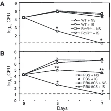
FcγRs, but not C3, are required for serum antibody–mediated clear-ance of B. pertussis.Antibodies may clear bacteria by neutralization, complement-mediated lysis, or opsonization for FcγR-mediated phagocytosis. We have previously shown that serum antibodies rapidly clear B. bronchiseptica from the lungs of mice and that the mechanism involved both complement and FcγRs (25, 26). Since
B. pertussis is very closely related to B. bronchiseptica but is cleared much more slowly by serum antibodies, we predicted that B. per-tussis has some mechanism to resist 1 or more of these antibody effector functions early in the infection. In order to test the impor-tance of complement in serum antibody–mediated bacterial clear-ance, we used mice lacking the central complement component C3, required for both classical and alternative complement cas-cades. Groups of wild-type C57BL/6 and congenic C3–/– mice were
inoculated with B. pertussis as described above. Immediately after inoculation, 200 µl of naive or B. pertussis–induced convalescent-phase serum was injected i.p. Fourteen days after inoculation, mice were sacrificed, and lungs were harvested to enumerate B. pertussis
CFU. Naive serum had no effect on bacterial numbers in the lungs of wild-type or C3–/– mice (Figure 2). Convalescent-phase serum
completely cleared the bacteria from the lungs of wild-type and C3–/– mice by day 14 (P < 0.001). These data indicate that C3 is not
required for serum antibody–mediated clearance of B. pertussis.
In order to test the importance of FcγRs in antibody-mediated clearance of B. pertussis, we used FcγR–/– mice, which lack all 3 Fc
receptors for IgG and 1 for IgE (35). Groups of FcγR–/– mice were
inoculated with B. pertussis andadoptively transferred naive serum or convalescent-phase serum as described above. As opposed to wild-type mice, in which convalescent-phase serum completely cleared bacteria from the lungs within 14 days, the same immune serum had no effect on bacterial numbers in the lungs of FcγR–/–
mice (P < 0.001; Figure 2). These data indicate that FcγRs are required for serum antibody–mediated clearance of B. pertussis. Together, these data showed that antibodies were not functioning by neutralization alone or via complement-mediated lysis. Anti-bodies appeared to clear B. pertussis from the lungs via phagocyto-sis by recruited FcγR-bearing cells.
Figure 1
Serum antibodies clear B. pertussis after 7 days of inoculation.
C57BL/6 mice were inoculated with 5 × 10⁵ CFU of B. pertussis and
adoptively transferred with 200 µl of naive serum (circles) or
convales-cent-phase serum (triangles). Lungs were harvested on the indicated days, and bacterial numbers were enumerated and expressed as
geo-metric mean ± SEM. Dotted line denotes limit of detection. n = 4 per
[image:3.585.330.516.544.654.2]group. *P < 0.001.
Figure 2
Adoptively transferred antibodies clear B. pertussis in the lungs of
wild-type and C3–/–, but not FcγR–/–, mice. C57BL/6 (WT), C3–/–, and
FcγR–/– mice were inoculated with 5 × 10⁵ CFU of B. pertussis and
adoptively transferred 200 µl of either naive serum (white bars) or
Neutrophils are required for antibody-mediated clearance of B. pertus-sis.We have previously shown that FcγR-bearing neutrophils are the primary inflammatory cells recruited to the lungs following infection with B. bronchiseptica and are required for rapid antibody-mediated bacterial clearance (26, 36). Since B. pertussis clearance required FcγRs, we sought to determine whether neutrophils are required for antibody-mediated clearance of this organism. Groups of C57BL/6 mice were injected i.p. with PBS or 1 mg anti– Ly-6 monoclonal antibodies (RB6-8C5). This treatment has been previously shown to deplete neutrophils for at least 2 weeks with no apparent effect on other cells such as macrophages or dendrit-ic cells (37, 38). Following treatment, mdendrit-ice were inoculated with
B. pertussis and adoptively transferred naive or immune serum as described above. The mice were sacrificed on day 14 after inocula-tion, and bacterial numbers in the lungs were enumerated (Figure 3). Naive serum had no effect on bacterial numbers in the lungs of mice treated with PBS or RB6-8C5. However, immune serum completely cleared bacteria from the lungs of mice treated with PBS, but not those treated with RB6-8C5 (P < 0.001), indicating that neutrophils are required for antibody-mediated bacterial clearance. The above-mentioned observations were verified using a recently isolated strain of B. pertussis, 6068 (a kind gift from Jeff Miller, UCLA, Los Angeles, California, USA).
PTx inhibits antibody-mediated clearance of B. pertussis.B. bronchi-septica and B. pertussis are very closely related and share a major-ity of the known virulence genes (22, 23). These 2 closely related subspecies both required similar antibody effector functions and phagocytic cells for their clearance from the lungs of mice (Figure 2 and ref. 26). Yet immune serum substantially reduces the num-bers of B. bronchiseptica in the lungs within 1 day (by more than 99%), whereas it has no effect on the numbers of B. pertussis for at least 7 days after inoculation (20). These data suggested that some
B. pertussis–specific virulence gene(s) delay antibody-mediated bacterial clearance. PTx is a B. pertussis–specific virulence factor that has been extensively studied in vitro and shown to inhibit the downstream effects of G protein–coupled receptors, includ-ing many chemokine receptors (27, 28). Furthermore, in vitro and in vivo experiments using purified PTx have shown that PTx can inhibit recruitment of cells such as neutrophils and lympho-cytes (29–31). One plausible explanation for the delayed
bacte-rial clearance may be that secreted PTx inhibits early neutrophil recruitment, which is necessary for antibody-mediated clearance. In support of this argument, we have previously observed that
B. bronchiseptica induces significantly more neutrophil infiltra-tion than B. pertussis on day 3 after inoculation (36). Therefore, we predicted that in the absence of PTx, B. pertussis would be more susceptible to antibody-mediated clearance. In order to test this hypothesis, we inoculated mice with 5 × 10⁵ CFU of B. pertussis or
the PTx mutant B. pertussis∆PTx as described aboveand adoptively transferred either naive or convalescent-phase serum. Lungs were harvested from these mice on days 3 and 7 after inoculation, and bacterial numbers in the lungs were enumerated. Convalescent-phase serum rapidly reduced numbers of B. pertussis∆PTx in the lungs as early as 3 days after inoculation (P < 0.05) and completely cleared this bacterium within 7 days (P < 0.05), but had no effect on the numbers of B. pertussis in this time frame (Figure 4). This was not due to the higher number of wild-type bacteria, since we have previously observed that adoptively transferred antibodies clear more than 106 bacteria when PTx is not present (G.S.
Kiri-manjeswara and E.T. Harvill, unpublished observations). These data suggest that PTx is involved in inhibiting antibody-mediated clearance of B. pertussis, perhaps by inhibiting neutrophil migra-tion to the lungs. Addimigra-tionally, analysis of the antibody-mediated clearance of B. pertussis in the trachea indicated a similar mecha-nism as that of the lungs was involved, and PTx inhibited rapid bacterial clearance in this organ as well. However, no significant effect of adoptively transferred serum antibodies was observed on the bacterial numbers in the nasal cavity on any tested days.
PTx reduces neutrophil recruitment to the lungs. B. bronchiseptica
induces the recruitment to the lungs of substantially higher numbers of neutrophils than does B. pertussis during the first week of infection (20, 26, 36). Since neutrophils are required for anti-body-mediated clearance of both B. bronchiseptica and B. pertussis, we predicted that B. pertussis has mechanisms to inhibit neutro-phil recruitment to the lungs in order to resist the effect of serum antibodies. Additionally, as previous studies have shown that PTx reduces the proportion of neutrophils recovered in the bron-cheoalveolar lavage fluid (39), we hypothesized that PTx inhibits the migration of neutrophils to the lungs, decreasing their
num-Figure 3
Depletion of neutrophils abrogates antibody-mediated clearance of
B. pertussis. C57BL/6 mice were treated with PBS or 1 mg of neu-trophil-depleting mAb RB6-8C5. Mice were subsequently inoculated
with 5 × 10⁵ CFU of B. pertussis and adoptively transferred 200 µl of
either naive (white bars) or convalescent-phase serum (black bars). On day 14 after inoculation, mice were sacrificed, and bacteria in the lungs were enumerated and expressed as geometric mean ± SEM.
[image:4.585.72.260.83.181.2]Dotted line denotes limit of detection. n = 4 per group. *P < 0.001.
Figure 4
Serum antibodies rapidly clear B. pertussis∆PTx but not B.
pertus-sis. C57BL/6 mice were inoculated with either B. pertussis (B.p) or
B. pertussis∆PTx (B.p∆PTx). Subsequently, these mice were
adop-tively transferred 200 µl of either naive serum (NS) or
convalescent-phase serum (IS). Mice were sacrificed on the indicated days, and bacterial numbers in the lungs were determined and expressed as
geometric mean ± SEM. Dotted line denotes limit of detection. n = 4
[image:4.585.306.532.540.645.2]research article
The Journal of Clinical Investigation http://www.jci.org Volume 115 Number 12 December 2005 3597
bers and thereby inhibiting antibody-mediated bacterial clear-ance. To test these hypotheses, groups of mice were inoculated with B. pertussis or B. pertussis∆PTx as described above, and leu-kocytes recruited to the lungs were enumerated. Approximately 5–6 × 10⁵ leukocytes were observed on day 1 after inoculation, and
7–8 × 10⁵ on days 3 and 7, in the lungs of mice inoculated with B. pertussis (Figure 5). On days 1, 3, and 7 after inoculation, the majority of leukocytes in the lungs of mice inoculated with B. per-tussis were macrophages (∼4 × 10⁵), a small percentage of cells were
lymphocytes (∼2 × 10⁵), and the rest were neutrophils (∼1 × 10⁵).
In contrast, the lungs of mice inoculated with B. pertussis∆PTx
harbored approximately 10–12 × 105 leukocytes on days 1, 3 and
7 after inoculation, and the majority of these cells were neutro-phils (5–6 × 10⁵). The absolute number of macrophages and
lym-phocytes recruited were similar between the 2 groups (P < 0.05). These data indicate that the higher proportion of neutrophils previously reported to be recruited by B. pertussis∆PTx (39) may be attributed to a large increase in absolute numbers of neutro-phils within the lungs. Thus, we concluded that PTx appears to inhibit neutrophil recruitment to the lungs during the first week of infection. As reported by other investigators (40), the number of leukocytes, particularly neutrophils, began to increase in the lungs of mice infected with B. pertussis on day 7 after inocula-tion, reaching a peak at day 10 after inoculation. On day 10, the lungs of mice inoculated with B. pertussis harbored approximately 10 × 105 leukocytes, of which 5–6 × 10⁵ cells were neutrophils and
2–3 × 10⁵ were lymphocytes, indicating that the effects of PTx are
ultimately overcome and neutrophils are recruited to the lungs later during infection. Interestingly, this delayed neutrophil recruitment correlates with the time frame in which antibodies begin to be effective against B. pertussis.
Antibody-mediated clearance of B. pertussis∆PTx requires FcγRs and neutrophils. The above data reveal a strong correlation between neu-trophil recruitment to the lungs and serum antibody–mediated bacterial clearance and suggest that neutrophils eliminate B. pertus-sis via FcγR-mediated opsonization and phagocytosis. However, it is possible that B. pertussis∆PTx may be cleared by serum antibodies by a mechanism different from that of wild-type B. pertussis.
There-fore, we sought to determine the mechanism of antibody-mediated clearance of B. pertussis∆PTx. Groups of C57BL/6 and FcγR–/– mice
were inoculated with B. pertussis∆PTx and adoptively transferred 200 µl of either naive or immune serum as described above. Naive serum had no significant effect on the bacterial numbers in the lungs of wild-type or FcγR–/– mice on either day 3 or day 7 (Figure
6A). In wild-type mice, immune serum rapidly reduced bacterial numbers in the lungs by day 3 after inoculation (P < 0.001) and completely cleared bacteria by day 7 (P < 0.001). However, no sig-nificant reduction in the numbers of bacteria was observed in the lungs of immune serum–treated FcγR–/– mice on days 3 and 7 after
inoculation, indicating that FcγRs are required for antibody-medi-ated clearance of B. pertussis∆PTx.
Based on these results, we tested the hypothesis that neu-trophils are involved in rapid antibody-mediated clearance of
B. pertussis∆PTx. Groups of C57BL/6 mice were inoculated with
B. pertussis∆PTx and adoptively transferred 200 µl of either naive or immune serum as described above. Half of the mice in each group were also given 1 mg RB6-8C5 injected i.p. to deplete neu-trophils, and the other half were given PBS as control. Among the mice given naive serum, RB6-8C5 treatment resulted in approxi-mately 10- to 100-fold higher numbers of B. pertussis∆PTx in the lungs compared with PBS treatment (Figure 6B). These numbers were indistinguishable from the numbers of wild-type B. pertussis
in the lungs at this time point (compare to Figure 4), indicating that PTx is not required for efficient colonization when neutro-phils are depleted. These data strongly suggest that PTx enables
B. pertussis to colonize the lung by inhibiting neutrophil recruit-ment, as proposed by Carbonetti et al. (39). Immune serum rapidly reduced the number of B. pertussis∆PTx in the lungs of mice treated with PBS on day 3 (P < 0.001) and completely cleared bacteria on day 7 after inoculation (P < 0.001). However, immune serum failed to reduce the number of bacteria on days 3 and 7 after
inocula-Figure 5
[image:5.585.48.278.82.231.2]PTx inhibits recruitment of neutrophils to the lungs. C57BL/6 mice were inoculated with 5 × 10⁵ CFU of B. pertussis (circles) or B. pertussis∆PTx (squares). Mice were sacrificed on the indicated days and their lungs perfused with 5 ml cold PBS, and total leukocytes per whole lung were enumerated. Individual cell types were determined by modified Giemsa staining of cells. n = 4 per group. *P < 0.001; **P < 0.05.
Figure 6
FcγRs and neutrophils are required for serum antibody–mediated
clearance of B. pertussis∆PTx. (A) C57BL/6 or FcγR–/– mice were
inoc-ulated with 5 × 10⁵ CFU of B. pertussis∆PTx and adoptively transferred
200 µl of either naive or convalescent-phase serum. (B) C57BL/6 mice
also received either PBS or 1 mg of mAb RB6-8C5 (n = 4 per group).
[image:5.585.323.511.463.650.2]tion in mice treated with RB6-8C5, indicating that neutrophils are required for rapid serum antibody–mediated clearance of
B. pertussis∆PTx. These data indicate that serum antibodies clear both B. pertussis and B. pertussis∆PTx by a similar mechanism involv-ing FcγR-dependent phagocytosis by neutrophils. These data were verified using an independently created PTx mutant of B. pertussis
(a kind gift from N. Carbonetti, University of Maryland, College Park, Maryland, USA).
PTx does not modulate chemokine and cytokine production by alveolar macrophages. PTx could affect neutrophil recruitment in various ways. It is known to influence chemokine and cytokine responses (41), interfere with G protein–coupled chemokine receptor sig-naling (42), and downregulate adhesion molecule expression on endothelial cells (43). To investigate whether PTx modulates che-mokine expression by alveolar macrophages, a confluent layer of murine alveolar macrophage (MH-S) cells was incubated with
B. pertussis or B. pertussis∆PTx at an MOI of 10 for 12 hours. The accumulated amounts of various chemokines, such as macrophage inflammatory protein-1α (MIP-1α), monocyte chemoattractant protein 1 (MCP-1), chemokine RANTES (RANTES), and chemo-kine KC (KC), were then analyzed by ELISA. Figure 7 shows that macrophages produced significant amounts of these chemokines when treated with 100 ng/ml of E. coli LPS as a positive control or infected with B. pertussis or B. pertussis∆PTx. However, no significant differences were observed between the number of chemokines pro-duced by macrophages incubated with these 2 strains. These data suggest that PTx does not inhibit neutrophil recruitment by block-ing chemokine production.
PTx inhibits neutrophil migration. In order to determine whether PTx acts directly on neutrophils or endothelial cells to inhibit neutrophil recruitment to the lungs, we used an in vitro neutro-phil migration assay to determine the number of neutroneutro-phils that migrate across endothelial cells toward chemokines produced by macrophages incubated with either B. pertussis or B. pertussis∆PTx
at an MOI of 10 for 12 hours. The average number of neutro-phils per field that migrated toward a media negative control was approximately 35 (Figure 8). More than 500 neutrophils per field were found to migrate across an endothelial cell barrier toward the supernatant from macrophages treated with E. coli LPS. The num-ber of neutrophils that migrated toward supernatant from macro-phages incubated with B. pertussis was slightly higher than the
nega-tive control at approximately 45 per field. However, the number of neutrophils that migrated toward supernatant from macrophages incubated with B. pertussis∆PTx was approximately 400 per field (P < 0.001). Since our previous data indicated that PTx does not modulate chemokine production by alveolar macrophages, we predicted that PTx in supernatants from macrophages incubated with B. pertussis may alter chemotaxis of neutrophils. In order to address this possibility, equal amounts of supernatants from cells incubated with B. pertussis and B. pertussis∆PTx were mixed and used for the in vitro neutrophil migration assay. The number of neutro-phils that migrated toward this mixture was approximately 50 per field, similar to the number of neutrophils that migrated toward supernatant from cells incubated with B. pertussis alone. These data suggest that culture supernatants from cells incubated with B. per-tussis have some components that inhibit chemotaxis of neutrophils toward supernatant from B. pertussis∆PTx–exposed macrophages.
Since PTx has been shown to affect G protein–coupled receptor signaling pathways that may influence chemotaxis, we hypoth-esized that the PTx present in the supernatant from macrophages incubated with B. pertussis may directly act on neutrophils to decrease their migration across endothelial cells. To test this, we mixed various amounts of purified PTx (obtained from N. Car-bonetti) with the supernatant from macrophages incubated with
B. pertussis∆PTx. Neutrophil migration toward supernatants from cells incubated with B. pertussis∆PTx was reduced by 90% when purified PTx was added (P < 0.001; Figure 8). However, adding the same amount of catalytically inactivated PTx (a kind gift of N. Carbonetti; described in refs. 39, 44–46) had no effect on the migration of neutrophils across endothelial cells. Mixing purified PTx with supernatant from macrophages treated with E. coli LPS also resulted in a significant reduction in neutrophil migration (P < 0.05). These data indicate that PTx inhibits the migration of neutrophils across endothelial cells and that this inhibition is dependent on its enzymatic activity.
[image:6.585.46.277.85.192.2]The inhibitory effect of PTx may be due to its action on endothelial cells or on neutrophils. In order to differentiate between
Figure 7
PTx does not modulate chemokine production by alveolar
macro-phages. MH-S cells were incubated with B. pertussis (black bars) or
B. pertussis∆PTx (dark gray bars) at MOI of 10 for 12 hours. Also shown are medium controls (white bars) and positive controls (100 ng/ml
[image:6.585.339.507.499.648.2]E. coli LPS, light gray bars). Culture supernatants were collected and analyzed for chemokines by ELISA and expressed as mean ± SEM.
Figure 8
PTx inhibits neutrophil migration. In 3-µM transwell chambers, 105
PMNs were allowed to migrate across endothelial cells toward
super-natant from alveolar macrophages (Mf) incubated with B. pertussis
research article
The Journal of Clinical Investigation http://www.jci.org Volume 115 Number 12 December 2005 3599
these 2 possibilities, endothelial cells growing on the upper cham-ber of the transwell system were preincubated with B. pertussis or
B. pertussis∆PTx. Subsequently, neutrophils were allowed to migrate across them toward supernatants from macrophages incubated with B. pertussis∆PTx. Preincubation of endothelial cells with either
B. pertussis or B. pertussis∆PTx did not alter neutrophil migration (data not shown). In fact, neutrophil migration assays carried out in the absence of endothelial cells showed that adding PTx to super-natant from macrophages incubated with B. pertussis∆PTx inhibited migration of neutrophils (data not shown). These data suggest that PTx acts directly on neutrophils and affects their chemotaxis across a membrane and their migration across endothelial cells.
Discussion
PTx has been studied extensively for its adjuvant properties (47) and inhibition of GTPase activities (48, 49). Although it has many effects on various cell types in vitro, its contributions to B. pertussis
infection and pathogenesis have not yet been determined. Recent-ly, Carbonetti et al. reported that PTx is required for efficient early colonization by B. pertussis in the lungs of mice (39). Our present data indicate that PTx facilitates B. pertussis colonization by inhib-iting early recruitment of neutrophils to the lungs. However, we also noted a much stronger effect of PTx on the ability of B. pertus-sis to resist antibody-mediated clearance that may be critical to the biology of this endemic human pathogen.
Based on its known in vitro activities, PTx could inhibit neutro-phil recruitment by modulating chemokine or cytokine produc-tion by epithelial cells and macrophages, directly interfering with the chemokine receptor signaling, and/or altering adhesion mol-ecule expression that may interfere with the diapedesis of blood leukocytes (30, 41, 45, 50, 51). In our study, both B. pertussis and
B. pertussis∆PTx induced production of similar levels of a range of chemokines by alveolar macrophages. However, only the superna-tant from macrophages incubated with B. pertussis∆PTx facilitated the migration of neutrophils across endothelial cells. Additionally, purified PTx, but not catalytically inactive PTx, was able to inhibit the migration of neutrophils in this assay. Together, these data indicate that PTx inhibits polymorphonuclear neutrophil (PMN) recruitment by acting directly on chemokine receptor signaling of neutrophils. However, other effects of PTx may also contribute to the inhibition of neutrophil recruitment in vivo. The migration of neutrophils across airway epithelium requires PTx-sensitive sig-nals (52), but the nature of these sigsig-nals is difficult to delineate during an active infection with B. pertussis, not only due to the mul-tiple possible inhibitory effects of PTx, but also due to the variety of pro- and antiinflammatory factors induced and released by the bacteria. Interestingly, the number of neutrophils recruited to the lungs increased after day 7 after inoculation in spite of the pres-ence of PTx. This increase in the number of neutrophils correlates with the kinetics of antibody-mediated bacterial clearance. We have observed that a T cell–dependent response was required to overcome the effect of PTx and allow antibody-mediated bacterial clearance during the second week of infection (data not shown). The implication of an inhibitory effect of PTx on neutrophil recruitment is 2-fold: it allows B. pertussis to evade innate immunity early in colonization and also provides a means to resist the effect of serum antibodies in the lungs. Although the former effect may be important to primary infection in naive, unvaccinated hosts, the latter may be more relevant to the persistence of B. pertussis as an endemic pathogen even within vaccinated populations.
Acute, highly contagious, immunizing pathogens face the sig-nificant epidemiological challenge of long-term persistence within the host population. Immunity results in depletion of susceptible hosts through the course of each epidemic; thereafter host replen-ishment requires births of susceptible individuals or loss of immu-nity— which is why pathogens that convey strong, long-lasting immunity result in “childhood diseases” (53). Rapid contagion, in turn, results in fast transmission among hosts, which is a short-term evolutionary benefit to the pathogen (32, 54). However, it also results in large-amplitude epidemics with intervening deep epidemic troughs. In small and medium host populations, the chain of transmission will be broken in the troughs so that the pathogen will go extinct. The most relevant theoretical models for childhood infections, the so-called realistic age-structured models (55, 56), predict an endemic threshold of around half a million hosts in order for transmission to be sustained through the epi-demic troughs of acute, immunizing infections. This prediction is closely matched by epidemiological surveillance data (57, 58). Pre-vious theoretical studies have highlighted 2 key adaptations that increase the height of the epidemic troughs to allow long-term endemism within smaller host communities: (a) reinfection of pre-viously immunized hosts and adult carriers and (b) prolongation of the infectious period (7). Our study is of wide epidemiological significance in showing that B. pertussis, through expression of PTx, slows migration of neutrophils and thereby extends the infection period (relative to B. pertussis∆PTx strains) and allows for transient reinfection of previously immunized hosts. PTx expression may, therefore, be a key adaptation by B. pertussis for interacting with the unique population dynamics of its human host.
Susceptibility of humans to B. pertussis infection, particularly among people with no history of disease, has been shown to cor-relate with low levels of anti-PTx antibodies (59). Other studies have recognized that PTx is one of the critical components nec-essary for higher efficacy of acellular pertussis vaccines (34, 60). Although antibody titer to pertactin positively correlates with the resistance to diagnosed disease symptoms, addition of pertactin or filamentous hemagglutinin to PTx increases the efficacy of acel-lular pertussis vaccines (15, 33). Interestingly, antibody titers to PTx decrease much faster than those to other B. pertussis antigens (61). Since PTx allows B. pertussis to largely avoid the effects of anti-bodies to other antigens, the most effective vaccination strategy to
B. pertussis may involve an increased focus on the induction of a long-lasting serum antibody response to PTx. Use of genetically inactivated mutant PTx in the current acellular vaccines, as pro-posed by Robbins et al. (62), may be a substantial improvement in achieving higher level of anti-PTx antibodies that reduce the rate of infection and severity of disease.
Methods
Glu 129 to Gly. This mutant PTx has been shown to be immunogenic but not toxigenic.
Inoculation and adoptive transfer protocols. C57BL/6 mice were obtained from Jackson ImmunoResearch Laboratories, and C3–/– mice backcrossed
extensively onto a C57BL/6 background have been described previously (64) and were kind gifts of R. Wetsel (University of Texas — Houston, Houston, Texas, USA). FcγR–/– (C57BL/6 background) were obtained from Taconic
and have been described previously (35). Mice were lightly sedated with isoflurane (IsoFlo; Abbott Laboratories), and 5 × 10⁵ CFU of bacteria in 50
µl of PBS were inoculated onto the tips of the external nares. Colonization levels were determined by homogenizing the lungs in 1 × PBS and plat-ing aliquots for colony counts. The homogenates and necessary dilutions were plated in 50-µl volumes onto BG agar with streptomycin. Colonies were counted after 3 days’ incubation at 37°C. For adoptive transfer experi-ments, mice were inoculated with B. pertussis or B. pertussis∆PTx as described above, immediately followed by i.p. injection of 200 µl convalescent-phase serum obtained from BP536-inoculated mice on day 28 after inoculation. Various batches of convalescent-phase sera were tested for anti–B. pertus-sis antibody levels and used if they matched the titers described by Kiri-manjeswara et al. (20). Animals were sacrificed on the indicated day after transfer (Figure Legends 1–6), and bacterial numbers were determined as described above. Animals were handled in accordance with institutional guidelines of The Pennsylvania State University. All animal experiments were approved by the Institutional Animal Care and Use Committee of The Pennsylvania State University.Statistical significance of data points was determined using a 2-tailed Student’s unpaired t test.
Enumeration of leukocytes in the lungs. Total leukocytes were isolated from the lungs after collagenase type I and DNAse I digestion as described previously (65). Briefly, lungs were perfused with PBS and finely sheared with scissors. This lung homogenate was subjected to collagenase type I and DNAse I treatment for about 3 hours. The enzymatically treated homogenate was laid over Histopaque 1119 (Sigma-Aldrich) and centrifuged for 30 minutes at 1,200 g. The leukocyte portion was collected, and the total number of cells was determined by hemocytometer. Individual cell types were deter-mined by staining the isolated cells with modified Giemsa stain by a certi-fied clinical laboratory technician.
Estimation of cytokines and chemokines. MH-S cells (66) were obtained from ATCC and cultured in DMEM medium supplemented with 10% FBS. Cells were grown to confluency and incubated with either unwashed B. pertussis
or B. pertussis∆PTx at MOI of 10 for 12 hours at 37°C. Culture supernatants were collected, filter sterilized, and stored at –80°C for further use. Con-centrations of KC, RANTES, MIP-1α, and MCP-1 were estimated by ELISA.
The percentage of cell death was measured by lactate dehydrogenaseassay and was below 15% at the tested MOI for both strains of bacteria.
Neutrophil migration assay. Primary murine aortic endothelial cells (105 cells;
a kind gift from L. Sordillo, Michigan State University, East Lansing, Michi-gan, USA) were cultured as a confluent monolayer on the upper chamber of 3-µM transwells with DMEM supplemented with 10% FCS for 24 hours. Peripheral blood PMNs were collected from C57BL/6 mice by differential density separation using Histopaque 1119 and 1077 (67) and resuspended in DMEM supplemented with 10% FCS. The percentage of PMNs in the cell suspension was estimated by Giemsa staining of isolated cells and was found to be approximately 90%. Approximately 105 PMNs in a total volume of 200
µl of DMEM were layered on the endothelial cells of the upper chamber of the transwell system. Two hundred microliters of supernatant from macro-phages treated with 100 ng/ml of E. coli LPS or cultured with B. pertussis or
B. pertussis∆PTx for 12 hours at MOI of 1:10 was used as a source of chemoat-tractants in the lower chamber of the transwell system. After 12 hours, the number of neutrophils that migrated to the lower chamber was measured by observing 10 random fields of the lower chamber under a light microscope.
Neutrophil depletion. RB6-8C5 is a mAb raised against Ly-6 present on neu-trophils and was a kind gift from G. Huffnagle (University of Michigan Medical School, Ann Arbor, Michigan, USA) (68). Previously, 1 mg of this mAb injected i.p. has been shown to deplete neutrophils for 7–14 days (37, 38). Peripheral PMNs were enumerated to determine the efficacy of the treatment and was below 2%.
Acknowledgments
We thank all the members of E.T. Harvill’s lab, Avery August, Andrew Henderson, and the members of the Center for Infectious Disease Dynamics, The Pennsylvania State University, for critical reading of this manuscript and discussion of this study. We thank Nicholas Carbonetti (University of Maryland), Jeff Miller (UCLA), and Drusilla Burns (FDA) for providing various strains of B. per-tussis and purified PTx. This work was supported by NIH grant 5-R01-A1053075-02.
Received for publication January 31, 2005, and accepted in revised form September 20, 2005.
Address correspondence to: Eric T. Harvill, 115 Henning Build-ing, The Pennsylvania State University, University Park, Pennsyl-vania 16802, USA. Phone: (814) 863-8522; Fax: (814) 863-6140; E-mail: eth10@psu.edu.
1. Cherry, J.D. 1984. The epidemiology of pertussis and pertussis immunization in the United King-dom and the United States: a comparative study.
Curr. Probl. Pediatr.14:1–78.
2. Cherry, J.D., Brunell, P.A., Golden, G.S., and Kar-son, D.T. 1988. Report of the task force on pertus-sis and pertuspertus-sis immunization-1988. Pediatrics.
81(Suppl.):939–984.
3. Cherry, J.D., and Heininger, U. 2004. Pertussis and other Bordetella infections. In Textbook of pediat-ric infectious diseases. W.B. Saunders. Philadelphia, Pennsylvania, USA. 1588–1608.
4. Gilberg, S., et al. 2002. Evidence of Bordetella per-tussis infection in adults presenting with persistent cough in a French area with very high whole-cell vaccine coverage. J. Infect. Dis.186:415–418. 5. Hodder, S.L., et al. 2000. Antibody responses to
Bordetella pertussis antigens and clinical correla-tions in elderly community residents. Clin. Infect. Dis.31:7–14.
6. Nennig, M.E., Shinefield, H.R., Edwards, K.M., Black, S.B., and Fireman, B.H. 1996. Prevalence and
incidence of adult pertussis in an urban popula-tion. JAMA.275:1672–1674.
7. Schmitt-Grohe, S., et al. 1995. Pertussis in German adults. Clin. Infect. Dis.21:860–866.
8. Wright, S.W., Edwards, K.M., Decker, M.D., and Zeldin, M.H. 1995. Pertussis infection in adults with persistent cough. JAMA.273:1044–1046. 9. Deville, J.G., et al. 1995. Frequency of unrecognized
Bordetella pertussis infections in adults. Clin. Infect. Dis.21:639–642.
10. Deen, J.L., et al. 1995. Household contact study of Bordetella pertussis infections. Clin. Infect. Dis.
21:1211–1219.
11. Cherry, J.D., et al. 1995. Comparison of values of antibody to Bordetella pertussis antigens in young German and American men. Clin. Infect. Dis.
20:1271–1274.
12. Crowcroft, N.S., et al. 2003. Severe and unrec-ognised: pertussis in UK infants. Arch. Dis. Child.
88:802–806.
13. Baron, S., et al. 1998. Epidemiology of pertussis in French hospitals in 1993 and 1994: thirty years
after a routine use of vaccination. Pediatr. Infect. Dis. J.17:412–418.
14. Bisgard, K.M., et al. 2004. Infant pertussis: who was the source? Pediatr. Infect. Dis. J.23:985–989. 15. Cherry, J.D., Gornbein, J., Heininger, U., and Stehr,
K. 1998. A search for serologic correlates of immu-nity to Bordetella pertussis cough illnesses. Vaccine.
16:1901–1906.
16. Storsaeter, J., Hallander, H.O., Gustafsson, L., and Olin, P. 1998. Levels of anti-pertussis antibodies related to protection after household exposure to Bordetella pertussis. Vaccine.16:1907–1916. 17. Long, S.S., Welkon, C.J., and Clark, J.L. 1990.
Wide-spread silent transmission of pertussis in families: antibody correlates of infection and symptomatol-ogy. J. Infect. Dis.161:480–486.
18. Leef, M., Elkins, K.L., Barbic, J., and Shahin, R.D. 2000. Protective immunity to Bordetella pertus-sis requires both B cells and CD4(+) T cells for key functions other than specific antibody production.
J. Exp. Med.191:1841–1852.
research article
The Journal of Clinical Investigation http://www.jci.org Volume 115 Number 12 December 2005 3601
and Mills, K.H. 1997. Atypical disease after Borde-tella pertussis respiratory infection of mice with targeted disruptions of interferon-gamma receptor or immunoglobulin mu chain genes. J. Exp. Med.
186:1843–1851.
20. Kirimanjeswara, G.S., Mann, P.B., and Harvill, E.T. 2003. Role of antibodies in immunity to Bordetella infections. Infect. Immun.71:1719–1724. 21. King, A.J., et al. 2001. Role of the polymorphic
region 1 of the Bordetella pertussis protein pertac-tin in immunity. Microbiology.147:2885–2895. 22. Musser, J.M., Hewlett, E.L., Peppler, M.S., and
Selander, R.K. 1986. Genetic diversity and relation-ships in populations of Bordetella spp. J. Bacteriol.
166:230–237.
23. van der Zee, A., Mooi, F., Van Embden, J., and Musser, J. 1997. Molecular evolution and host adaptation of Bordetella spp.: phylogenetic analy-sis using multilocus enzyme electrophoreanaly-sis and typing with three insertion sequences. J. Bacteriol.
179:6609–6617.
24. Parkhill, J., et al. 2003. Comparative analysis of the genome sequences of Bordetella pertussis, Borde-tella parapertussis and BordeBorde-tella bronchiseptica.
Nat. Genet.35:32–40.
25. Pishko, E.J., et al. 2004. Antibody-mediated bacte-rial clearance from the lower respiratory tract of mice requires complement component C3. Eur. J. Immunol.34:184–193.
26. Kirimanjeswara, G.S., et al. 2005. The complex mech-anism of antibody-mediated clearance of Bordetella from the lungs requires TLR4. J. Immunol. In press. 27. Moss, J., et al. 1983. Activation by thiol of the latent
NAD glycohydrolase and ADP-ribosyltransferase activities of Bordetella pertussis toxin (islet-acti-vating protein). J. Biol. Chem.258:11879–11882. 28. Murayama, T., Katada, T., and Ui, M. 1983.
Gua-nine nucleotide activation and inhibition of adenylate cyclase as modified by islet-activating protein, pertussis toxin, in mouse 3T3 fibroblasts.
Arch. Biochem. Biophys.221:381–390.
29. Spangrude, G.J., Sacchi, F., Hill, H.R., Van Epps, D.E., and Daynes, R.A. 1985. Inhibition of lympho-cyte and neutrophil chemotaxis by pertussis toxin.
J. Immunol.135:4135–4143.
30. Brito, G.A., et al. 1997. Role of pertussis toxin A subunit in neutrophil migration and vascular per-meability. Infect. Immun.65:1114–1118.
31. Meade, B.D., Kind, P.D., and Manclark, C.R. 1984. Lymphocytosis-promoting factor of Bordetella pertussis alters mononuclear phagocyte circula-tion and response to inflammacircula-tion. Infect. Immun.
46:733–739.
32. Hewlett, E.L. 1999. A commentary on the patho-genesis of pertussis. Clin. Infect. Dis.28(Suppl. 2): S94–S98.
33. Cherry, J.D. 1997. Comparative efficacy of acellular pertussis vaccines: an analysis of recent trials. Pedi-atr. Infect. Dis. J.16(4 Suppl.):S90–S96.
34. Taranger, J., et al. 2001. Mass vaccination of chil-dren with pertussis toxoid--decreased incidence in both vaccinated and nonvaccinated persons. Clin. Infect. Dis.33:1004–1010.
35. Ravetch, J.V. 1997. Fc receptors. Curr. Opin. Immu-nol.9:121–125.
36. Harvill, E.T., Cotter, P.A., and Miller, J.F. 1999. Pregenomic comparative analysis between borde-tella bronchiseptica RB50 and Bordeborde-tella
pertus-sis tohama I in murine models of respiratory tract infection. Infect. Immun.67:6109–6118.
37. Conlan, J.W. 1997. Critical roles of neutrophils in host defense against experimental systemic infec-tions of mice by Listeria monocytogenes, Salmo-nella typhimurium, and Yersinia enterocolitica.
Infect. Immun.65:630–635.
38. Seiler, P., et al. 2000. Rapid neutrophil response controls fast-replicating intracellular bacteria but not slow-replicating Mycobacterium tuberculosis.
J. Infect. Dis.181:671–680.
39. Carbonetti, N.H., Artamonova, G.V., Mays, R.M., and Worthington, Z.E. 2003. Pertussis toxin plays an early role in respiratory tract colonization by Bordetella pertussis. Infect. Immun.71:6358–6366. 40. McGuirk, P., and Mills, K.H. 2000. A regulatory
role for interleukin 4 in differential inflammatory responses in the lung following infection of mice primed with Th1- or Th2-inducing pertussis vac-cines. Infect. Immun.68:1383–1390.
41. Belcher, C.E., et al. 2000. The transcriptional responses of respiratory epithelial cells to Borde-tella pertussis reveal host defensive and pathogen counter-defensive strategies. Proc. Natl. Acad. Sci. U. S. A.97:13847–13852.
42. Agarwal, R.K., Sun, S.H., Su, S.B., Chan, C.C., and Caspi, R.R. 2002. Pertussis toxin alters the innate and the adaptive immune responses in a pertussis-dependent model of autoimmunity. J. Neuroimmu-nol.129:133–140.
43. Shen, W., Bendall, L.J., Gottlieb, D.J., and Bradstock, K.F. 2001. The chemokine receptor CXCR4 enhanc-es integrin-mediated in vitro adhenhanc-esion and facili-tates engraftment of leukemic precursor-B cells in the bone marrow. Exp. Hematol.29:1439–1447. 44. Pizza, M., et al. 1989. Mutants of pertussis toxin
suit-able for vaccine development. Science.246:497–500. 45. Carbonetti, N.H., et al. 2004. Suppression of serum
antibody responses by pertussis toxin after respira-tory tract colonization by Bordetella pertussis and identification of an immunodominant lipoprotein.
Infect. Immun.72:3350–3358.
46. Nencioni, L., et al. 1990. Characterization of geneti-cally inactivated pertussis toxin mutants: candi-dates for a new vaccine against whooping cough.
Infect. Immun.58:1308–1315.
47. Ausiello, C.M., et al. 2002. Native and genetically inactivated pertussis toxins induce human den-dritic cell maturation and synergize with lipopoly-saccharide in promoting T helper type 1 responses.
J. Infect. Dis.186:351–360.
48. Bokoch, G.M., and Gilman, A.G. 1984. Inhibition of receptor-mediated release of arachidonic acid by pertussis toxin. Cell.39:301–308.
49. Katada, T., Tamura, M., and Ui, M. 1983. The A protomer of islet-activating protein, pertussis toxin, as an active peptide catalyzing ADP-ribosyl-ation of a membrane protein. Arch. Biochem. Biophys.
224:290–298.
50. Boldrick, J.C., et al. 2002. Stereotyped and spe-cific gene expression programs in human innate immune responses to bacteria. Proc. Natl. Acad. Sci. U. S. A.99:972–977.
51. Smith, M.L., Olson, T.S., and Ley, K. 2004. CXCR2- and E-selectin-induced neutrophil arrest during inflammation in vivo. J. Exp. Med.200:935–939. 52. Miller, L.A., Usachenko, J., McDonald, R.J., and
Hyde, D.M. 2000. Trafficking of neutrophils
across airway epithelium is dependent upon both thioredoxin- and pertussis toxin-sensitive signaling mechanisms. J. Leukoc. Biol.68:201–208. 53. Lawrence, G.L., MacIntyre, C.R., Hull, B.P., and
McIntyre, P.B. 2003. Measles vaccination cover-age among five-year-old children: implications for disease elimination in Australia. Aust. N. Z. J. Public Health.27:413–418.
54. Glass, K., and Grenfell, B.T. 2004. Waning immu-nity and subclinical measles infections in England.
Vaccine.22:4110–4116.
55. Bolker, B.M., and Grenfell, B.T. 1993. Chaos and biological complexity in measles dynamics. Proc. Biol. Sci.251:75–81.
56. Keeling, M.J., and Grenfell, B.T. 1997. Disease extinction and community size: modeling the per-sistence of measles. Science.275:65–67.
57. Bjornstad, O.N., Peltonen, M., Liebhold, A.M., and Baltensweiler, W. 2002. Waves of larch bud-moth outbreaks in the European alps. Science.
298:1020–1023.
58. Finkenstadt, B.F., Bjornstad, O.N., and Grenfell, B.T. 2002. A stochastic model for extinction and recurrence of epidemics: estimation and inference for measles outbreaks. Biostatistics.3:493–510. 59. Storsaeter, J., Hallander, H.O., Gustafsson, L., and
Olin, P. 2003. Low levels of antipertussis antibodies plus lack of history of pertussis correlate with sus-ceptibility after household exposure to Bordetella pertussis. Vaccine.21:3542–3549.
60. Taranger, J., et al. 2001. Immunologic and epide-miologic experience of vaccination with a mono-component pertussis toxoid vaccine. Pediatrics.
108:E115.
61. Le, T., et al. 2004. Immune responses and anti-body decay after immunization of adolescents and adults with an acellular pertussis vaccine: the APERT Study. J. Infect. Dis.190:535–544. 62. Robbins, J.B., et al. 2005. The diphtheria and
per-tussis components of diphtheria-tetanus toxoids-pertussis vaccine should be genetically inactivated mutant toxins. J. Infect. Dis.191:81–88.
63. Hausman, S.Z., and Burns, D.L. 2000. Use of per-tussis toxin encoded by ptx genes from Bordetella bronchiseptica to model the effects of antigenic drift of pertussis toxin on antibody neutralization.
Infect. Immun.68:3763–3767.
64. Circolo, A., et al. 1999. Genetic disruption of the murine complement C3 promoter region generates deficient mice with extrahepatic expression of C3 mRNA. Immunopharmacology.42:135–149. 65. Hodge, L.M., and Simecka, J.W. 2002. Role of upper
and lower respiratory tract immunity in resistance to Mycoplasma respiratory disease. J. Infect. Dis.
186:290–294.
66. Mbawuike, I.N., and Herscowitz, H.B. 1989. MH-S, a murine alveolar macrophage cell line: morpho-logical, cytochemical, and functional characteris-tics. J. Leukoc. Biol.46:119–127.
67. Rubin-Bejerano, I., Fraser, I., Grisafi, P., and Fink, G.R. 2003. Phagocytosis by neutrophils induces an amino acid deprivation response in Saccharomyces cerevisiae and Candida albicans. Proc. Natl. Acad. Sci. U. S. A.100:11007–11012.