Endoplasmic reticulum stress: cell life and
death decisions
Chunyan Xu, … , Beatrice Bailly-Maitre, John C. Reed
J Clin Invest.
2005;
115(10)
:2656-2664.
https://doi.org/10.1172/JCI26373
.
Disturbances in the normal functions of the ER lead to an evolutionarily conserved cell
stress response, the unfolded protein response, which is aimed initially at compensating for
damage but can eventually trigger cell death if ER dysfunction is severe or prolonged. The
mechanisms by which ER stress leads to cell death remain enigmatic, with multiple
potential participants described but little clarity about which specific death effectors
dominate in particular cellular contexts. Important roles for ER-initiated cell death pathways
have been recognized for several diseases, including hypoxia, ischemia/reperfusion injury,
neurodegeneration, heart disease, and diabetes.
Review Series
Find the latest version:
Endoplasmic reticulum stress:
cell life and death decisions
Chunyan Xu, Beatrice Bailly-Maitre, and John C. Reed
The Burnham Institute for Medical Research, La Jolla, California, USA.
Disturbances in the normal functions of the ER lead to an evolutionarily conserved cell stress response, the
unfold-ed protein response, which is aimunfold-ed initially at compensating for damage but can eventually trigger cell death if ER
dysfunction is severe or prolonged. The mechanisms by which ER stress leads to cell death remain enigmatic, with
multiple potential participants described but little clarity about which specific death effectors dominate in
particu-lar celluparticu-lar contexts. Important roles for ER-initiated cell death pathways have been recognized for several diseases,
including hypoxia, ischemia/reperfusion injury, neurodegeneration, heart disease, and diabetes.
Introduction
The ER fulfills multiple cellular functions (reviewed in refs. 1–4). The lumen of the ER is a unique environment, containing the highest concentration of Ca2+ within the cell because of active
transport of calcium ions by Ca2+
ATPases. The lumen is an oxi-dative environment, critical for formation of disulfide bonds and proper folding of proteins destined for secretion or display on the cell surface. Because of its role in protein folding and transport, the ER is also rich in Ca2+-dependent molecular chaperones, such
as Grp78, Grp94, and calreticulin, which stabilize protein folding intermediates (reviewed in refs. 1, 5–7).
Many disturbances, including those of cellular redox regulation, cause accumulation of unfolded proteins in the ER, triggering an evolutionarily conserved response, termed the unfolded pro-tein response (UPR). Glucose deprivation also leads to ER stress, by interfering with N-linked protein glycosylation. Aberrant Ca2+
regulation in the ER causes protein unfolding, because of the Ca2+
- dependent nature of Grp78, Grp94, and calreticulin (6). Viral infec- tion may also trigger the UPR, representing one of the ancient evo-lutionary pressures for linking ER stress to cell suicide in order to avoid spread of viruses. Further, because a certain amount of basal protein misfolding occurs in the ER, normally ameliorated by ret- rograde transport of misfolded proteins into the cytosol for protea-some-dependent degradation, situations that impair proteasome function can create a veritable protein traffic jam and can even cause inclusion body diseases associated with neurodegeneration.
The initial intent of the UPR is to adapt to the changing environ- ment, and reestablish normal ER function. These adaptive mecha-nisms involve transcriptional programs that induce expression of genes that enhance the protein folding capacity of the ER, and promote ER-associated protein degradation to remove misfolded proteins. Translation of mRNAs is also initially inhibited, reduc-ing the influx of new proteins into the ER for hours until mRNAs encoding UPR proteins are produced. When adaptation fails,
ER-initiated pathways signal alarm by activating NF-κ B, a tran- scription factor that induces expression of genes encoding media-tors of host defense. Excessive and prolonged ER stress triggers cell suicide, usually in the form of apoptosis, representing a last resort of multicellular organisms to dispense of dysfunctional cells. Prog-ress in understanding the mechanisms underlying these 3 phases of adaptation, alarm, and apoptosis has improved our knowledge of ER stress, and its role in disease.
Adaptation to ER stress: mechanisms to restore homeostasis
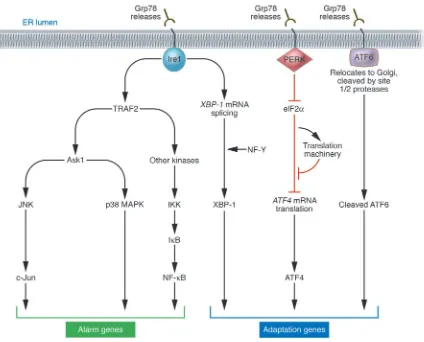
When unfolded proteins accumulate in the ER, resident chap-erones become occupied, releasing transmembrane ER proteins involved in inducing the UPR. These proteins straddle ER mem-branes, with their N-terminus in the lumen of the ER and their C-terminus in the cytosol, providing a bridge that connects these 2 compartments. Normally, the N-termini of these transmembrane ER proteins are held by ER chaperone Grp78 (BiP), preventing their aggregation. But when misfolded proteins accumulate, Grp78 releases, allowing aggregation of these transmembrane signaling proteins, and launching the UPR. Among the critical transmem-brane ER signaling proteins are PERK, Ire1, and ATF6 (Figure 1) (reviewed in refs. 1, 2, 8).
PERK (PKR-like ER kinase) is a Ser/Thr protein kinase, the cata-lytic domain of which shares substantial homology to other kinases of the eukaryotic initiation factor 2α (eIF2α) family (9, 10). Upon removal of Grp78, PERK oligomerizes in ER membranes, inducing its autophosphorylation and activating the kinase domain. PERK phosphorylates and inactivates eIF2α, thereby globally shutting off mRNA translation and reducing the protein load on the ER. However, certain mRNAs gain a selective advantage for translation under these conditions, including the mRNA encoding transcrip-tion factor ATF4. The ATF4 protein is a member of the bZIP family of transcription factors, which regulates the promoters of several genes implicated in the UPR. The importance of PERK-initiated signals for protection against ER stress has been documented by studies of perk–/–
cells and of knock-in cells that express non-phos-phorylatable eIF2α(S51A), both of which display hypersensitivity to ER stress (11, 12).
Ire1 similarly oligomerizes in ER membranes when released by Grp78. The Ire1α protein is a type I transmembrane protein, which contains both a Ser/Thr kinase domain and an endoribonuclease domain; the latter processes an intron from X box protein-1 (XBP-1)
Nonstandard abbreviations used: AD, Alzheimer disease; AβP, amyloid β-peptide; DED-L, death effector domain–like; eIF2α, eukaryotic initiation factor 2α; Htt, Huntingtin; IP3, inositol triphosphate; IP3R, IP3 receptor; NOS, nitric oxide synthase; PD, Parkinson disease; PERK, PKR-like ER kinase; polyQ, polyglutamine; PS-1, presenilin-1; SERCA, sarcoplasmic/endoplasmic reticulum Ca2+ ATPase; UPR, unfolded protein response; XBP-1, X box protein-1.
Conflict of interest: The authors have declared that no conflict of interest exists.
review series
mRNA, rendering it competent for translation to produce the 41-kDa XBP-1 protein, a bZIP-family transcription factor. XBP-1 binds to promoters of several genes involved in retrograde trans-port of misfolded proteins from ER to cytosol and in ER-induced protein degradation (reviewed in ref. 8). XBP-1 heterodimerizes with protein NF-Y and binds at least 2 types of cis-acting elements in gene promoters, including the ER stress enhancer (ERSE) and unfolded protein response element (UPRE) (13). Ablation of Ire1α in mice produces an embryonic lethal phenotype. Fibroblasts from
Ire1α–/–
embryos are defective in activation of UPRE-driven report-er genes, thus showing a cause-and-effect linkage of Ire1α to this
cis-acting element (14).
Release of Grp78 from the N-terminus of ATF6 triggers a differ-ent mechanism of protein activation, compared with PERK and Ire1. Instead of oligomerizing, release of Grp78 frees ATF6 to trans-locate to the Golgi apparatus, where resident proteases (site 1 and site 2 protease) cleave ATF6 at a juxtamembrane site, releasing this transcription factor into the cytosol and allowing it to migrate into the nucleus to regulate gene expression (15). ATF6 collaborates
with Ire1, where ATF6 induces transcription to increase XBP-1 mRNA, and Ire1’s endoribonuclease activity then processes that mRNA so that XBP-1 protein is produced.
Sounding the alarm in response to ER stress: NF-κB activation mechanisms
[image:3.585.89.513.80.422.2]Given the massive glycoprotein production associated with many viral infections, it is not surprising that ER stress activates some of the same signal transduction pathways associated with innate immunity. In this regard, Ire1 shares in common with many mem- bers of the TNF receptor family the ability to bind adapter pro-tein TRAF2. TRAF2 is an E3 ligase that binds Ubc13, resulting in non-canonical polyubiquitination of substrates involving lysine 63 rather than the canonical lysine 48 as a linking site (16). TRAF2 activates protein kinases implicated in immunity and inflamma-tion, including Ask1, which activates JNK, and kinases linked to NF-κB activation. Also recruited to Ire1 is the c-Jun N-terminal inhibitory kinase (JIK), responsible for posttranslational modifica-tion of components of the Ire1α/TRAF2/Ask1 complex (17, 18). Figure 1
Apoptosis induced by ER stress: so many mechanisms, so little clarity
The adaptive responses to misfolded proteins in the ER provide protection from cell death, inasmuch as gene transfer–mediated overexpression of Grp78 or protein-disulfide isomerase (PDI) reduces cell death induced by oxidative stress, Ca2+ disturbances,
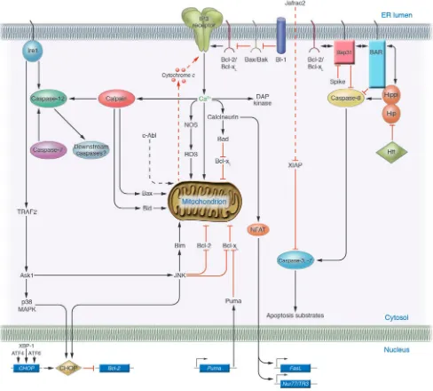
and hypoxia (19, 20). However, when protein misfolding is per- sistent or excessive, ER stress triggers cell death, typically apop-tosis. Several mechanisms, described below, have been proposed for linking the distressed ER to cell death (Figure 2), including direct activation of proteases, kinases, transcription factors, and Bcl-2–family proteins and their modulators.
Proteases . Caspases are required for apoptosis, and certain mem-bers of this family of cysteine proteases associate with the ER (reviewed in ref. 21). In rodents, caspase-12 associates with acti-vated Ire1, resulting in proteolytic processing of caspase-12. Mice lacking caspase-12 genes display partial resistance to pharmaco-logical inducers of ER stress, such as tunicamycin (inhibitor of N-linked protein glycosylation) and thapsigargin (inhibitor of sar-coplasmic/endoplasmic reticulum Ca2+ ATPases [SERCAs], which
pump Ca2+ into the ER) (22). Because proteolytic activity has been
difficult to demonstrate for caspase-12 (23), it is unknown whether the proteolytic processing of caspase-12 that occurs during ER stress results in its activation. Also, the mechanisms responsible for proteolysis of caspase-12 may be indirect, involving calpains activated by Ca2+ released in the vicinity of the ER (24), instead of
an induced proximity mechanism where oligomers of Ire1 provide a scaffold for clustering caspase-12 zymogens. Caspase-7 also may activate caspase-12 by translocating from cytosol to ER (25). How-ever, the relevance of caspase-12 to ER-induced apoptosis has been questioned because of an absence of caspase-12 in most humans. In this regard, the ancestral human CASPASE-12 gene is disrupt-ed by a termination codon and thus is inactive (26). For persons with hereditary polymorphisms that leave the open reading frame intact (estimated at ∼ 1% of African populations), caspase-12 oper-ates as a trans-dominant inhibitor of proinflammatory caspases, lacking conserved residues required for catalytic activity (23).
Human caspase-4, one of the closest paralogs of rodent caspase-12, may associate with ER (27), raising the possibility that this protease can perform the functions normally ascribed to rodent caspase-12 in the context of ER stress. But caspase-4 belongs to the group of proinflammatory caspases responsible for proteo-lytic activation of cytokines, rather than the apoptotic caspases. Nevertheless, small interfering RNA–mediatedknock down of caspase-4 in human neuroblastoma cells partially reduces cell death caused by the ER stress inducers thapsigargin and amyloid
β-peptide (AβP), but not inducers of mitochondria-dependent cell death (e.g., UV irradiation, DNA-damaging drugs). However, caspase-4 knock down in HeLa cells had little effect on apoptosis induced by ER stress, implying that the relevance of this protease to ER stress is tissue-specific.
The ER resident protein Bap31 contains 3 predicted transmem-brane domains, followed by a leucine zipper and a death effector domain–like (DED-L) region that associates with certain isoforms of procaspase-8 in the cytosol (28). Bap31 can display either pro- or antiapoptotic phenotypes, depending on whether its cytosolic tail is removed by cleavage by caspases. Overexpression of full-length Bap31 blocks apoptosis induced by anti-Fas antibody and cyclohex-imide, while expression of the truncated 20-kDa protein induces apoptosis (29, 30). A mutant of Bap31 in which the caspase-8
cleavage site was mutated suppressed Fas-induced apoptosis (29). Since proximal steps in Fas signaling were not blocked by mutant Bap31, this suggests that the ER participates as an intermediary in death receptor–induced apoptosis in some cells.
The DED-L domain of Bap31 also binds a homologous DED-L domain in BAR, another ER-associated apoptosis regulator (31). The 52-kDa BAR protein contains a RING domain that binds ubiquitin-conjugating enzymes, followed by an α-helical region that binds Bcl-2 and Bcl-xL, the DED-L domain, and a C-terminal
membrane-anchoring domain (32). Like the DED-L domain of Bap31, the DED-L domain of BAR binds caspase-8, sequestering it and thwarting apoptosis induction initiated by TNF/Fas-family death receptors (32, 33). BAR also binds the apoptosis regulators Hip and Hippi, which contain DED-L domains homologous to those found in BAR and Bap31. Hip associates with Huntingtin (Htt), the protein implicated in Huntington disease that causes degeneration of neurons containing Htt polyglutamine (polyQ) expansions (34). Hippi is a DED-L domain–containing Hip-inter-acting protein that binds procaspase-8. Htt with polyQ expansion has reduced affinity for Hip compared with the normal Htt pro-tein, a circumstance under which it has been proposed that Hip is free to bind Hippi and trigger caspase-8 activation (35). Inter- actions of BAR, Bap31, Hip, and Hippi deserve further investiga- tion on a number of fronts, including whether these proteins rep-resent substrates for the E3 ligase activity of BAR, elucidation of their agonistic and antagonistic relations among each other, and evaluation of effects of these protein interactions on nonapoptotic functions of Htt and its interacting proteins.
The ability of BAR and Bap31 to bind procaspase-8 prompts speculation that perhaps these ER proteins could promote rather than inhibit caspase-8 activation, if induced to aggregate in ER membranes, thereby constituting a novel ER-associated “apopto-some.” If so, then the parallel ability of BAR and Bap31 to bind Bcl-2 and Bcl-xL through domains separate from the DED-L
domain might supply a mechanism for preventing caspase acti-vation, providing a long-sought analogy to the paradigm for caspase regulation seen in Caenorhabditis elegans, where the Bcl-2 ortholog Ced9 binds caspase activator Ced4, preventing activa-tion of Ced3 protease (36).
Kinases . The kinase Ask1 has been implicated in apoptosis induc-tion in the context of signaling by TNF-family receptors (reviewed in ref. 37). During ER stress, Ask1 is recruited to oligomerized Ire1 complexes containing TRAF2, activating this kinase and causing downstream activation of JNK and p38 MAPK. Consistent with a key role for Ask1 in apoptosis induced by ER stress, studies of ask1–/–
neurons subjected to inducers of ER stress indicate a requirement for this kinase for JNK activation and cell death (38). The down-stream death effectors of Ask1 are not clear. The kinase pathway initiated by Ask1 leads to JNK activation, and JNK-mediated phos-phorylation activates the proapoptotic protein Bim (39–41), while inhibiting the antiapoptotic protein Bcl-2 (42).
Thus, Ire1 plays roles in all 3 of the ER responses to unfolded proteins (adaptation, alarm, and apoptosis), through its actions upon XBP-1 (adaptation), TRAF2 (alarm [NF-κB]), and apoptosis effectors caspase-12 and Ask1. How these 3 functions of Ire1 are integrated remains unclear.
The protein tyrosine kinase c-Abl can translocate from the ER surface to mitochondria in response to ER stress (43). More-over, a functional role for c-Abl has been suggested by studies of
review series
by Ca2+ ionophores, brefeldin A, and tunicamycin (43). How c-Abl
promotes apoptosis is unknown at present.
Transcription factors. CHOP (GADD153) is a member of the C/EBP family of bZIP transcription factors, and its expression is induced to high levels by ER stress (reviewed in ref. 44). The chop
gene promoter contains binding sites for all of the major inducers of the UPR, including ATF4, ATF6, and XBP-1, and these tran-scription factors play causative roles in inducing chop gene tran-scription. Cause-and-effect roles in chop gene induction have been demonstrated for signaling molecules involved in ER stress by genetic manipulation of mice, showing that perk–/– and atf4–/–cells
and eIF2α(S51A) knock-in cells fail to induce chop during ER stress (11, 12, 45). Cross-talk between the PERK/eIF2α pathway and the Ire1/TRAF2/Ask1 pathway may also enhance CHOP activity at a posttranscriptional level, given that Ask1 activates both JNK and p38 MAPKs, and phosphorylation of the CHOP protein on ser-ine 78 and serine 81 by p38 MAPKs increases its transcriptional and apoptotic activity (46). In addition to the aforementioned regulators, upstream activators of chop also include ATF2, which is induced by hypoxia and which is required for chop induction dur-ing amino acid starvation (47).
Overexpression of CHOP protein induces apoptosis, through a Bcl-2–inhibitable mechanism (48, 49). Moreover, chop–/– mice are
resistant to kidney damage induced by tunicamycin and to brain injury resulting from cerebral artery occlusion, demonstrating a role for CHOP in cell destruction when ER stress is involved (48, 50). How CHOP induces apoptosis is unclear. CHOP forms het- erodimers with other C/EBP-family transcription factors via bZIP-domain interactions, which suppresses their binding to C/EBP sites in DNA, while promoting binding to alternative DNA sequences for target gene activation (51). Consequently, CHOP inhibits expression of genes responsive to C/EBP-family transcription factors, while enhancing expression of other genes containing the consensus motif 5′-(A/G)(A/G)(A/G)TGCAAT(A/C)CCC-3′ . One relevant tar-get may be bcl-2, whose expression is suppressed by CHOP, at least in some cellular contexts (49). CHOP may also have nontranscriptional actions, still poorly defined (44). While capable of inducing apopto-sis and contributing to cell death in several scenarios involving ER stress, CHOP is not essential for cell death induced by ER stress, as demonstrated by the observation that perk–/– and eIF2α(S51A)
knock-in cells are hypersensitive to ER stress–induced apoptosis but fail to induce chop gene expression (12, 45).
Scotin is another ER-targeted apoptosis inducer (52). The gene encoding Scotin is a direct target of p53, suggesting a way to link DNA damage to ER-mediated cell death mechanisms.
Bcl-2–family proteins and their modulators. Association of certain Bcl-2/ Bax–family proteins with ER membranes dates back to the initial discovery of Bcl-2 (53). Though better known for their actions upon mitochondria, Bcl-2/Bax–family proteins also integrate into ER membranes, where they modulate ER Ca2+
homeostasis and con-trol cell death induced by ER stress agents, including tunicamycin, brefeldin A (an inhibitor of ER-Golgi transport), thapsigargin, and oxidants (reviewed in ref. 54). Experiments in which the normal C-terminal transmembrane domain of Bcl-2 was swapped with membrane-targeting domains from ER resident proteins suggested that Bcl-2 targeted exclusively to the ER (as opposed to both ER and mitochondria) is more restricted in its antiapoptotic actions, suppressing cell death induced by ER stress agents and by c-Myc. Recent findings that apoptosis induced by c-Myc may be attribut-able to its induction of Bim suggest that ER-targeted Bcl-2 may
sequester this BH3-only protein, preventing it from interacting with other members of the Bcl-2/Bax family (55).
Spike is a BH3-only protein anchored in the ER (56). The BH3-like domain of Spike is required for apoptosis induction, but dimerization partners among Bcl-2/Bax–family proteins have yet to be found. Several other Bcl-2/Bax–family proteins reside at least in part in association with or integrated into ER membranes, with some, such as the antiapoptotic protein Mcl-1 and proapoptotic Bik, found predominantly in the ER (57, 58). Given the preferences of certain BH3 domains for interactions with particular members of the Bcl-2/Bax family (59), it seems likely that a network of inter-actions among a subset of this family of apoptosis regulators takes place on ER membranes, the functional consequences of which are not yet fully understood. Recently, expression of at least 1 of the Bcl-2/Bax–family genes was linked to ER stress. The BH3-only protein Puma is induced by tunicamycin and thapsigargin in a p53-independent manner, with Puma–/– cells showing resistance
to apoptosis induced by ER stress (60).
The BI-1 protein contains 6 transmembrane domains, resides in the ER (61), interacts functionally or physically with Bcl-2–fam-ily members, and is induced by hypoxia (62). This protein blocks cell death induced by oxidative stress in yeast, plants, and animals (63). Mice lacking BI-1 display increased sensitivity to tunicamy-cin-induced kidney damage and to stroke injury, implying a role for BI-1 in protection from insults known to trigger ER stress. In cultured cells, overexpression of BI-1 selectively reduces, while BI-1 deficiency selectively increases, sensitivity to cell death induced by agents that trigger ER stress, while having far less effect on apop-tosis induced by agents that trigger cell death pathways linked to mitochondria (intrinsic pathway) or TNF/Fas-family death receptors (extrinsic pathway) (64). BI-1 associates with the anti-apoptotic proteins Bcl-2 and Bcl-xL, but not proapoptotic Bax and
Bak (61). Nevertheless, BI-1 inhibits cell death induced by Bax overexpression, in animal cells, plants, and yeast.
The ER protein Bap31 lacks homology with Bcl-2/Bax–family proteins and contains no recognizable BH3 dimerization domain, but it binds Bcl-2 and Bcl-xL and regulates apoptosis. BAR also
binds Bcl-2 and Bcl-xL, and the responsible domain is required for
BAR-mediated suppression of cell death (32). Interestingly, BAR is capable of suppressing Bax-induced death of yeast, implying cas-pase-independent functions for this protein, given that yeast lack bona fide caspases. This suggests that, mechanistically, BAR may share something in common with Bcl-2 and BI-1, which also sup-press Bax-induced killing of yeast.
Other apoptosis regulators . Given that mitochondria release apop- togenic proteins into the cytosol, the ER might use similar mech-anisms for linking ER stress to cell death. In insect cells, at least one example has been uncovered of a protein, called Jafrac2, that is normally sequestered in the ER but is released into the cytosol during apoptosis induced by certain stimuli (65). Like most pro-teins imported into the ER, the N-terminal leader peptide of Jafrac2 is removed by proteolysis. This proteolytic processing exposes an IAP-binding motif in Jafrac2, poising it to attack antiapoptotic IAP- family proteins upon accessing the cytosol, thereby freeing caspas-es. It remains to be determined whether examples of apoptogenic protein release from the ER of mammalian cells will be discovered. Ca2+ and apoptosis induced by ER stress
Release of Ca2+
(IP3) and cytosolic ADP-ribose and other regulators via effects on IP3 receptors (IP3Rs) and ryanodine receptors (66, 67). Oppos-ing these gated Ca2+ channels are the SERCA-family proteins,
Ca2+ ATPases that pump Ca2+ into the ER, which are regulated by
phosphorylation and interactions with other proteins (e.g., phos-pholamban and sarcolipin). Various stimuli that cause the ER to dump Ca2+ precipitate cell death, including hypoxia, oxidants,
stimulators of IP3 production, and pharmacological antagonists of SERCA. The downstream effectors of Ca2+-induced cell death
are potentially myriad and could minimally include (a) induction of mitochondrial permeability transition, induced upon entry of excessive amounts of Ca2+ into the matrix of mitochondria (68,
69); (b) local activation near the ER of calpains, a family of Ca2+
-dependent cysteine proteases implicated in pathological cell death
(70, 71), whose substrates include Bax and Bid (which are activat-ed) (72–74), Bcl-2 and Bcl-xL
(which are inhibited), and several cas-pases (reviewed in ref. 4); (c) alterations in Ca2+
-dependent phos-pholipid scramblases, which alter membrane biology to promote apoptosis or necrosis, including transferring phosphatidylserine to the outer leaflet of the plasma membrane (a signal for clearance of cells by phagocytosis) and transferring cardiolipin from the inner to outer membrane of mitochondria (a signal for targeted insertion of proapoptotic Bcl-2–family proteins Bid and Bax into membranes) (75–77); (d) Ca2+/calmodulin–mediated activation
of the protein phosphatase calcineurin, which dephosphorylates the proapoptotic protein Bad, allowing it to dimerize with and antagonize Bcl-xL (78), and which dephosphorylates NFAT-family
transcription factors, allowing entry into the nucleus and trans-Figure 2
Cell death mechanisms induced by ER stress. Several of the proposed pathways linking ER stress to cell death are depicted. Dashed lines indi-cate protein translocation events (c-Abl, Jafrac2). The mitochondrial permeability transition pore complex, which is Ca2+-sensitive, is not shown
[image:6.585.49.537.85.525.2]review series
activation of proapoptotic genes encoding FasL and Nur77/TR3 (79); (e) stimulation of Ca2+
-sensitive isoforms of nitric oxide syn-thase (NOS), exacerbating oxidative stress (reviewed in ref. 5); (f) activation of death-associated protein kinase (DAP kinase) and its relative DRP-1, which contain calmodulin-binding domains (reviewed in ref. 80); (g) activation of Ca2+-sensitive mitochondrial
fission protein DRP-1 (81), which has been implicated in Bax-induced release of cytochrome c from mitochondria; and possibly (h) alterations in the Ca2+
-binding protein TCTP (fortilin), a puta-tive modulator of antiapoptotic Bcl-2/Bax–family proteins such as Mcl-1 (82). In addition, ectopic expression of the proapoptotic mammalian protein Bak in yeast induces cell death through a cal-nexin-dependent pathway, correlating with Bak binding to this Ca2+-dependent ER chaperone (83).
A role for Bcl-2 in modulating intracellular Ca2+
was first dem-onstrated over a decade ago (84), but only recently have clues about the mechanisms involved begun to emerge. Based on data from a variety of techniques, it appears that overexpression of anti-apoptotic proteins Bcl-2 and Bcl-xL lowers the basal amounts of
Ca2+ in the ER, because of increased leakage of Ca2+
under rest-ing conditions. The consequence of this is that upon exposure to stimuli that precipitously dump Ca2+ from internal stores, less
Ca2+ enters the cytosol, resulting in lower peak concentrations of
cytosolic Ca2+ and less overall cytosolic Ca2+ accumulation (54,
85–88). Downstream, less Ca2+
enters mitochondria, which pos-sibly explains the inhibition of mitochondrial depolarization and the suppression of cytochrome c release. Like overexpression of Bcl-2 or Bcl-xL, ablation of the expression of proapoptotic Bax and
Bak also reduces basal Ca2+ in the ER, implying a role for these
proapoptotic proteins in setting cellular ER Ca2+ concentrations
(89). Interestingly, Bcl-2 remains competent in its ability to reduce ER Ca2+, even in cells lacking Bax and Bak, implying that Bcl-2
operates downstream of Bax/Bak with respect to ER Ca2+
regula-tion, unlike the situation with mitochondria-dependent cell death, where genetic evidence indicates that Bcl-xL and Bcl-2 function
upstream of Bax/Bak (90).
Attempts to establish whether these changes in ER Ca2+
han-dling mediated by Bcl-2/Bax–family proteins are causally linked to cell death regulation have failed to provide firm answers, but sup-porting evidence has been obtained from a variety of experimental approaches, including genetic manipulations of Ca2+-regulating
proteins in the ER (88, 89, 91, 92).
Because several Bcl-2/Bax–family proteins share structural simi-larity with the pore-forming domains of bacterial toxins, they may function as ion channels, thus explaining the ability of Bcl-2/ Bax–family proteins to modulate ER Ca2+ (reviewed in ref. 93).
However, mutations designed to impair the putative pore-forming regions of Bcl-2 do not affect its ability to regulate ER Ca2+ (94);
this suggests alternative mechanisms. In this regard, Bcl-2 was reported to bind IP3Rs several years ago (95), and recently Bcl-2 has been implicated in regulating IP3R activity (96). IP3R knock down inhibits the ability of Bcl-2 to promote leakage of Ca2+ from
the ER, suggesting that Bcl-2 relies on IP3Rs to reduce luminal ER Ca2+. The mechanism by which Bcl-2 modulates IP3Rs has yet to
be defined, particularly the issue of whether this is a direct effect of these proteins on IP3Rs or an indirect effect on unidentified IP3R-interacting proteins present in ER membranes. Interest-ingly, cytochrome c, an apoptosis-inducing protein released from mitochondria, binds IP3Rs and induces Ca2+ release from ER,
thereby triggering ER stress (97) and providing another potential
link between IP3Rs and cell death regulation. Also, reduction in or ablation of expression of certain IP3Rs (e.g., IP3R1 and IP3R3) decreases sensitivity of some types of cells (e.g., lymphocytes, neu-rons) to apoptosis (98–100), suggesting further links between Ca2+
dysregulation by IP3Rs and apoptosis induction.
Curiously, the antiapoptotic protein BI-1 also regulates ER Ca2+
homeostasis in a manner analogous to that of Bcl-2 and Bcl-xL.
Overexpression of BI-1 reduces basal ER Ca2+ concentrations, while
ablation of the genes encoding BI-1 increases amounts of thapsigargin-releasable Ca2+ from internal stores (64). Since BI-1 associates with
Bcl-2 and Bcl-xL
in ER membranes (61), it will be interesting to deter-mine whether BI-1 also interacts with and regulates IP3Rs.
The truncated Bap31, resembling the caspase-cleavage product, induces Ca2+
efflux from the ER and induces apoptosis (30), pro-viding further correlative connections between modulation of ER Ca2+ dynamics and cell death regulation. Interestingly, Bap31 was
reported to bind an ER-associated putative ion channel called A4, but the relevance of this protein interaction to regulation of ER Ca2+ remains unclear (101).
ER stress and diseases
ER stress is associated with a range of diseases, including ischemia/ reperfusion injury, neurodegeneration, and diabetes (reviewed in ref. 44), making ER stress a probable instigator of pathological cell death and dysfunction.
ER stress and neurodegeneration. AβP is a proteolytic product of amyloid β-precursor protein that is causally associated with Alzheimer disease (AD). Mice lacking caspase-12 are partially resis-tant to apoptosis induced by exposure to Aβ P (22), raising the pos-sibility of a functional link between ER stress and Aβ P-induced tox-icity. Mutant versions of the AβP-interacting protein presenilin-1 (PS-1), previously associated with AD, interfere with the UPR (102) and may render neurons more susceptible to cell death induced by ER stress (103). The brains of mice harboring AD mutants of PS-1 also have increased CHOP protein (104). Interestingly, PS-1 induc-es cleavage of Ire1α, releasing the cytosolic domain to translocate to the nucleus, suggesting further interactions between molecules involved in AD and ER stress responses (105).
Hereditary mutations in the ER-associated E3 ubiquitin ligase Parkin have also been associated with ER stress–induced cell death and are found in patients with familial Parkinson disease (PD) (106, 107). Overexpression of wild-type Parkin suppresses cell death induced by several ER stress–inducing agents, and by
α-synuclein, the principal component of pathological Lewy bodies seen in PD (107). Parkin expression is induced by ER stress, sug- gesting a role for it in adaptation to ER stress, presumably func-tioning in the ER-associated protein degradation pathway to clear misfolded proteins.
Neurodegenerative diseases associated with inclusion body for-mation and protein aggregation have also been linked to ER stress, including amyotrophic lateral sclerosis, PD, Huntington disease, and others (reviewed in refs. 44, 108). Htt variants with polyQ expansions, for example, induce classical signal transduction events associated with the UPR and cause proteolytic processing of caspase-12 (109), as well as cause global reductions in proteasome activity (110). Thus, by exhausting the cytosolic protein degrada-tion machinery, inclusion body diseases probably cause a back-up of misfolded proteins in the ER, triggering ER stress.
hypoxia and hypoglycemia, which cause protein misfolding and ER stress. Reperfusion of the affected tissues then triggers oxidative stress, with production of NO, and other reactive oxygen species that result in protein misfolding. NO and other reactive molecules also may modify oxidizable residues (cysteines) in ER-associated Ca2+
channels, including ryanodine receptors and SERCAs, caus-ing ER Ca2+ depletion, yet another cause of protein misfolding.
Brain ischemia/reperfusion injury activates the PERK/eIF2α pathway and induces chop expression in rodents (111, 112). Moreover, chop–/–
mice suffer less tissue loss after stroke, imply-ing a causal role for this mediator of ER stress in neuronal cell death (113). NO, a known mediator of brain injury during stroke, induces chop expression in cultured neurons. Furthermore, a NOS inhibitor is protective in a rodent model of brain ischemia (114), and mice lacking the gene encoding iNOS display decreased sensi-tivity to brain ischemia (115), suggesting a causal role for this ER stress inducer in stroke damage.
A role for the antiapoptotic protein BI-1 in protection from cerebral ischemia has been demonstrated by studies of bi-1–/– mice,
which suffer larger infarcts following cerebral artery occlusion (64). Given that hypoxia has been implicated in bi-1 gene induction (62), these findings imply a role for endogenous bi-1 in survival of cells traumatized by ER stress.
ER stress and heart disease. The role of ER stress in heart disease has not been extensively studied, but Ask1 kinase activity increases in mice following myocardial infarction or aortic constriction, and
ask1–/– mice showed reduced cardiomyocyte apoptosis, in addition
to better preservation of left ventricular function, compared with wild-type animals (116).
ER stress in diabetes. Pancreatic β cells have a well-developed ER, reflecting their role in secreting large amounts of insulin and various glycoproteins. This function of β cells may explain why mice lacking PERK are susceptible to diabetes, showing apoptosis of their β cells and progressive hyperglycemia with aging (117). Moreover, PERK gene mutations in association with infant-onset diabetes occur in humans with the autosomal recessive disor-der Wolcott-Rallison syndrome (118). At autopsy, these patients show massive β cell loss, resembling the pathology of perk–/– mice.
Similarly, eIF2α(S51A) knock-in mice suffer from β cell depletion, which begins in utero, suggesting a more rapid course than that in
perk–/– mice (12). The failure of perk–/– to phenocopy eIF2α(S51A)
raises the possibility that other kinases besides PERK inhibit eIF2α during ER stress. Pancreatic β cell apoptosis induced by NO, a mediator of inflammation relevant to autoimmune diabetes, is CHOP-dependent, further implicating ER stress as an instigator of β cell death (50). Also, in rodent models of diabetes caused by a nonsecreted insulin mutant, homozygous deletion of chop delays disease onset (119), implying a role for this gene in β cell depletion in vivo. Recently, xbp-1+/– heterozygous mice have been shown to
be more sensitive to diabetes caused by obesity and high-fat diet (120). The underlying mechanism is related to the requirement of XBP-1 for dampening of JNK activation caused by ER stress, which correlates with phosphorylation of IRS-1 and reduced tyrosine phosphorylation of IRS-1 in insulin-stimulated cells. Interestingly, signs of ER stress were found in liver and adipose tissue of obese mice and mice fed high-fat diets, indicating that the metabolic abnormalities associated with obesity and unhealthy diets cause ER stress in vivo.
Other cells that secrete proteins in large quantities may also be at risk for ER stress–induced apoptosis. For example, studies of perk–/–
mice indicate a requirement for differentiation (or survival) of plas-ma cells, known for their production of immunoglobulins (121).
Therapeutic targets. Several mediators of ER-initiated cell death are candidates for drug discovery efforts, though some are better validated than others. Gene ablation studies in mice argue that agents inhibiting Ask1 and CHOP are attractive, because mice lacking these genes are phenotypically normal but exhibit reduced sensitivity to cell death induced by ER stressors, such as stroke and polyQ-expanded proteins associated with neurodegeneration (reviewed in refs. 38, 44). Presumably Ask1 is also responsible for the hyperactivity of JNK associated with insulin resistance in the context of ER stress caused by high-fat diet. Ask1 theoretically could be attacked by small molecules targeting the ATP-binding site of the kinase domain, analogous to other kinase inhibitors recently approved for other indications. The CHOP protein may be difficult to attack with small-molecule drugs. However, since p38 MAPK augments CHOP activity, small-molecule antago-nists of this kinase currently in development for inflammatory diseases might find utility as cytoprotective agents in clinical sce-narios involving ER stress. Also, c-Abl inhibitors such as imatinib (Gleevec) could be examined for cytoprotective activity in ischemic and degenerative diseases, given recent evidence that c-Abl may relay death signals from ER to mitochondria (43).
Compounds that augment the PERK/eIF2α pathway may also protect against cell death by ER stress. Indeed, a recent screen for inhibitors of neuronal death induced by tunicamycin identified compounds that suppress protein phosphatases responsible for dephosphorylation of eIF2α on serine 51, thus increasing accu-mulation of phosphorylated eIF2α and providing protection from apoptosis induced by several inducers of ER stress (122). Interest- ingly, the prototype compound characterized (Salubrinal) appar-ently is not an active-site inhibitor of the phosphatase but rather specifically disrupts complexes containing GADD35 and protein phosphatase-1 (PP1), thereby preventing GADD34-mediated tar-geting of PP1 onto substrate phospho-eIF2α.
It remains to be determined whether broad-spectrum inhibi-tors of caspase-family cell death proteases would preserve cell survival in the face of ER stress, given that culture experiments have shown that nonapoptotic cell death still occurs in the pres- ence of compounds such as benzoyl-valinyl-alaninyl-aspartyl-flu- oromethylketone (zVAD-fmk), at least when strong pharmaco-logical inducers of ER stress are used (64). However, mice lacking various individual caspases, including caspase-1, caspase-2, and caspase-11 (a probable caspase-4 or -5 ortholog), are resistant to stroke injury (123), a condition in which ER stress participates in the cell death mechanism.
Provided that side effects from vascular instability are not an issue, inhibitors of NOS are also attractive, since mice that lack iNOS show decreased sensitivity to brain ischemia and reduced CHOP expression (115). Finally, inhibitors of proapoptotic Bcl-2/Bax–fam-ily proteins that operate upon ER membranes could be useful for ameliorating tissue loss due to stimulators of ER stress.
Conclusions
review series
preserve cell survival. Further studies of genes and gene products involved in ER-initiated cell death are needed to fully validate tar-gets for drug discovery. Acknowledgments We thank J. Valois for manuscript preparation, M. Hanaii for artwork, and the California Breast Cancer Research Program, the Fondation pour la Recherche Médicale, and the NIH (grants NS047855 and AG15393) for their generous support. Address correspondence to: John C. Reed, The Burnham Institute for Medical Research, 10901 North Torrey Pines Road, La Jolla, California 92037, USA. Phone: (858) 646-3140; Fax: (858) 646-3194; E-mail: reedoffice@burnham.org. 1. Schroder, M., and Kaufman, R.J. 2005. ER stress and the unfolded protein response. Mutat. Res.569:29–63.
2. Shen, X., Zhang, K., and Kaufman, R.J. 2004. The unfolded protein response: a stress signaling path-way of the endoplasmic reticulum [review]. J. Chem. Neuroanat. 28:79–92.
3. Rao, R.V., Ellerby, H.M., and Bredesen, D.E. 2004. Coupling endoplasmic reticulum stress to the cell death program. Cell Death Differ. 11:372–380. 4. Breckenridge, D.G., Germain, M., Mathai, J.P.,
Nguyen, M., and Shore, G.C. 2003. Regulation of apoptosis by endoplasmic reticulum pathways.
Oncogene. 22:8608–8618.
5. Orrenius, S., Zhivotovsky, B., and Nicotera, P. 2003. Regulation of cell death: the calcium-apoptosis link [review]. Nat. Rev. Mol. Cell Biol. 4:552–565. 6. Ma, Y., and Hendershot, L.M. 2004. ER chaperone
functions during normal and stress conditions.
J. Chem. Neuroanat. 28:51–65.
7. Rizzuto, R., Duchen, M.R., and Pozzan, T. 2004. Flirting in little space: the ER/mitochondria Ca2+ liaison [review]. Sci. STKE. 2004:re1.
8. Rao, R.V., and Bredesen, D.E. 2004. Misfolded pro- teins, endoplasmic reticulum stress and neurode-generation. Curr. Opin. Cell Biol. 16:653–662.
9. Shi, Y., et al. 1998. Identification and character-ization of pancreatic eukaryotic initiation factor 2 alpha-subunit kinase, PEK, involved in transla-tional control. Mol. Cell. Biol. 18:7499–7509. 10. Harding, H.P., Zhang, Y., and Ron, D. 1999. Protein
translation and folding are coupled by an endoplas-mic-reticulum-resident kinase. Nature. 397:271–274. 11. Harding, H.P., Zhang, Y., Bertolotti, A., Zeng, H., and Ron, D. 2000. Perk is essential for translational regulation and cell survival during the unfolded protein response. Mol. Cell. 5:897–904.
12. Scheuner, D., et al. 2001. Translational control is required for the unfolded protein response and in vivo glucose homeostasis. Mol. Cell. 7:1165–1176. 13. Yoshida, H., Matsui, T., Yamamoto, A., Okada,
T., and Mori, K. 2001. XBP1 mRNA is induced by ATF6 and spliced by IRE1 in response to ER stress to produce a highly active transcription factor. Cell.
107:881–891.
14. Lee, K., et al. 2002. IRE1-mediated unconventional mRNA splicing and S2P-mediated ATF6 cleavage merge to regulate XBP1 in signaling the unfolded protein response. Genes Dev. 16:452–466. 15. Ye, J., et al. 2000. ER stress induces cleavage of
membrane-bound ATF6 by the same proteases that process SREBPs. Mol. Cell. 6:1355–1364.
16. Habelhah, H., et al. 2004. Ubiquitination and trans-location of TRAF2 is required for activation of JNK but not of p38 or NF-kappaB. EMBO J. 23:322–332. 17. Urano, F., et al. 2000. Coupling of stress in the ER
to activation of JNK protein kinases by transmem-brane protein kinase IRE1. Science. 287:664–666. 18. Yoneda, T., et al. 2001. Activation of caspase-12,
an endoplastic reticulum (ER) resident caspase, through tumor necrosis factor receptor-associated factor 2-dependent mechanism in response to the ER stress. J. Biol. Chem. 276:13935–13940.
19. Liu, H., et al. 1997. Endoplasmic reticulum chap-erones GRP78 and calreticulin prevent oxidative stress, Ca2+ disturbances, and cell death in renal epithelial cells. J. Biol. Chem. 272:21751–21759. 20. Tanaka, S., Uehara, T., and Nomura, Y. 2000.
Up-regulation of protein-disulfide isomerase in response to hypoxia/brain ischemia and its protec-tive effect against apoptotic cell death. J. Biol. Chem.
275:10388–10393.
21. Momoi, T. 2004. Caspases involved in ER stress-mediated cell death. J. Chem. Neuroanat. 28:101–105. 22. Nakagawa, T., et al. 2000. Caspase-12 mediates
endoplasmic-reticulum-specific apoptosis and cytotoxicity by amyloid-β. Nature. 403:98–103. 23. Saleh, M., et al. 2004. Differential modulation of
endotoxin responsiveness by human caspase-12 polymorphisms. Nature. 429:75–79.
24. Nakagawa, T., and Yuan, J. 2000. Cross-talk between two cysteine protease families. Activation of caspase-12 by calpain in apoptosis. J. Cell Biol.
150:887–894.
25. Rao, R.V., et al. 2001. Coupling endoplasmic reticu-lum stress to the cell death program: mechanism of caspase activation. J. Biol. Chem. 276:33869–33874. 26. Fischer, H., Koenig, U., Eckhart, L., and Tschachler,
E. 2002. Human caspase-12 has acquired del-eterious mutations. Biochem. Biophys. Res. Commun.
293:722–726.
27. Hitomi, J., et al. 2004. Involvement of caspase-4 in endoplasmic reticulum stress-induced apoptosis and Abeta-induced cell death. J. Cell Biol. 165:347–356. 28. Ng, F.W.H., et al. 1997. p28 Bap31, a Bcl-2/Bcl-XL-
and procaspase-8-associated protein in the endo-plasmic reticulum. J. Cell Biol. 139:327–338. 29. Nguyen, M., Breckenridge, D.G., Ducret, A., and
Shore, G.C. 2000. Caspase-resistant BAP31 inhibits fas-mediated apoptotic membrane fragmentation and release of cytochrome c from mitochondria.
Mol. Cell. Biol. 20:6731–6740.
30. Breckenridge, D.G., Stojanovic, M., Marcellus, R., and Shore, G.C. 2003. Caspase cleavage product of BAP31 induces mitochondrial fission through endoplasmic reticulum calcium signals, enhancing cytochrome c release to the cytosol. J. Cell Biol. 160:1115–1127.
31. Roth, W., et al. 2003. Bifunctional apoptosis inhibi-tor (BAR) protects neurons from diverse cell death pathways. Cell Death Differ. 10:1178–1187. 32. Zhang, H., et al. 2000. BAR: an apoptosis regulator
at the intersection of caspase and bcl-2 family pro-teins. Proc. Natl. Acad. Sci. U. S. A. 97:2597–2602. 33. Stegh, A.H., et al. 2002. Inactivation of caspase-8
on mitochondria of Bcl-xL expressing MCF7-Fas cells. J. Biol. Chem. 277:4351–4360.
34. Kalchman, M.A., et al. 1997. HIP1, a human homo- logue of S. cerevisiae Sla2p, interacts with mem-brane-associated huntingtin in the brain. Nat. Genet. 16:44–53.
35. Gervais, F.G., et al. 2002. Recruitment and activa-tion of caspase-8 by the Huntingtin-interacting protein Hip-1 and a novel partner Hippi. Nat. Cell Biol. 4:95–105.
36. Hengartner, M.O. 2000. The biochemistry of apop-tosis. Nature. 407:770–776.
37. Matsukawa, J., Matsuzawa, A., Takeda, K., and Ichijo, H. 2004. The ASK1-MAP kinase cascades in mamma-lian stress response. J. Biochem. (Tokyo). 136:261–265. 38. Nishitoh, H., et al. 2002. ASK1 is essential for
endoplasmic reticulum stress-induced neuronal cell death triggered by expanded polyglutamine repeats. Genes Dev. 16:1345–1355.
39. Lei, K., and Davis, R.J. 2003. JNK phosphorylation of Bim-related members of the Bcl2 family induces Bax-dependent apoptosis. Proc. Natl. Acad. Sci. U. S. A.
100:2432–2437.
40. Putcha, G.V., et al. 2003. JNK-mediated BIM phos-phorylation potentiates BAX-dependent apoptosis.
Neuron. 38:899–914.
41. Luciano, F., et al. 2003. Phosphorylation of Bim-EL by Erk1/2 on serine 69 promotes its degradation via the proteasome pathway and regulates its pro-apoptotic function. Oncogene. 22:6785–6793. 42. Yamamoto, K., Ichijo, H., and Korsmeyer, S.J.
1999. BCL-2 is phosphorylated and inactivated by an ASK1/Jun N-terminal protein kinase path-way normally activated at G(2)/M. Mol. Cell. Biol.
19:8469–8478.
43. Ito, Y., et al. 2001. Targeting of the c-Abl tyrosine kinase to mitochondria in endoplasmic reticu-lum stress-induced apoptosis. Mol. Cell. Biol.
21:6233–6242.
44. Oyadomari, S., and Mori, M. 2004. Roles of CHOP/ GADD153 in endoplasmic reticulum stress. Cell Death Differ. 11:381–389.
45. Harding, H.P., et al. 2003. An integrated stress response regulates amino acid metabolism and resistance to oxidative stress. Mol. Cell. 11:619–633. 46. Wang, X.Z., and Ron, D. 1996. Stress-induced
phosphorylation and activation of the transcrip-tion factor CHOP (GADD153) by p38 MAP Kinase.
Science. 272:1347–1349.
47. Bruhat, A., et al. 2000. Amino acids control mam-malian gene transcription: activating transcription factor 2 is essential for the amino acid respon-siveness of the CHOP promoter. Mol. Cell. Biol.
20:7192–7204.
48. Zinszner, H., et al. 1998. CHOP is implicated in programmed cell death in response to impaired function of the endoplasmic reticulum. Genes Dev.
12:982–995.
49. McCullough, K.D., Martindale, J.L., Klotz, L.O., Aw, T.Y., and Holbrook, N.J. 2001. Gadd153 sensitizes cells to endoplasmic reticulum stress by down-regulating Bcl2 and perturbing the cellular redox state. Mol. Cell. Biol. 21:1249–1259.
50. Oyadomari, S., et al. 2001. Nitric oxide-induced apoptosis in pancreatic beta cells is mediated by the endoplasmic reticulum stress pathway. Proc. Natl. Acad. Sci. U. S. A. 98:10845–10850.
51. Ubeda, M., et al. 1996. Stress-induced binding of the transcriptional factor CHOP to a novel DNA control element. Mol. Cell. Biol. 16:1479–1489. 52. Bourdon, J.C., Renzing, J., Robertson, P.L., Fernandes,
K.N., and Lane, D.P. 2002. Scotin, a novel p53-induc-ible proapoptotic protein located in the ER and the nuclear membrane. J. Cell Biol. 158:235–246. 53. Tsujimoto, Y., and Croce, C. 1986. Analysis of the
structure, transcripts, and protein products of Bcl-2, the gene involved in human follicular lym-phoma. Proc. Natl. Acad. Sci. U. S. A. 83:5214–5218. 54. Thomenius, M.J., and Distelhorst, C.W. 2003.
Bcl-2 on the endoplasmic reticulum: protecting the mitochondria from a distance [review]. J. Cell Sci.
116:4493–4499.
55. Egle, A., Harris, A.W., Bouillet, P., and Cory, S. 2004. Bim is a suppressor of Myc-induced mouse B cell leu-kemia. Proc. Natl. Acad. Sci. U. S. A. 101:6164–6169. 56. Mund, T., Gewies, A., Schoenfeld, N., Bauer, M.K.,
and Grimm, S. 2003. Spike, a novel BH3-only pro- tein, regulates apoptosis at the endoplasmic reticu-lum. FASEB J. 17:696–698.