Pax3 is required for enteric ganglia formation
and functions with Sox10 to modulate
expression of c-ret
Deborah Lang, … , Min Min Lu, Jonathan A. Epstein
J Clin Invest.
2000;106(8):963-971. https://doi.org/10.1172/JCI10828.
Hirschsprung disease and Waardenburg syndrome are human genetic diseases
characterized by distinct neural crest defects. Patients with Hirschsprung disease suffer
from gastrointestinal motility disorders, whereas Waardenburg syndrome consists of
defective melanocyte function, deafness, and craniofacial abnormalities. Mutations
responsible for Hirschsprung disease and Waardenburg syndrome have been identified,
and some patients have been described with characteristics of both disorders. Here, we
demonstrate that
PAX3
, which is often mutated in Waardenburg syndrome, is required for
normal enteric ganglia formation. Pax3 can bind to and activate expression of the
c-RET
gene, which is often mutated in Hirschsprung disease. Pax3 functions with Sox10 to
activate transcription of
c-RET
, and
SOX10
mutations result in Waardenburg-Hirschsprung
syndrome. Thus, Pax3, Sox10, and c-Ret are components of a neural crest development
pathway, and interruption of this pathway at various stages results in neural crest–related
human genetic syndromes.
Article
Introduction
Neural crest cells arise from the dorsal neural tube and migrate over precise pathways to populate multiple organs and tissues in the developing embryo (1). They differentiate into many cell types including melanocytes, neurons, and glia. In humans, various syndromes have been described that are characterized by deficiencies of neural crest derivatives (2). In recent years, some of the genes responsible for these disorders have been identified, and relationships among these genes and syndromes have begun to emerge.
A subset of neural crest precursors gives rise to the enteric nervous system (3). The intestine is colonized by a rostral to caudal wave of migrating cells originat-ing from the vagal crest region, although truncal and sacral crest cells also contribute. Enteric ganglia pre-cursors must both migrate and proliferate in order to provide the necessary expansion of the precursor pool required for the population of the entire intestinal tract. This process is critically dependent on several growth factors and their cognate receptors. For instance, enteric ganglia precursors express the c-Ret tyrosine kinase receptor that is required for their sur-vival and proliferation (4–7). The ligand for c-Ret, glial cell–derived neurotrophic factor (GDNF), is expressed by mesenchymal cells surrounding enteric ganglia and is required for proper enteric plexus development (8–11). The endothelin B receptor (EDNRB) and its lig-and endothelin 3 (EDN3) are also required (12–16), suggesting that multiple growth factor pathways con-verge to regulate enteric neuron development and
mat-uration. In addition, several transcription factors, including Sox10, Phox2b, and Mash1 regulate various aspects of enteric neuron differentiation (17–23).
In humans, abnormalities of the enteric nervous sys-tem result in Hirschsprung disease (aganglionic mega-colon) and gastrointestinal motility disorders. The most common mutations found in patients with Hirschsprung disease are in the c-RETgene (5, 6). Fam-ilies with Hirschsprung disease have also been described in which heterozygous mutations in EDNRB or EDN3have been identified (14, 16, 24). Interesting-ly, homozygous mutations of either of these genes result in additional phenotypes characteristic of Waar-denburg syndrome that include pigmentation abnor-malities, craniofacial defects, and deafness (16, 24, 25). Heterozygous mutations in SOX10also result in fea-tures of both Waardenburg syndrome and Hirschsprung disease (19). Sox10 is a member of the high-mobility group (HMG) of nuclear factors relat-ed to the sex-determining factor Sry (26). Sox10 is capable of sequence-specific DNA binding (26), whereas HMG proteins in general function as tran-scriptional coactivators by acting as architectural pro-teins and synergizing with other transcriptional regu-lators (27, 28). Mutations in Sox10 in the mouse account for the Domphenotype that has been studied as a model for Hirschsprung disease (20).
Patients with Waardenburg syndrome without fea-tures of Hirschsprung disease most often harbor mutations in the PAX3gene (29, 30). Pax3 is a mem-ber of the paired-box–containing family of nuclear
Pax3 is required for enteric ganglia formation and
functions with Sox10 to modulate expression of c-ret
Deborah Lang, Fabian Chen, Rita Milewski, Jun Li, Min Min Lu, and Jonathan A. Epstein
Cardiovascular Division, Department of Medicine, University of Pennsylvania, Philadelphia, Pennsylvania, USA
Address correspondence to: Jonathan A. Epstein, 954 BRB II, 421 Curie Boulevard, Philadelphia, Pennsylvania 19104, USA. Phone: (215) 898-8731; Fax: (215) 573-2094; E-mail: epsteinj@mail.med.upenn.edu.
Deborah Lang and Fabian Chen contributed equally to this work.
Received for publication July 20, 2000, and accepted in revised form September 5, 2000.
Hirschsprung disease and Waardenburg syndrome are human genetic diseases characterized by distinct neural crest defects. Patients with Hirschsprung disease suffer from gastrointestinal motility disorders, whereas Waardenburg syndrome consists of defective melanocyte function, deafness, and craniofacial abnormalities. Mutations responsible for Hirschsprung disease and Waardenburg syndrome have been identified, and some patients have been described with char-acteristics of both disorders. Here, we demonstrate that PAX3, which is often mutated in Waar-denburg syndrome, is required for normal enteric ganglia formation. Pax3 can bind to and acti-vate expression of the c-RET gene, which is often mutated in Hirschsprung disease. Pax3 functions with Sox10 to activate transcription of c-RET, and SOX10mutations result in Waar-denburg-Hirschsprung syndrome. Thus, Pax3, Sox10, and c-Ret are components of a neural crest development pathway, and interruption of this pathway at various stages results in neural crest–related human genetic syndromes.
transcription factors (31). Affected individuals are heterozygous for PAX3mutations; homozygous loss of function is lethal (32). Waardenburg syndrome can also be caused by mutations in the MITFgene (33), and Pax3 probably directly regulates MITF tran-scription during melanocyte development (34). Enteric ganglia defects have not been reported in patients with Waardenburg syndrome with PAX3or MITFmutations.
In the mouse, Pax3mutations result in the Splotch phenotype (35, 36), characterized by a white belly spot in the heterozygous state (37). Homozygous-deficient embryos die during midgestation with neural tube and cardiac defects. Abnormalities of the enteric nervous system have not been previously identified.
Here, we demonstrate that Pax3-expressing neural crest precursors give rise to enteric ganglia. We show that Pax3 is required for formation of the enteric nervous system. Furthermore, Pax3 synergizes with Sox10 to activate an enhancer in the c-RETgene, and Pax3 and Sox10 bind to adjacent sites within this enhancer. Pax3, Sox10, and c-Ret are components of a molecular cascade regulating neural crest and enteric nervous system development.
Methods
Transgenic mice. P3pro-Cre consisted of the 1.6-kb prox-imal Pax3regulatory region upstream of Cre recombi-nase (38). RET-lacZ was generated by replacement of a HindIII-BamHI fragment of RET-luc (see below) con-taining the luciferase gene with a HindIII-BamHI frag-ment from pSV–β-gal (Promega Corp., Madison, Wis-consin, USA) encoding β-galactosidase. Splotchmice and embryos were genotyped as described elsewhere (39).
Immunohistochemistry. Immunohistochemistry for detection of neural cell specific neurofilament was per-formed on paraffin-embedded sections of tissue fixed in 4% paraformaldehyde after exposing antigen with Antigen Unmasking Solution (Vector Laboratories, Burlingame, California, USA) and incubation with 2H3 mAb (Developmental Studies Hybridoma Bank, Iowa City, Iowa, USA) diluted 1:200. Antibody was detected using either peroxidase or FITC conjugated goat anti-mouse secondary antibody. Antibodies to c-Ret (Immuno-Biological Laboratories, Tokyo, Japan), neu-ron-specific enolase (DAKO Corp., Carpinteria, Cali-fornia, USA), GFAP (DAKO Corp.), and S100 (Novo-castra, Newcastle-upon-Tyne, United Kingdom) were used at dilutions of 1:50, 1:200, 1:500, and 1:200, respectively. Paraformaldehyde-fixed (10 minutes) frozen sections were used with the c-Ret antibody and paraffin sections of paraformaldehyde-fixed (24 hours) tissue were used for the other antibodies.
In situ hybridization. Radioactive (40) and whole mount (41) in situ hybridization studies were per-formed as described elsewhere. Specific sequences used as templates for synthesis of RNA probes are list-ed as follows with gene name followlist-ed by GenBank accession number and specific base pairs amplified:
c-ret, X67812, bp 2142-2608; Sox10, AF047043, bp 1151-1450; ednrb, U32329, bp 1506-1794; edn3, U32330, bp 737-1041; SM22α, L41154, bp 696-975. Northern blot analysis. RNA was prepared 48 hours after transfection of P19 cells, denatured with glyox-al and dimethyl sulfoxide, electrophoresed on a 1.5% agarose/0.01M phosphate buffer gel, transferred to a nylon membrane (Genescreen Plus; NEN Life Science Products, Boston, Massachusetts, USA), and hybridized with a c-retprobe (bp 2142–2608 of Gen-bank no. X67812).
Luciferase assays. NIH 3T3 and 293T cells were main-tained in DME with 10% FCS and transfected at 50% confluency in 35-mm dishes. A total of 2 µg of plasmid DNA was mixed with 40 µl Effectene reagent and used to transfect quadruplicate wells (QIAGEN Inc., Valen-cia, California, USA). Each reaction, used for four equal transfections, contained 0.5 µg of reporter/luciferase plasmid, 0.5 µg of pCMV–β -galac-tosidase, and variable amounts of pCMV-Pax3 (42), pCMV-Pax2 (provided by R. Maas), pCMV-Pax6 (pro-vided by R. Maas), pCDNA-Pax9 (derived from pcPax9-K5, provided by H. Peters), and/or pCMV-Sox10 (derived from dcgs10-1, provided by M. Southard-Smith and W. Pavan). Total amount of DNA was kept constant by the addition of pBS plasmid. After 72 hours, luciferase assays (Promega Corp.) and β -galac-tosidase assays using ONTG substrate (Promega Corp.) were performed. Luciferase activity was nor-malized for β-galactosidase transfection efficiency. RET-luc was prepared by PCR using forward primer 5′ -CCGCTCGAGCTTCCACTCTCCACGTTGCTTand reverse primer 5′-CCCAAGCTTCGGTCGCGGGACTGAAGCTAG with TaKaRa DNA polymerase (PanVera, Madison, Wisconsin, USA). The PCR product was cloned into the XhoI and HindIII sites of pGL2-basic (Promega Corp.). Initial transfections determined the optimal ratios of expression plasmid to reporter plasmid and established a dose-response curve (data not shown). At high concentrations, Pax3 inhibited luciferase reporter activity, as has been described previously (43).
Electrophoretic mobility shift assay. Electrophoretic mobility shift assay (EMSA) was performed as described previously (44) in the presence of 250 µg/ml poly-dIdC and 50 mM KCl. Nonradioactive competi-tor DNA, when included, was present at 100-fold molar excess over labeled probe. The Pax3 protein used was GST-P3PdHd (45) that includes the Pax3 paired and homeodomains. In vitro transcribed and translated Sox10 (TNT kit, Promega) was prepared from ClaI linearized Sox10 plasmid (dgs10-1) as tem-plate. Products of control in vitro transcription/trans-lation reactions performed in the absence of Sox10 plasmid resulted in no detectable electrophoretic mobility shift with any probe used. Primers used for EMSA are listed as follows; engineered mutations are underlined. Probe A: 5′-CAT GCA GAG TCT GTG CTT CCA ACC ACC ATG TCA CAC TGC CCA TG; APaxmut: 5′ -CAT GCA GAG TCT GTG CTT CCA ACC ACC ATA GGC GCG CCC CCA TG; ASoxmut: 5′-CAT GCA GAG TGG CGC GCC CCA ACC ACC ATG TCA CAC TGC CCA TG.
Results
Pax3-expressing precursors are fated to become enteric gan-glia. We have recently demonstrated that the proximal 1.6-kb upstream regulatory region of the murine Pax3 gene is sufficient to direct transgene expression to the dorsal neural tube and to neural crest cells (46). This transgene recapitulates a subdomain of endogenous Pax3expression (31) and omits expression in all myo-genic precursors. We have created transmyo-genic mice using the 1.6-kb Pax3regulatory element directing Cre recombinase expression in neural crest progenitors so that we could determine the fate of Pax3expressing
neural crest cells (38, 47, 48). The pattern of Cre expression in these mice is consistent with the previ-ously characterized activity of the Pax3promoter ele-ment used (46, 49), which directs expression that ini-tiates at approximately embryonic day 9 (E9) and is restricted to dorsal neural tube and neural crest. Trans-genic embryos resulting from a cross to R26R Cre reporter mice in which β-galactosidase is constitutive-ly expressed onconstitutive-ly in cells that are subjected to Cre-mediated recombination (48) demonstrate prominent β-galactosidase activity at E14.5 in the stomach and intestine (Figure 1a). Cross sections through labeled loops of intestine indicate that cells expressing β -galac-tosidase represent enteric ganglia (Figure 1b, arrows) and express neurofilament (Figure 1, c and d). We uti-lized three independent transgenic Cre lines in these and subsequent experiments to ensure that transgene insertion sites were not confounding results. Pax3 expression is not detectable in enteric neurons between E11.5 and E16.5, suggesting that Pax3 functions in migrating or premigratory cells. Our fate mapping data indicate that enteric ganglia are among the tissues derived from Pax3–expressing neural crest cells.
[image:4.612.105.499.453.624.2]Pax3 is required for normal enteric ganglia development. We have confirmed and extended these results using a different transgenic mouse that also expresses β -galac-tosidase in neural crest derivatives and we have used this transgenic line to track neural crest cell fate in Pax3-deficient Splotchembryos. The 6.5-kb proximal connexin 43 (Cx43)promoter directs expression in a subset of the endogenous Cx43expression domain that includes dorsal neural tube, dorsal root ganglia, cardiac neural crest, and other neural crest–derived tissues (50).
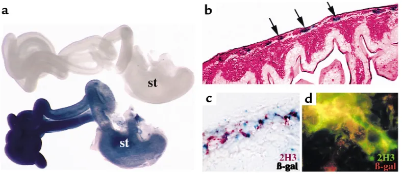
Figure 1
A subset of Pax3-expressing cells are fated to become enteric ganglia. Neural crest–specific regulatory elements of the Pax3 promoter were used to make transgenic mice expressing Cre recombinase. (a) After mating with Cre reporter mice, Pax3-expressing neural crest cells express β-galactosidase (stained blue) and populate the stomach (st) and intestines. The gastrointestinal tract from an E14.5 trans-genic embryo is blue (bottom), whereas tissue from a Cre reporter embryo without the P3pro-Cre transgene processed in parallel is not stained (top). (b) Transverse section through a labeled loop of bowel reveals β-galactosidase–expressing cells in the region populated by enteric ganglia (arrows). The section is counterstained with eosin. (c) Coimmunohistochemistry of an adjacent section to that shown in
We have crossed Cx43-lacZ transgenic mice to Splotch mice, and we have examined enteric ganglia develop-ment at E10.5–E12.5. Litters from pregnant Sp+/–
females derived from matings with transgenic Sp+/–
males were stained for β-galactosidase activity. In wild-type embryos, positively staining cells were seen lining the foregut at E10.5 (Figure 2a) and formed a conflu-ent ring encircling each loop of bowel by E12.5 (Figure 2c). In Sp–/–embryos, few positively staining cells were
seen surrounding the gut at E10.5 (Figure 2b). At E12.5, some labeled cells were identified in the stom-ach of Splotch embryos, but few or no labeled cells were seen in the intestine distal to the stomach (Figure 2d). Enteric ganglia are thought to populate the gut in a
rostral to caudal wave of migration. Our results in Splotch embryos suggest delayed or deficient neural crest colonization of the developing intestine.
We confirmed that differences in transgene expres-sion in Sp–/–versus wild-type embryos represented
dif-ferences in enteric ganglia formation by costaining for expression of neural cell–specific neurofilament using a mAb (2H3). Neurofilament expression was similar to Cx43 transgene expression in the intestinal wall of wild-type embryos (Figure 2e) and was deficient in Splotchembryo intestines (Figure 2f). Costaining of wild-type tissue with antibodies specific for β -galac-tosidase and neurofilament demonstrated colocaliza-tion (Figure 2, g and h), confirming transgene expres-sion by enteric ganglia cells.
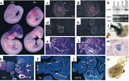
The c-Ret tyrosine kinase receptor is required for enteric ganglia development in the intestine (5–7). At E9.5 and E10.0, whole mount in situ hybridization reveals c-ret–expressing cells (Figure 3a). In Splotch embryos, c-ret expression is reduced (Figure 3a). By E12.5, c-ret is expressed in the bowel wall of wild-type embryos (WT, Figure 3, b and c) in a pattern similar to Cx43-lacZand neurofilament. Expression of c-retat the RNA (Figure 3f) and protein (Figure 3g) level is defi-cient in Sp–/–embryos, although some expression is
retained in the stomach (st, Figure 3f). Immunohisto-chemistry using antibodies specific for neuron specif-ic enolase and for glial markers GFAP and S-100 showed similar differences between wild-type and Splotchembryos, suggesting that most or all neural crest derivatives in the intestine are affected by the absence of Pax3 (see Methods; data not shown).
Cell-autonomous function for Pax3 in enteric ganglia pre-cursors. Given that expression and fate mapping data indicate that Pax3 is required for enteric ganglia for-mation, we asked whether this represented a cell-autonomous function of Pax3 in neural crest cells. We have previously reported that transgenic Splotch mice in which Pax3 expression is reconstituted using the 1.6-kb proximal Pax3 promoter that is specific for neural tube and neural crest are rescued with respect to car-diac neural crest (but not skeletal muscle) development (46). We analyzed enteric ganglia development in these rescued embryos at E12.5. Enteric ganglia expressing c-retwere identified in all segments of bowel in trans-genic Splotch embryos (Sp/Tg, Figure 3j) in contrast to homozygous mutant Splotch littermates, indicating suc-cessful rescue of enteric plexus development by expres-sion of Pax3 in neural crest progenitors.
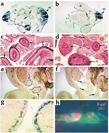
[image:5.612.61.291.242.522.2]The genes encoding the endothelin receptor B (EDNRB) and its ligand endothelin 3 (EDN3) have also been implicated in Hirschsprung’s disease (12, 14) and are required for distal hindgut ganglia formation. Ednrbis normally expressed by enteric ganglia and also by surrounding mesenchyme (51). In Splotch, ednrb expression is specifically missing in the region where enteric ganglia reside, but it is retained in the sur-rounding mesenchyme (Figure 3, d and h). Expression of edn3in the intestinal mesenchyme is not affected by Figure 2
Enteric ganglia are deficient in Splotchintestine. (aand b) Transverse sections of E10.5 wild-type (a) and Sp–/–(b) Cx43-LacZtransgenic
embryos are shown after staining for β-galactosidase activity (blue).
β-Galactosidase–positive cells are seen lining the primitive gut in wild-type (arrows, a) but not Sp–/–(b) embryos, although a small number
of blue cells are seen near the proximal foregut (arrow, b). (cand d)
β-Galactosidase detection in E12.5 wild-type (c) and Sp–/–(d)
trans-verse sections at the abdominal level reveals expression in the wall of the stomach (st) and all loops of bowel (arrows, c) in wild-type embryos. No expression is seen in the gut distal to the stomach in
Sp–/–embryos (arrows, d). (eand f) Neurofilament
immunohisto-chemistry of E12.5 wild-type (e) and Sp–/–(f) sections shows loss of
neurofilament expression in Sp–/–embryos in a pattern similar to that
seen for β-galactosidase (compare arrows, eand f). (g) Costaining for β-galactosidase (blue) and neurofilament (brown) shows over-lapping expression patterns in the bowel wall of wild-type embryos. (h) Higher power of nuclear β-galactosidase (red) and neurofilament (2H3, green) stained wild-type intestine confirms that Cx43-lacZ
the absence of Pax3 (data not shown). A smooth mus-cle marker, SM22α, was expressed normally in Splotch embryos (Figure 3, e and i).
Pax3 and Sox10 activate c-ret. Deficiency of enteric gan-glia in Splotch embryos could be a direct result of decreased expression of the c-Ret receptor by enteric ganglia precursors, as c-Ret is required for survival and proliferation of these cells (4–7). Therefore, we tested the ability of Pax3 to activate expression of the c-ret gene in cultured cells by transfecting P19 cells with expression plasmids encoding Pax3 and testing for enhanced c-retexpression. P19 cells can be induced by retinoic acid to express endogenous Pax3 and can dif-ferentiate into neuronal cell types (52). At the same time, we tested the effect of cotransfecting expression plasmids encoding Sox10. Compared with control transfected cells, Pax3 and Pax3 with Sox10 induced significant increases in endogenous c-ret mRNA
expression (Figure 3k). Sox10 alone resulted in very slight activation of c-retin this system. RT-PCR exper-iments (data not shown) confirmed mild upregulation of c-retafter Sox10 transfection.
[image:6.612.74.529.54.341.2]Using available sequence data, we cloned the human c-RETpromoter to determine whether Pax3 and Sox10 are able to specifically regulate transcriptional activi-ty of proximal upstream regulatory regions of the c-RETgene. Transient transgenic mouse embryos con-taining a 5.1-kb upstream genomic region of c-RET linked to lacZ were examined at E13.5 to evaluate in vivo activity of this promoter. β-Galactosidase activity was detected in portions of the stomach and intestine in four transgenic embryos (Figure 3, part l) but not in 26 wild-type littermates. Sections through transgenic intestines revealed β-galactosidase staining in enteric ganglia (Figure 3m). Nontransgenic intestines processed in parallel never revealed staining. However, Figure 3
Pax3 is required for normal c-retexpression. (a) Whole-mount in situ hybridization reveals c-retexpression in wild-type (WT) E9.5 (top) and E10 (bottom) embryos. c-retexpression is reduced in Splotch(Sp) embryos (arrows). (b) Radioactive in situ hybridization reveals c-ret
retpromoter to address these possibilities. Transgene expression was consistently detected in the ureter of the developing kidney (Figure 3n), although it did not appear to be confined to the ureteric buds, as is endogenous c-retexpression (4). β-Galactosidase activ-ity was not detected in wild-type littermates.
Figure 4
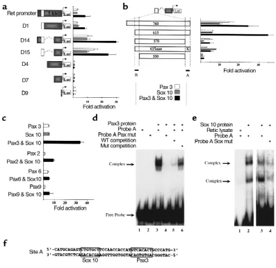
A Pax3- and Sox10- responsive enhancer in the c-RETgene. (a) Cotransfection of 293T cells with reporter constructs composed of portions of the c-RETupstream genomic sequence (see Methods) cloned upstream of a luciferase gene with pCMV-Pax3, pCMV-Sox10, or both reveals a 750-bp enhancer element (open box) and a more proximal repressor element (compare full-length Ret promoter to construct D15). All trans-fections were normalized for transfection efficiency (see Methods) and are expressed as fold activation compared with transfection without Pax3 or Sox10 (mean ±SD; n =12 for each condition). (b) Further analysis of the 750-bp enhancer was performed by modifying construct D15. Deletion of 45 bp at the 3′end significantly reduced activity (compare constructs 615 and 570). Mutation in a putative Pax3 binding site (indi-cated by “X” in construct 615mut) had a similar effect, although activation by Sox10 alone was not affected by this mutation. Residual Pax3 responsiveness present in construct 570 is eliminated by deletion of 20 bp at the 5′end of the enhancer (compare constructs 570 and 550). The 3′and 5′response elements are indicated by dotted lines and filled bars labeled A and B, respectively. Cotransfection results are expressed as in a, n=12 for each condition. (c) The response of construct 615 (depicted in b) is specific for Pax3 and is activated to a much lesser degree by Pax2, Pax6, and Pax9. Although low-level activation is apparent after cotransfection with Pax2 or Pax6, no synergistic activation with cotrans-fected Sox10 is evident. Cotransfection results are expressed as in a; n=8 for each condition. Pax3 and Sox10 bind to the c-RETenhancer. (d) EMSA using a 45-bp probe derived from the 3′response element in the 750-bp c-RETenhancer (site A in part b) reveals specific binding by Pax3 (lane 3). Pax3 does not bind to a similar probe in which the putative Pax3 binding site has been replaced by an AscI restriction site (lane 4; and see Methods). Unlabeled competitor wild-type probe is able to compete for binding (lane 5), whereas mutated probe competes far less effi-ciently (lane 6), indicating binding specificity. (e) Sox10 also binds to site A, and mutation in the putative Sox10 binding site significantly reduces binding. In vitro translated Sox10 forms two complexes in EMSA experiments with probe A (lanes 2 and 3), whereas the reticulocyte lysate with-out Sox10 does not bind (lane 1). Mutation in the putative Sox10 binding site (see Methods) reduces Sox10 binding (lane 4). Lanes 1 and 2 are from a different gel than lanes 3 and 4. (f) The sequence of probe A is shown with Sox10 and Pax3 binding sites boxed.
c-Identification of Pax3- and Sox10-responsive elements in the c-RET promoter. In cotransfection assays, Pax3 and Sox10 were able to activate expression of a 5.1-kb c-RETpromoter-luciferase reporter both in NIH-3T3 cells (data not shown) and in 293T cells, and the pres-ence of Pax3 and Sox10 together produced an approx-imately ninefold activation (Figure 4a). Analysis of serial deletions of the c-RET promoter–luciferase reporter construct, depicted in Figure 4a, defined an internal repressor element that, when removed, revealed a 30-fold activation of transcription by Pax3 with Sox10 (construct D14, Figure 4a). The enhancer element responsible for this activity was localized to a 750-bp fragment (construct D15, open box, Figure 4a) that was analyzed further.
A series of deletion constructs (Figure 4b and data not shown) composed of portions of the 750-bp enhancer linked to the proximal c-RETpromoter were cotransfected with Pax3, Sox10, or both. These experi-ments resulted in the identification of a 45-bp element at the 3′end of the enhancer (site A, Figure 4b) that is required for synergistic activation by Sox10 and Pax3 (filled bars, Figure 4b; compare constructs 615 and 570). This site contains Sox10 and Pax3 binding sequences (see below). Mutation of the putative Pax3 paired domain binding site (construct 615mut, Figure 4b; and see Methods) results in elimination of syner-gistic activation reproducing the effect of deleting the entire 45-bp site. Note that construct 615mut retains responsiveness to Sox10 (shaded bars, Figure 4b), sug-gesting that Sox10 binding has not been impaired by the engineered mutation. The residual Pax3 respon-siveness retained by construct 570 (open bar, Figure 4b) is completely eliminated by deletion of 20 bp at the 5′ end of this enhancer (site B, Figure 4b; compare con-structs 570 and 550). In summary, cotransfection experiments identified a critical 45-bp element (site A) required for the majority of c-RETpromoter/enhancer activation by Sox10 and Pax3. Synergistic activation of reporter activity mediated by the c-RETenhancer was specific for Pax3 and was not reproduced by cotrans-fection of Sox10 with other Pax family members including Pax2, Pax6, or Pax9 (Figure 4c).
Pax3 and Sox10 bind to the c-RET enhancer. The region identified as site A in Figure 4b, which is responsible for the majority of Pax3 and Sox10 responsiveness, contains a putative Sox10 binding sequence (CTGT-GCT) that contains five of seven nucleotides found in a previously identified Sox10 binding site (CTTTGTT) (26). This site is followed 12 bp downstream by a puta-tive Pax3-paired domain binding site (GTCACACT) that is identical at seven of eight positions to the opti-mal Pax3-paired domain binding sequence identified in binding site selection assays (42, 53). We tested the ability of Pax3 and Sox10 to bind to these sequences using EMSAs with endogenous and mutated sequences (see Methods for primer sequences). We used a GST-fusion protein encoding both the paired and homeodomains of Pax3, and in vitro translated
Sox10, for these experiments. As shown in Figure 4d, Pax3 is able to bind to site A under stringent binding conditions (50 mM KCl, 250 µg/ml poly-dIdC), and binding is markedly reduced if the putative paired domain binding site is altered (compare lanes 3 and 4, Figure 4d). Addition of unlabeled wild-type probe effectively competes for specific binding (lane 5, Fig-ure 4d), whereas the unlabeled mutant probe does so far less efficiently (lane 6, Figure 4d). Hence, Pax3 binds to site A in a sequence specific fashion. In vitro translated Sox10 also binds to site A, producing two shifted complexes in EMSA experiments, suggesting that Sox10 may dimerize on this site (lane 2, Figure 4e). Proteins present in the reticulocyte lysate used to prepare Sox10 did not bind to probe A as demonstrat-ed in lane 1, Figure 4e. Mutation of the putative Sox10 binding sequence significantly reduced Sox10 binding (compare lanes 4 and 3, Figure 4e). Mutations in the paired domain binding sequence had no effect on Sox10 binding (data not shown). The sequence of probe A and the location of the Sox10 and Pax3 bind-ing sites identified in these experiments are depicted in Figure 4f. HMG box transcription factors of the Sox10 type are known to bind to the minor groove of DNA (27), whereas Pax proteins bind predominantly to the major groove (54). It is worth noting that the 12-bp spacing between Sox10 and Pax3 sites within the c-RETpromoter allows for a half turn of the DNA spiral and predicts that both proteins could dock on the same side of DNA, potentially enabling protein-pro-tein interactions. Hence, the c-RETenhancer contains a 45-bp element that is required for synergistic Pax3 and Sox10 activation and contains adjacent Pax3 and Sox10 binding sites.
Discussion
We suggest that deficient c-Ret expression in the absence of Pax3 is related to the ability of Pax3 to regu-late the c-RET promoter directly. However, we cannot rule out the possibility that Pax3 is required for survival or proliferation of enteric ganglia precursors in a man-ner unrelated to its ability to modulate c-retexpression in vitro. These possibilities will prove difficult to differ-entiate because inappropriate apoptosis of enteric neu-rons occurs in the absence of c-ret (7, 55). Thus, it will be difficult to determine whether Pax3 is required for cell survival via c-ret–dependent or –independent pathways. Our demonstration of adjacent Pax3 and Sox10 bind-ing sites in the c-RETpromoter, and the ability of Pax3 and Sox10 to activate c-retexpression in P19 cells, sug-gest that the decrease in c-retexpression evident in Splotchembryos is at least partially attributable to the loss of Pax3 transcriptional regulation of c-ret.
There is precedent for Pax proteins to act in con-junction with other transcriptional regulators in a manner analogous to what we propose here for Pax3 and Sox10. Pax5 is capable of recruiting ETS domain transcription factors to DNA and both factors bind to adjacent sites (56). X-ray crystallographic analysis and computer modeling has suggested that the paired domain binds to the major groove of DNA in a fash-ion that resembles two linked helix-turn-helix motifs docking on opposite faces of DNA (54, 57). Spatial and structural considerations allow for the docking of ETS domain factors to adjacent minor groove positions and suggest possible protein-protein interactions (57). A similar model may apply to Sox and Pax proteins binding to adjacent sites on DNA, and the observation that Sox and Pax binding sites are located 12 bp apart in the c-RETpromoter is consistent with this propos-al. To date, we have been unable to demonstrate spe-cific protein-protein interactions between purified Pax3 and Sox10 proteins. However, we have previous-ly demonstrated that the paired domain adopts a sta-ble αhelical conformation only upon binding to DNA (58), and hence appropriate stable conformations to allow Pax3-Sox10 interactions may only occur in the presence of specific DNA binding sites.
On the basis of our results, we speculate that other combinations of Pax and Sox transcription factors may synergize to regulate important developmental processes in other systems. For instance, Pax5 and Sox4 are both expressed in pre–B cells (59–61), and inactiva-tion of either gene in the mouse results in arrest of B-cell development at similar or identical stages (60, 61). Pax5 binds to and activates the promoter for the CD19 cell-surface receptor (62), and it would be interesting to test whether Sox4, which is closely related to Sox10, can modify the ability of Pax5 to activate CD19 or other downstream targets in B cells.
In summary, in both humans and mice, PAX3, SOX10, and c-RET mutations result in neural crest–related defects. In some cases, there is phenotypic overlap between the resulting syndromes. Here we provide evi-dence that Pax3-expressing neural crest precursors give
rise to enteric ganglia and that Pax3 is required for this developmental process. Pax3 functions with Sox10 to modulate expression of c-RET,and Pax3 and Sox10 bind to adjacent sites within a c-RETenhancer. Pax3, Sox10, and c-Ret are components of a molecular cas-cade regulating neural crest development.
Acknowledgments
We are grateful to R. Mass, H. Peters, M. Southard-Smith, and W. Pavan for providing plasmids. This work was supported by NIH grants RO1HL62974, RO1HL61475, RO1DK57050 and a pilot project of Center grant P30DK50306 to JAE.
1. LeDouarin, N.M. 1982. The neural crest. Cambridge University Press. London, United Kingdom. 259 pp.
2. McKusick, V.A. 1998. Online mendelian inheritance in man, OMIM™. http://www.ncbi.nlm.nih.gov/Omim. Johns Hopkins University and National Center for Biotechnology Information, National Library of Medicine. Baltimore, Maryland, USA, and Bethesda, Maryland, USA. 3. Gershon, M.D. 1999. Disorders of enteric neuronal development:
insights from transgenic mice. Am. J. Physiol. 277:G262–G267. 4. Pachnis, V., Mankoo, B., and Costantini, F. 1993. Expression of the
c-ret proto-oncogene during mouse embryogenesis. Development.
119:1005–1017.
5. Romeo, G., et al. 1994. Point mutations affecting the tyrosine kinase domain of the RET proto-oncogene in Hirschsprung’s disease. Nature.
367:377–378.
6. Edery, P., et al. 1994. Mutations of the RET proto-oncogene in Hirschsprung’s disease. Nature. 367:378–380.
7. Durbec, P.L., Larsson-Blomberg, L.B., Schuchardt, A., Costantini, F., and Pachnis, V. 1996. Common origin and developmental dependence on c-ret of subsets of enteric and sympathetic neuroblasts. Development.
122:349–358.
8. Durbec, P., et al. 1996. GDNF signalling through the Ret receptor tyro-sine kinase. Nature. 381:789–793.
9. Moore, M.W., et al. 1996. Renal and neuronal abnormalities in mice lacking GDNF. Nature. 382:76–79.
10. Pichel, J.G., et al. 1996. Defects in enteric innervation and kidney devel-opment in mice lacking GDNF. Nature. 382:73–76.
11. Sanchez, M.P., et al. 1996. Renal agenesis and the absence of enteric neu-rons in mice lacking GDNF. Nature. 382:70–73.
12. Baynash, A.G., et al. 1994. Interaction of 3 with endothelin-B receptor is essential for development of epidermal melanocytes and enteric neurons. Cell. 79:1277–1285.
13. Hosoda, K., et al. 1994. Targeted and natural (piebald-lethal) mutations of endothelin-B receptor gene produce megacolon associated with spot-ted coat color in mice. Cell. 79:1267–1276.
14. Puffenberger, E.G., et al. 1994. A missense mutation of the endothelin-B receptor gene in multigenic Hirschsprung’s disease. Cell.
79:1257–1266.
15. Gariepy, C.E., Cass, D.T., and Yanagisawa, M. 1996. Null mutation of endothelin receptor type B gene in spotting lethal rats causes agan-glionic megacolon and white coat color. Proc. Natl. Acad. Sci. USA.
93:867–872.
16. Edery, P., et al. 1996. Mutation of the endothelin-3 gene in the Waar-denburg-Hirschsprung disease (Shah-Waardenburg syndrome). Nat. Genet. 12:442–444.
17. Pattyn, A., Morin, X., Cremer, H., Goridis, C., and Brunet, J.F. 1999. The homeobox gene Phox2b is essential for the development of autonomic neural crest derivatives. Nature. 399:366–370.
18. Southard-Smith, E.M., Kos, L., and Pavan, W.J. 1998. Sox10 mutation disrupts neural crest development in Dom Hirschsprung mouse model.
Nat. Genet. 18:60–64.
19. Pingault, V., et al. 1998. SOX10 mutations in patients with Waarden-burg-Hirschsprung disease. Nat. Genet. 18:171–173.
20. Herbarth, B., et al. 1998. Mutation of the Sry-related Sox10 gene in Dominant megacolon, a mouse model for human Hirschsprung dis-ease. Proc. Natl. Acad. Sci. USA. 95:5161–5165.
21. Lo, L.C., Johnson, J.E., Wuenschell, C.W., Saito, T., and Anderson, D.J. 1991. Mammalian achaete-scute homolog 1 is transiently expressed by spatially restricted subsets of early neuroepithelial and neural crest cells.
Genes Dev. 5:1524–1537.
23. Blaugrund, E., et al. 1996. Distinct subpopulations of enteric neuronal progenitors defined by time of development, sympathoadrenal lineage markers and Mash-1-dependence. Development. 122:309–320. 24. Hofstra, R.M., et al. 1996. A homozygous mutation in the
endothelin-3 gene associated with a combined Waardenburg type 2 and Hirschsprung phenotype (Shah-Waardenburg syndrome). Nat. Genet.
12:445–447.
25. Attie, T., et al. 1995. Mutation of the endothelin-receptor B gene in Waardenburg-Hirschsprung disease. Hum. Mol. Genet. 4:2407–2409. 26. Kuhlbrodt, K., Herbarth, B., Sock, E., Hermans-Borgmeyer, I., and Weg-ner, M. 1998. Sox10, a novel transcriptional modulator in glial cells. J. Neurosci. 18:237–250.
27. Giese, K., Cox, J., and Grosschedl, R. 1992. The HMG domain of lym-phoid enhancer factor 1 bends DNA and facilitates assembly of func-tional nucleoprotein structures. Cell. 69:185–195.
28. Grosschedl, R., Giese, K., and Pagel, J. 1994. HMG domain proteins: architectural elements in the assembly of nucleoprotein structures.
Trends Genet. 10:94–100.
29. Baldwin, C.T., Hoth, C.F., Amos, J.A., da Silva, E.O., and Milunsky, A. 1992. An exonic mutation in the HuP2 paired domain gene causes Waardenburg’s syndrome. Nature. 355:637–638.
30. Tassabehji, M., et al. 1992. Waardenburg’s syndrome patients have mutations in the human homologue of the Pax-3 paired box gene.
Nature. 355:635–636.
31. Goulding, M.D., Chalepakis, G., Deutsch, U., Erselius, J.R., and Gruss, P. 1991. Pax-3, a novel murine DNA binding protein expressed during early neurogenesis. EMBO J.10:1135–1147.
32. Ayme, S., and Philip, N. 1995. Possible homozygous Waardenburg syn-drome in a fetus with exencephaly. Am. J. Med. Genet.59:263–265. 33. Tassabehji, M., Newton, V., and Read, A. 1994. Waardenburg syndrome
type 2 caused by mutations in the human microphthalmia (MITF) gene. Nat. Genet. 8:251–255.
34. Watanabe, A., Takeda, K., Ploplis, B., and Tachibana, M. 1998. Epistat-ic relationship between Waardenburg syndrome genes MITF and PAX3.
Nat. Genet. 18:283–286.
35. Epstein, D.J., Vekemans, M., and Gros, P. 1991. Splotch (Sp2H), a muta-tion affecting development of the mouse neural tube, shows a delemuta-tion within the paired homeodomain of Pax-3. Cell. 67:767–774. 36. Epstein, D.J., Vogan, K.J., Trasler, D.G., and Gros, P. 1993. A mutation
within intron 3 of the Pax-3 gene produces aberrantly spliced mRNA transcripts in the splotch (Sp) mouse mutant. Proc. Natl. Acad. Sci. USA.
90:532–536.
37. Auerbach, R. 1954. Analysis of the developmental effects of a lethal mutation in the house mouse. J. Exp. Zool.127:305–329.
38. Li, J., Chen, F., and Epstein, J.A. 2000. Neural crest expression of Cre recombinase directed by the proximal Pax3 promoter in transgenic mice. Genesis. 26:162–164.
39. Tajbakhsh, S., Rocancourt, D., Cossu, G., and Buckingham, M. 1997. Redefining the genetic hierarchies controlling skeletal myogenesis: Pax-3 and myf-5 act upstream of myoD. Cell. 89:127–138.
40. Wawersik, S., and Epstein, J.A. 2000. Gene expression analysis by in situ hybridization. In Methods in molecular biology. Volume 137. Developmental biology protocols.R.S. Tuan and C.W. Lo, editors. Humana Press Inc. Totowa, New Jersey, USA. 87–96.
41. Riddle, R.D., Johnson, R.L., Laufer, E., and Tabin, C. 1993. Sonic hedge-hog mediates the polarizing activity of the ZPA. Cell. 75:1401–1416. 42. Epstein, J.A., Shapiro, D.N., Cheng, J., Lam, P.Y., and Maas, R.L. 1996. Pax3 modulates expression of the c-Met receptor during limb muscle development. Proc. Natl. Acad. Sci. USA. 93:4213–4218.
43. Chalepakis, G., Jones, F.S., Edelman, G.M., and Gruss, P. 1994. Pax-3 contains domains for transcription activation and transcription inhi-bition. Proc. Natl. Acad. Sci. USA. 91:12745–12749.
44. Epstein, J.A., et al. 1994. Two independent and interactive DNA bind-ing subdomains of the PAX6 paired domain are regulated by alterna-tive splicing. Genes Dev.8:2022–2034.
45. Epstein, J.A., Lam, P., Jepeal, L., Maas, R.L., and Shapiro, D.N. 1995. Pax3 inhibits myogenic differentiation of cultured myoblast cells. J. Biol. Chem.270:11719–11722.
46. Li, J., Liu, K.C., Jin, F., Lu, M.M., and Epstein, J.A. 1999. Transgenic res-cue of congenital heart disease and spina bifida in Splotch mice. Devel-opment. 126:2495–2503.
47. Epstein, J.A., et al. 2000. Migration of cardiac neural crest cells in Splotch embryos. Development. 127:1869–1878.
48. Soriano, P. 1999. Generalized lacZ expression with the ROSA26 Cre reporter strain. Nat. Genet. 21:70–71.
49. Natoli, T.A., Ellsworth, M.K., Wu, C., Gross, K.W., and Pruitt, S.C. 1997. Positive and negative DNA sequence elements are required to establish the pattern of Pax3 expression. Development. 124:617–626.
50. Waldo, K.L., Lo, C.W., and Kirby, M.L. 1999. Connexin 43 expression reflects neural crest patterns during cardiovascular development. Dev. Biol. 208:307–323.
51. Wu, J.J., Chen, J.X., Rothman, T.P., and Gershon, M.D. 1999. Inhibition of in vitro enteric neuronal development by endothelin-3: mediation by endothelin B receptors. Development. 126:1161–1173.
52. Pruitt, S.C. 1992. Expression of Pax-3- and neuroectoderm-inducing activities during differentiation of P19 embryonal carcinoma cells.
Development. 116:573–583.
53. Chalepakis, G., and Gruss, P. 1995. Identification of DNA recognition sequences for the Pax3 paired domain. Gene. 162:267–270.
54. Xu, W., Rould, M.A., Jun, S., Desplan, C., and Pabo, C.O. 1995. Crystal structure of a paired domain-DNA complex at 2.5 A resolution reveals structural basis for Pax developmental mutations. Cell. 80:639–650. 55. Taraviras, S., et al. 1999. Signalling by the RET receptor tyrosine kinase and its role in the development of the mammalian enteric nervous sys-tem. Development. 126:2785–2797.
56. Fitzsimmons, D., et al. 1996. Pax-5 (BSAP) recruits Ets proto-oncogene family proteins to form functional ternary complexes on a B-cell-spe-cific promoter. Genes Dev.10:2198–2211.
57. Xu, H.E., et al. 1999. Crystal structure of the human Pax6 paired domain-DNA complex reveals specific roles for the linker region and carboxy-terminal subdomain in DNA binding. Genes Dev.
13:1263–1275.
58. Epstein, J., Cai, J., Glaser, T., Jepeal, L., and Maas, R. 1994. Identification of a Pax paired domain recognition sequence and evidence for DNA-dependent conformational changes. J. Biol. Chem.269:8355–8361. 59. Adams, B., et al. 1992. Pax-5 encodes the transcription factor BSAP and
is expressed in B lymphocytes, the developing CNS, and adult testis.
Genes Dev.6:1589–1607.
60. Urbanek, P., Wang, X.Q., Fetka, I., Wagner, E.F., and Busslinger, M. 1994. Complete block of early B cell differentiation and altered pat-terning of the posterior midbrain in mice lacking Pax5/BSAP. Cell.
79:901–912.
61. Schilham, M.W., et al. 1996. Defects in cardiac outflow tract formation and pro-B-lymphocyte expansion in mice lacking Sox-4. Nature.
380:711–714.