150-kD oxygen-regulated protein is expressed
in human atherosclerotic plaques and allows
mononuclear phagocytes to withstand cellular
stress on exposure to hypoxia and modified low
density lipoprotein.
Y Tsukamoto, … , S Ogawa, Y Kitamura
J Clin Invest.
1996;
98(8)
:1930-1941.
https://doi.org/10.1172/JCI118994
.
The 150-kD oxygen-regulated protein (ORP150) was initially characterized based on its
selective expression in astrocytes subjected to oxygen deprivation (Kuwabara, K., M.
Matsumoto, J. Ikeda, O. Hori, S. Ogawa, Y. Maeda, K. Kitagawa, N. Imuta, K. Kinoshita,
D.M. Stern, et al. 1996. J. Biol. Chem. 279:5025-5032). We have found that exposure of
cultured human aortic smooth muscle cells and mononuclear phagocytes (MPs) to hypoxia
(pO2 approximately 12-14 torr) induces ORP150 transcripts and production of the antigen,
whereas incubation with either hydrogen peroxide, sodium arsenite, heat shock, or
2-deoxyglucose was without effect. Tissue extracts prepared from human atherosclerotic
lesions demonstrated expression of ORP150 mRNA and antigen, vs lack of ORP150 in
samples from nonatherosclerotic areas. In situ hybridization using ORP150 riboprobes
showed the mRNA to be predominantly [correction of predominately] present in
macrophages in in atherosclerotic plaques. Furthermore, autoantibody to ORP150 was
demonstrated in the serum of patients with severe atherosclerosis, consistent with inducible
in vivo expression of ORP150. Introduction of antisense oligonucleotide for ORP150
selectively diminished hypoxia-mediated induction of ORP150 antigen and reduced the
viability of hypoxic MPs, especially in the presence of modified (oxidized/acetylated) LDL. In
support of a role for ORP150 in the MPs' response to the microenvironment of an atheroma,
the presence of oxidized LDL enhanced by approximately 10-fold ORP150 expression in
hypoxic cultures. These data indicate […]
Research Article
Find the latest version:
J. Clin. Invest.
© The American Society for Clinical Investigation, Inc. 0021-9738/96/10/1930/12 $2.00
Volume 98, Number 8, October 1996, 1930–1941
150-kD Oxygen-regulated Protein Is Expressed in Human Atherosclerotic Plaques
and Allows Mononuclear Phagocytes to Withstand Cellular Stress on Exposure to
Hypoxia and Modified Low Density Lipoprotein
Yoshitane Tsukamoto,*‡ Keisuke Kuwabara,‡ Seiichi Hirota,* Jun Ikeda,§ David Stern,i Hideki Yanagi,§ Masayasu Matsumoto,‡
Satoshi Ogawa,‡ and Yukihiko Kitamura*
*Department of Pathology, and ‡First Department of Medicine, Osaka University Medical School, Suita City, 565 Japan; §HSP Research
Institute, Kyoto 600, Japan; and iDepartment of Physiology and Cellular Biophysics, College of Physicians and Surgeons, Columbia
University, New York, New York 10032
Abstract
The 150-kD oxygen-regulated protein (ORP150) was
ini-tially characterized based on its selective expression in
as-trocytes subjected to oxygen deprivation (Kuwabara, K., M.
Matsumoto, J. Ikeda, O. Hori, S. Ogawa, Y. Maeda, K.
Kitagawa, N. Imuta, K. Kinoshita, D.M. Stern, et al. 1996.
J. Biol. Chem.
279:5025–5032). We have found that
expo-sure of cultured human aortic smooth muscle cells and
mononuclear phagocytes (MPs) to hypoxia (pO
2z
12–14
torr) induces ORP150 transcripts and production of the
an-tigen, whereas incubation with either hydrogen peroxide,
sodium arsenite, heat shock, or 2-deoxyglucose was without
effect. Tissue extracts prepared from human atherosclerotic
lesions demonstrated expression of ORP150 mRNA and
an-tigen, vs lack of ORP150 in samples from
nonatheroscle-rotic areas. In situ hybridization using ORP150 riboprobes
showed the mRNA to be predominately present in
mac-rophages in atherosclerotic plaques. Furthermore,
autoanti-body to ORP150 was demonstrated in the serum of patients
with severe atherosclerosis, consistent with inducible in vivo
expression of ORP150. Introduction of antisense
oligonucle-otide for ORP150 selectively diminished hypoxia-mediated
induction of ORP150 antigen and reduced the viability of
hypoxic MPs, especially in the presence of modified
(oxi-dized/acetylated) LDL. In support of a role for ORP150 in
the MPs’
response to the microenvironment of an atheroma,
the presence of oxidized LDL enhanced by
z
10-fold ORP150
expression in hypoxic cultures. These data indicate that
cells of the atherosclerotic vessel wall express ORP150 as
part of a protective mechanism, potentially triggered by
lo-cal hypoxia/hypoxemia and augmented by modified
lipo-proteins. The presence of antibody to ORP150 in sera of
pa-tients with severe atherosclerosis emphasizes the possibility
that ORP150 may be a marker of vascular pathology. (
J.
Clin. Invest.
1996. 98:1930–1941.) Key words:
atherosclero-sis
•smooth muscle cell
•macrophage
•stress response
Introduction
The cellular response to oxygen deprivation involves redirec-tion of biosynthetic mechanisms with expression of a set of polypeptides termed oxygen-regulated proteins (ORPs)1 (1).
Such stress proteins induced by hypoxia can overlap with those synthesized in response to glucose deprivation and heat shock. For example, a 78-kD polypeptide induced by hypoxia/reoxy-genation in cultured astrocytes is identical to glucose-regulated protein 78, and functions as a molecular chaperone to facilitate elaboration of a neurotrophic cytokine, interleukin 6 (2, 3). In-duction of heat shock protein (HSP) 70, shown to have a pro-tective function in a gerbil forebrain ischemia model (4), also occurs in response to hypoxia followed by replacement into normoxia.
Recently, we isolated and cloned a novel 150-kD polypep-tide termed oxygen-regulated protein (ORP150) that is in-duced in astrocytes by hypoxia. This polypeptide is localized in the endoplasmic reticulum, suggesting that it may participate in protein folding and/or translocation in response to environ-mental stress. In contrast with many other ORPs, which over-lap with HSPs and glucose-regulated protein, synthesis of the 150-kD polypeptide was only triggered by hypoxia, not by glu-cose deprivation, heat shock, or multiple other stimuli (5). This leads us to propose that expression of ORP150 is more closely tied to oxygen depletion or cellular events initiated in this situ-ation. In the current study, we demonstrate expression of this novel stress protein in human atherosclerotic plaques, espe-cially in mononuclear phagocytes. Biosynthesis of ORP150 in cultured monocyte-derived macrophages exposed to hypoxia is potentiated by z 10-fold in the presence of modified lipo-proteins. Furthermore, suppression of ORP150 expression markedly attenuated survival of mononuclear phagocytes un-der hypoxic conditions in the presence of modified lipopro-teins. Consistent with the presence of ORP150 in abnormal vasculature, sera from patients with severe atherosclerosis showed the presence of IgG reactive with ORP150 by immu-noblotting. These data lead us to suggest that ORP150 is a component of the protective response of macrophages to envi-ronmental stress, in this instance the combination of modified lipoproteins and possible concomitant hypoxia.
Address correspondence to Satoshi Ogawa, Department of Anatomy and Neuroscience, Osaka University Medical School, 2-2 Yamada-oka Suita City, 565 Japan. Phone: 3221; FAX: 81-6-879-3229; E-mail: QZA03417@niftyserve.or.jp
Received for publication 15 May 1996 and accepted in revised form 14 August 1996.
1. Abbreviations used in this paper: 1A4, monoclonal antibody to
Methods
Cell culture and induction of hypoxia. Human peripheral blood– derived mononuclear phagocytes (MPs) were prepared as described (6). In brief, the mononuclear cell fraction was separated by density gradient centrifugation (Histopaque 1077; Sigma Chemical Co., St. Louis, MO) followed by adherence to tissue culture plasticware for 4 h at 378C. Adherent cells were cultured for 10–14 d in RPMI 1640 (Gibco Laboratories, Grand Island, NY) containing human serum (10%) and penicillin/streptomycin (100 U/ml, 100 mg/ml). Human aortic smooth muscle cells (SMCs) were purchased from KURABO (Osaka, Japan) and cultured in S-BM medium (KURABO) contain-ing recombinant human basic fibroblast growth factor (2 ng/ml), re-combinant human EGF (10 ng/ml), gentamicin (50 mg/ml), ampho-terin-B (amphotericin B, 50 ng/ml), dexamethasone (0.39 mg/ml), and FCS (5%). SMCs used for experiments were at passage 5. When cul-tures achieved confluence, they were exposed to hypoxia using an in-cubator attached to a hypoxia chamber that maintained a humidified atmosphere with low oxygen tension (pO2, 12–14 torr; Coy
Labora-tory Products, Ann Arbor, MI) as described previously (7). Where in-dicated, after exposure to hypoxia, cultures were returned to the am-bient atmosphere (reoxygenation), at which time the conditioned medium was rapidly exchanged with fresh medium. Oxygen tension in the medium was measured using a blood gas analyzer (ABL-2; Ra-diometer, Sweden). Cell viability was assessed by morphological cri-teria, trypan blue exclusion, lactate dehydrogenase (LDH) release, and general protein synthesis measured by the incorporation of [3H]leucine to trichloroacetic acid–precipitable material (8).
Modification of LDL. Modification of native LDL was per-formed as described. In brief, acetylated LDL (Ac-LDL) was pre-pared by incubating human LDL with acetic anhydride at 48C over-night as described (9). Oxidized LDL (Ox-LDL) was prepared by incubating LDL at 180 mg/ml in 5 mM CuSO4 for 24 h at 378C (10).
Ac-LDL and Ox-LDL were characterized by SDS-PAGE (11), aga-rose gel electrophoresis, immunoblotting, and quantification of thiobarbituric acid–reactive substances. Based on these criteria, our preparations of Ac-LDL and Ox-LDL conformed to what has been observed previously (12, 13).
Preparation of anti–ORP150 antibody and immunoblotting. A pep-tide was synthesized based on the NH2-terminal 15 amino acids
ob-tained from purified rat ORP150 (5). This sequence is identical to the predicted NH2-terminal amino acid sequence of human ORP150
de-duced from the human cDNA (Ikeda, J., H. Yanagi, K. Kuwabara, S. Ogawa, M. Matsumoto, and Y. Yura, manuscript in preparation [Genbank No. U41853]). Synthetic peptide was aggregated by the Multiple Antigen Peptide method (Sawadie Technology Inc., Tokyo, Japan; reference 14) and used to immunize rabbits as described (15). Animals received a second immunization at 4 wk, 1 mo later, immune serum was harvested and IgG was prepared by affinity chromatogra-phy using immobilized protein A (Econopack; Bio-Rad Laboratories, Hercules, CA). The titer of antiserum raised against the NH2
-termi-nal ORP150 synthetic peptide was assessed by the ELISA. Protein concentration was measured by the protein assay (Bio-Rad Labora-tories) after overnight dialysis vs PBS (z 1 mg/ml in each case).
Induction of ORP150 antigen was analyzed by immunoblotting with anti–human ORP150 IgG by the method of Towbin et al. (16). In brief, U373 cells (a human glioma cell line, kindly provided by Prof. Hirano, Osaka University, Japan), Hela cells (American Tissue Culture Collection, Rockville, MD), human aortic SMCs and MPs were cultured to a density of z 106 cells, and then exposed to
hy-poxia. At the indicated time points, cells were harvested, pelletted by centrifugation, and lysed in phosphate-buffered saline containing NP-40 (1%) and EDTA (5 mM). Then, after determination of pro-tein content (17), the indicated amount of each sample was subjected to SDS-PAGE (11) and immunoreactive material was detected by in-cubation with anti–human ORP150 IgG (5 mg/ml) followed by horse-radish peroxidase–conjugated secondary antibody (Sigma Chemical Co.). Where indicated, either native LDL, Ac-LDL, Ox-LDL, LPS,
or g-interferon (Sigma Chemical Co.) was added to cultures media with no serum, and cells were further incubated in either hypoxic or normoxic condition for 8 h before harvest (18).
To assess the effect of chemical stress on induction of ORP150, cultured MPs were either exposed to 2-deoxyglucose, heat shock, hy-drogen peroxide, or cobalt chloride for the indicated times at nor-moxic condition. Cells were washed with PBS and subjected to West-ern blotting as above.
To determine the specificity of anti–human ORP150 IgG raised to the synthetic peptide for detection of intact ORP150, immunoad-sorption using anti–ORP150 IgG raised against purified rat ORP150 was employed as described (5). In brief, protein extract was prepared from U373 cells (z 5 3 108) exposed to hypoxia for 48 h by treatment
with Tris-buffered saline (z 12 ml) containing NP-40 (1%), EDTA
(5 mM), and PMSF (1 mM) for 12 h at 48C with either anti–rat ORP150 IgG or preimmune IgG (1:50 dilution, 20 mg/ml in each case). Then, a suspension of Staphylococcus aureus protein A (0.4 ml/ tube, 10% suspension of IgGSorb; The Enzyme Center, Malden, MA) was added to each tube and incubated for 1 h at 48C. After cen-trifugation (4,000 rpm for 10 min), the supernatant was collected, concentrated 50 fold by ultrafiltration, an aliquot (20 ml containing
z 10 mg protein) was subjected to Western blotting, and membranes
were reacted with anti–human ORP150 IgG raised to the human ORP150 synthetic peptide.
Northern analysis of cultured vascular cells. To assess expres-sion of ORP150 transcripts, Northern analysis was performed using a
32P-labeled probe comprising a partial rat ORP150 cDNA
corre-sponding to basepairs 151–381 (deduced amino acid residues 51–127) (5). Total RNA (z 10 mg) was extracted from either aortic SMCs or
MPs exposed to hypoxia or hypoxia/reoxygenation for the indicated times as described (19). RNA was separated by agarose gel (1%) electrophoresis, transferred overnight onto Hybond N1 (Amersham International, Little Chalfont, UK), and then fixed to the membrane by ultraviolet irradiation before hybridization. The membrane was prehybridized for 3 h at 508C in hybridization buffer (0.9 M NaCl, 90 mM sodium citrate, pH 7.0) containing 5 3 Denhardt’s solution, SDS (0.5%), and heat-denatured salmon sperm DNA (100 mg/ml). A rat ORP150 cDNA was radiolabeled with [32P]dCTP (DuPont-NEN,
Boston, MA) by the random primer procedure (Megaprime DNA Labelling System, UK). After hybridization overnight at 508C in hy-bridization buffer containing radiolabeled cDNA probe, filters were washed twice with 2 3 SSC/0.1% SDS and 0.2 3 SSC/0.1% SDS for 30 min at 508C, exposed to x-ray film (Eastman Kodak Co., Roches-ter, NY), and subjected to autoradiography. The level of ORP150 mRNA was evaluated by the comparison with b-actin mRNA. To as-sess the effect of oxidized LDL or lipopolysaccharide on MPs, cul-tures were exposed to normoxia or hypoxia, and these agents were added 8 h before harvest. Total RNA was then extracted and sub-jected to Northern blotting as described above.
Preparation of human tissues. In accordance with our approved human investigation protocol from the Osaka University Hospital Ethics Review Board (Osaka, Japan), all specimens of human aortae and coronary arteries were obtained from autopsy cases within 1 to 4 h of death (18 individuals aged 1–83-yr-old). Specimens were divided into two categories based on macroscopic inspection: either nonath-erosclerotic lesions (early lesions but no atheromatous plaques), or atherosclerotic lesions (advanced lesions with apparent atheromatous plaques) (20). Tissue samples for RNA extraction were frozen at
2808C until use, and those for in situ hybridization and immunohis-tochemistry were fixed with 4% paraformaldehyde in 0.1 M phos-phate buffer, pH 7.0, and embedded in paraffin. Serial sections (3 mm) were cut from either thoracic or coronary artery and subjected to fur-ther experiments.
extracts were resuspended in PBS containing NP-40 (1%), and immu-noblotting was performed with monoclonal antibody to a-smooth muscle actin (1A4) (21). Protein content was adjusted such that ap-proximately the same amount of a-smooth muscle actin was detected in each sample (total protein content was also comparable between samples), and the presence of immunoreactive ORP150 was assessed by Western blotting using anti–human ORP150 IgG (5 mg/ml).
Total RNA was extracted from aortic tissue as above (19). After the purification of RNA, the quality of RNA was examined by North-ern blot analysis and hybridization with a human b-actin probe. RNA samples that failed to show a b-actin band were excluded from fur-ther experiments. For Norfur-thern blotting, total RNA (10 mg) was frac-tionated in agarose gel (1%) electrophoresis and transferred to Hy-bond N1 nylon membrane. Membranes were prehybridized and then hybridized with the a-[32P]dCTP–labeled rat ORP150 cDNA
frag-ment. After hybridization, the membranes were washed and signals were detected by autoradiography.
Laser densitometric analysis. Where indicated, laser densitomet-ric analysis was performed to standardize results of Western and Northern blots. Autoradiograms from either Western or Northern blots were scanned with a laser densitometer and density of the corre-sponding bands was further analyzed with Quality One software (pdi, Huntington Station, NY).
In situ hybridization analysis of ORP150 in human atherosclerotic plaques. A rat ORP150 cDNA fragment (151–381) was subcloned into the EcoRV site of pBluescript KS(2) vector (Stratagene Inc., La Jolla, CA) and the sequence was confirmed as described (22). After linearization of the plasmid, digoxigenin-labeled single-stranded RNA was synthesized by T7 and T3 RNA polymerase with digoxigenin-UTP and unlabeled ATP, GTP, and CTP. Details of the in situ hy-bridization protocol have been described (23). Hyhy-bridization of ORP150 mRNA was performed at 508C for 16 h, and signals were lo-calized using a Nucleic Acid Detection Kit (Boehringer Mannheim, Mannheim, Germany). Controls included: (a) hybridization with sense probe; (b) RNase treatment before hybridization; and (c) use of neither the antisense RNA probe nor the antidigoxigenin antibody.
Immunohistochemical analysis of ORP150 in aortic samples. Im-munohistochemistry was carried out as described previously (23). Sections were incubated in H2O2 (0.3%) in methanol for 30 min,
fol-lowed by washing in phosphate-buffered saline (0.01 M) and treat-ment with normal mouse serum (1%) for 30 min at room temperature to block nonspecific binding. Slides were then incubated with the pri-mary antibodies for 18 h at 48C; mouse mAbs 1A4 (21) and PG-M1 (anti–CD68) (24) (DAKO SA, Glostrup, Denmark). Binding of mAbs was demonstrated using Vectostain ABC KIT (Vector Labora-tories, Inc., Burlingame, CA). Sections were incubated with either nonimmune mouse serum instead of the primary antibody or with phosphate-buffered saline instead of the secondary antibody as nega-tive controls.
Effect of human ORP150 antisense oligonucleotide on the viability of MPs. Three antisense 20-mer phosphorothioate oligonucleotides corresponding to three different structures around the initiation codon (named BK-59, 60, and 61) were synthesized (Yuki Gosei Ko-gyo Co., Ltd., Japan) and used as described (25). The effect of these oligonucleotides on expression of ORP150 was assessed by immuno-blotting of MP extracts after exposure to hypoxia for 24 h in the pres-ence of either oligonucleotides (20 mM in each case). Human MPs plated on 24-well plates (z 105 cells/well) were transferred to the
hy-poxia chamber followed by the exchange of the medium with RPMI 1640 containing human serum (0.5%), and then incubated for 24 h in the presence or absence of the antisense oligonucleotides. The selec-tivity of the antisense for suppressing expression of ORP150 was con-firmed by performing two lines of experiments: comparing their ef-fect (antisense and sense, BK-609) on induction of HSP72 (3), and evaluating the effect of the sense oligonucleotide (BK-609) on the in-duction of ORP150 by hypoxia.
Where indicated, MP cultures were exposed to hypoxia for 24 h in the presence of either antisense or sense oligonucleotides (20 mM in
both cases) and then Ox-LDL (50 mg/ml) was added to the culture 8 h before harvest. Viability of MPs was assessed by measuring the re-lease of LDH activity (Sigma Chemical Co.) into the culture superna-tant at the end of the experiment. Where indicated, either probucol (10 mM) or N-acetylcysteine (10 mM, both reagents were from Sigma Chemical Co.) was added to the culture at the beginning of hypoxia.
Detection of human endogenous antibody to ORP150 in the serum of patients with severe atherosclerosis. The existence of endogenous anti–ORP150 antibody in the serum of patients with severe athero-sclerosis was evaluated by immunoblotting using patient serum. Pro-tein extract of human MPs (corresponding to z 5 mg protein)
ex-posed to hypoxia (24 h) was subjected to SDS-PAGE (10%), transferred to polyvinylidene difluoride paper, and reacted with test serum (1:50 dilution) obtained from patients with severe atheroscle-rosis (see Results for definition of this patient group) or healthy age-matched volunteers. To confirm that patient serum specifically re-acted with human ORP150, membranes with immobilized extract of hypoxic MPs were pretreated with excess rabbit anti–rat ORP150 an-tibody. The membranes were then washed and stained with goat anti– human IgG (Organon-Teknika, West Chester, PA) as described above.
To better compare the level of anti–ORP150 autoantibody in pa-tient sera, an ELISA was developed as described previously (26) us-ing fractions from hypoxic U373 cells. In brief, U373 cells (z 108
cells) were exposed to hypoxia for 48 h and ORP150 was partially pu-rified by ion exchange chromatography on fast protein liquid chroma-tography Mono Q as described (5). The ORP150-enriched fraction (z 20 mg protein) was then absorbed onto microtiter wells of ELISA
plates to which rabbit anti–human ORP150 IgG (z 5 mg; derived
from immunizing rabbits with ORP150-derived peptides; see above) had already been bound. After incubation at 378C for 1 h, plates were washed three times with PBS containing Tween-20 (0.05%) and incu-bated for 1 h at 378C with patients’ serum at indicated dilution in PBS with Tween-20 (0.05%) and casein (0.1%). Plates were then washed four times with PBS containing Tween 20 (0.05%), sites of primary antibody were visualized after addition of peroxidase-conjugated anti–human IgG (Boehringer Mannheim) and OD495 was measured
by Titerteck (Dainippon Co., Japan). Background OD520 in each
se-rum was obtained by preincubating anti–rat ORP150 IgG (10 mg in each well) 1 h at 378C before the addition of patient serum. Anti– ORP150 autoantibody in patient serum was titrated by the diluting until OD4955 0.2 was obtained.
Statistical analyses were performed by either Newman-Keul’s method for the multiple comparisons, employing analysis of variance, or nonpaired t test, as indicated.
Results
Figure 1. Expression of ORP150 antigen in cultured human cells exposed to hypoxia and specificity of the anti–human ORP150 antibody and cultured human vascular cells. (A) HeLa or U373 cells (human glioma cell line) were cultured and exposed to hypoxia (H) or normoxia (N) for 24 h. Cell extracts (3 mg protein each) were subjected to SDS-PAGE (7.5%) and immunoblotting. Immunoreactive protein was visualized by re-acting blots with anti–human ORP150 antibody followed by secondary antibody. (B) Lysate of hypoxic U373 cells (5 3 108 cells) was
pread-sorbed with either anti–ORP150 IgG raised against purified rat ORP150 (aORP150) or nonimmune IgG (Nonimmune) and protein A was added. IgG–protein A complexes were collected by centrifugation, supernatant was concentrated (to z 20 ml in each case) and subjected to
re-duced SDS-PAGE (7.5%) followed by immunoblotting. Other samples applied to the gel were lysate of normoxic (Normoxia) or hypoxic ( Hy-poxia) U373 cells (z 3 mg protein) without further processing. After Western blotting, membranes were reacted with anti–ORP150 IgG (5 mg/
ml) raised against the peptide synthesized to the NH2-terminal sequence of human ORP150. (C and D) Human peripheral blood–derived
mononuclear phagocytes (MPs, C) or vascular smooth muscle cells (SMCs, D) were plated in 10-cm dishes (106 cells in each case) and exposed to
indi-cated times, cells were harvested as above, and each sample (3 mg) was subjected to immunoblotting using anti–ORP150 IgG. The migration of simultaneously run molecular weight markers is indicated on the left side of the gel (ovalbumin [46 kD], bovine serum albumin [66 kD], phos-phorylase b [97.4 kD], b-galactosidase [116 kD], and myosin [200 kD]). (E) Laser densitometric analysis of Western blots of mononuclear pha-gocytes (open column) derived from six different donors and human aortic SMCs (closed column) derived from three different donors. The den-sitometric value of each band was expressed as the percentage of maximal induction of ORP150 in each cell type. Mean6SD is shown. These experiments were repeated a minimum of four times.
Figure 2.Induction of ORP150 antigen in cultured human peripheral blood–derived MPs under normoxic (A), hypoxic (B) conditions, and laser densitometric analysis (C). Effect of modified LDL, li-popolysaccharide (LP), g-interferon (IN), 2-deoxyglucose (2D), heat shock (HT), hydrogen peroxide (HP), and cobalt (Co). (A and
B) MPs were plated in 10-cm dishes (106 cells in each case) and
ex-posed to either normoxia (N) or hypoxia (H) alone for 24 h, or in the same environment (normoxia or hypoxia) in the presence of one of the following added 8 h before harvesting of cells: native LDL (Na, 50 mg/ml), acetylated LDL (Ac, 50 mg/ml), oxidized LDL (Ox, 50 mg/ml), lipopolysaccharide (LP, 10 mg/ml), or g-interferon (IN, 5 ng/ml). MPs in normoxia were also subjected to either heat shock (438C for 3 h, HT), exposure to hydrogen peroxide (5 mM for 10 min,
HP) followed by a 6-h incubation period, 2-deoxyglucose (25 mM,
2D) or cobalt chloride (1 mM, Co) for 24 h. Cells were then har-vested as described in Fig. 1, and 5 mg from each sample were sub-jected to immunoblotting using the same antibody preparations. The migration of simultaneously run molecular weight markers is shown on the left side of the gel. (C) Induction of ORP150 in MPs obtained from six different donors was quantified by the laser densitometric analysis. Values are expressed as percent induction of that observed in MPs exposed to hypoxia alone. Mean6SD is shown. These experi-ments were repeated a minimum of three times.
after replacement in normoxia or reoxygenation (Fig. 1, C
and D, respectively). More detailed studies in MPs demon-strated that hypoxia was unique in eliciting expression of ORP150 compared with multiple other stimuli, including hy-drogen peroxide, 2-deoxyglucose, oxidized/acetylated LDLs,
Substitu-tion of native LDL for oxidized/acetylated LDLs resulted in no enhancement of ORP150 expression (Fig. 2, B and C). The enhancement of ORP150 in MPs’ expression by the combina-tion of hypoxia and the latter stimuli was not observed in cul-tured human SMCs under the same conditions (data not shown). Experiments with human MPs employed cells from a total of 10 donors (6 donors for Western analysis as in Fig. 1 C, and 4 donors for Northern analysis as in Fig. 3 A, see below). With cells from each donor, hypoxia enhanced ORP150 induc-tion, although the absolute level of increased ORP150 expres-sion varied somewhat from donor to donor.
Expression of ORP150 antigen was preceded by an in-crease in transcripts in both MPs and SMCs, compared with virtually undetectable mRNA levels in normoxic cultures or hypoxic cultures incubated in normoxia for 24 h (Fig. 3, A–C). The band to which the rat cDNA for ORP150 hybridized in RNA extracted from these human cell types displayed similar migration to the 4.2 kb corresponding to the size of the full-length cDNA for human ORP150. At the mRNA level, hy-poxia 1 either oxidized LDL or lipopolysaccharide appeared to augment the level of transcripts in MPs compared with hy-poxia alone (no band was evident in samples from cultures ex-posed to oxidized LDL under normoxic condition, data not shown). Native LDL at the same concentration had no effect on the level of ORP150 mRNA (Fig. 3 A).
Expression of ORP150 antigen and message in human vas-cular tissue. These data suggested that the presence of modi-fied lipoproteins might promote ORP150 induction, leading us to assess its expression in human aortae. Anti–human
ORP150-derived peptide antibody visualized a single band with Mrz 150 kD in the samples from segments of aorta with
severe atherosclerosis. In contrast, slight or no corresponding band was detectable in the tissue extract obtained from aortae without atherosclerotic changes beyond early lesions (Fig. 4,
A and C). Northern analysis showed marked induction of ORP150 mRNA in samples from severe atherosclerotic le-sions (Fig. 4, B and C). In contrast, slight or no ORP150 mes-sage was detected in RNA harvested from portions of aorta with or without only minimal atherosclerosis, or in apparently normal aortic tissue from younger individuals (Fig. 4, B and C). These data suggest that ORP150 is selectively induced in se-vere atherosclerotic lesions of human aorta.
To further assess the localization of ORP150 message in atherosclerotic lesions, in situ hybridization using rat ORP150 riboprobes was undertaken. In aortic samples, ORP150 tran-scripts were detected in neointima and close to atheromatous debris (Fig. 5, A, C, and E) of an atherosclerotic lesion in tho-racic aorta (Fig. 5 B shows hematoxylin and eosin staining of an adjacent section to A for orientation). When sections were hybridized with sense probe in place of antisense probe, no sig-nificant staining was observed. Other controls, in which sam-ples were pretreated with RNase or either the antisense ribo-probe or antibody to digoxigenin was omitted, similarly showed no staining (data not shown). Adjacent sections stained with antibodies to the smooth muscle cell a-actin (Fig. 5 D) and the macrophage marker CD68 (Fig. 5 F) indicated that the predominant cells colocalized with ORP150 mRNA were macrophages. Additional studies in sections of
athero-Figure 3. Induction of ORP150 transcripts in cultured human MPs and vascular SMCs. Either human MPs (A) or SMCs (B) (z 108 cells in each
[image:7.612.58.554.59.297.2]sclerotic coronary arteries in which few infiltrating macro-phages were present demonstrated the presence of ORP150 mRNA (Fig. 6, B and E) in a subpopulation of SMCs (Fig. 6, C
and F, these panels demonstrate anti-smooth muscle a-actin staining of sections adjacent to B and E, respectively; Fig. 6, A
and D show hematoxylin and eosin staining of adjacent sec-tions to B and E, respectively, for orientation). Comparing SMC and MP expression of ORP150 in these atherosclerotic lesions suggested that MP production of ORP150 was strongly inducible, probably as a result of the microenvironment of the atherosclerotic plaque.
Effect of ORP150 antisense nucleotides on the viability of MPs subjected to hypoxia. To understand functional effects of ORP150 in hypoxic mononuclear phagocytes, studies were performed using three antisense constructs corresponding to regions around the initiation codon of human ORP150 (Fig. 7 A). Of these, BK-60 most effectively suppressed induction of ORP150, as was found previously when these antisense con-structs were employed in cultured astrocytes (25). MPs ex-posed to hypoxia expressed ORP150 (Fig. 7 B, lane 2), which was suppressed to virtually undetectable levels on addition of BK-60 (Fig. 7 B, lane 3), comparable to what was observed in normoxic MPs (Fig. 7 B, lane 1). In contrast, the correspond-ing sense oligonucleotide (BK-609) had no effect on ORP150 expression in hypoxic MPs (Fig. 7 B, lane 4). The specificity of
the effects of BK-60 for suppression of ORP150 expression were confirmed by experiments demonstrating its lack of ef-fect on induction of HSP72 after exposure of MPs to heat shock (Fig. 7 B, lanes 6 and 7; note that lane 5 shows control MPs not exposed to heat shock and lane 8 shows hypoxic MPs exposed to the sense oligonucleotide).
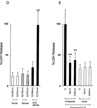
A functional role for ORP150 in the adaptation of cultured MPs to hypoxia was suggested by the impairment of cell viabil-ity in cultures subject to oxygen deprivation, especially in the presence of oxidized LDL. Although MPs exposed to hypoxia for 24 h showed no significant release of LDH into the me-dium, when BK-60 was added to suppress ORP150 expression, cell viability was diminished to a small, but significant extent. Addition of oxidized LDL to the medium of hypoxic MPs in-cubated with BK-60 markedly diminished cell viability com-pared with cultures in which ORP150 expression was not sup-pressed (Fig. 7 C). Addition of Ac-LDL diminished viability of MPs in a manner analogous to Ox-LDL (data not shown). In contrast, native LDL did not affect the viability of hypoxic MPs, even in the presence of antisense oligonucleotide (Fig. 7 D).
[image:8.612.56.555.74.273.2]To further assess mechanisms through which ORP150 ex-erted its cytoprotective effect in hypoxic MPs incubated with oxidized LDL, we evaluated the effect of antioxidants, probu-col, and N-acetylcysteine. Cultures in which ORP150 expres-sion was suppressed by the antisense oligonucleotide (BK-60)
were exposed to hypoxia and oxidized LDL (50 mg/ml); both probucol and N-acetylcysteine had strong protective effects (Fig. 7 E).
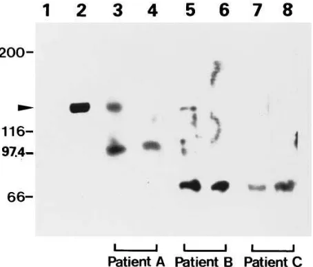
The presence of anti–ORP150 antibody in plasma of pa-tients with severe atherosclerosis. In view of the association of ORP150 expression with cellular stress, as in severe
[image:9.612.58.413.59.428.2]athero-sclerosis, we speculated that patients with extensive vascular disease might develop autoantibodies. Western blotting was performed using cell lysates from cultured human MPs ex-posed to hypoxia as the source of ORP150, and patient sera at 1:50 dilution (Fig. 8). Sera from two patients with angiographi-cally demonstrated severe atherosclerotic vascular disease of
Figure 5. Localization of ORP150 mRNA in atheromatous plaque of aorta. Thoracic aorta was prepared from the 77-yr-old male who died of cardiac rupture after acute myocar-dial infarction (Fig. 4 B, lanes 5 and
6) within 3 h of death and fixed with 4% paraformaldehyde in 0.1 M phos-phate buffer, pH 7.0. Tissue speci-mens were subjected to in situ hy-bridization using rat ORP150 probe (A, 203; C and E, 803). Adjacent sections were stained with hematoxy-lin and eosin (B, 203), monoclonal antibody 1A4 (D, 803), or mono-clonal antibody PG-M1 (anti–CD68 IgG; F, 803).
Figure 6. Localization of ORP150 mRNA in coronary artery. Coronary artery was obtained from the 83-yr-old male who died of cerebral infarction (Fig. 4 B, lanes
7 and 8), and subjected to in situ hybrid-ization using rat ORP150 probe (B, 403
[image:9.612.59.394.532.738.2]Figure 7. Effect of ORP150 antisense oligonucleotide BK-60 on the induc-tion of stress proteins and viability of MPs in hypoxia. (A) Schematic de-piction of ORP150 antisense (BK-59, 60, 61) and sense (BK-609) oligonu-cleotides. (B) Human MPs (z 106 cells) were cultured and exposed to
hypoxia for 24 h either in the absence (lane 2) or presence of ORP150 an-tisense (BK-60, 20 mM, lane 3) or the sense oligonucleotide (BK-609, 20
mM, lane 4). Cells were harvested and 3 mg protein was subjected to im-munoblotting using anti–ORP150 IgG as described (lanes 1–4). Normoxic control sample is shown in lane 1. For detection of HSP72 (lanes 5–8), cul-tured MPs were exposed to elevated temperature (438C) for 3 h in the ab-sence (lane 6) or presence of ORP150 antisense (BK-60, 20 mM, lane 7) or sense oligonucleotide (BK-609, 20 mM, lane 8). Protein was extracted from each sample (z 3 mg) and subjected to immunoblotting with anti–HSP72
antibody. A control experiment was also performed with MPs without ex-posure to elevated temperature (lane 5). The migration of simultaneously run molecular weight markers is shown on both sides of the gel. (C) Hu-man MPs (z 2 3 105 cells) were cultured under either normoxic (N) or
hy-poxic (H) condition for 24 h either in the absence (open bars) or presence of sense oligonucleotide (BK-609, 20 mM, hatched bars) or antisense oligonucleotide (BK-60, 20 mM, closed bars). Where indicated, oxidized LDL (50 mg/ml) was added to the culture 8 h before harvest (OxLDL). LDH activity in the culture supernatant was then measured and ex-pressed as a percentage of maximal release (arbitrarily defined as 100% in the presence of hypoxia 1 oxidized LDL 1 BK-60). Mean6SD is shown (n5 6). *P, 0.01 by Newman-Keul’s analysis compared with H in the presence of BK-60. (D) Human MPs (z 2 3 105 cells) were
cul-tured under hypoxic (H) conditions for 24 h either in the absence (open bars) or presence of sense oligonucleotide (BK-609, 20 mM, hatched bars) or antisense oligonucleotide (BK-60, 20 mM, closed bars). Either oxidized LDL (OxLDL, 50 mg/ml) or native LDL (NaLDL, 50 mg/ml) was added to the culture 8 h before harvest. LDH activity in each culture was measured and expressed as a percentage of maximal release (that observed in cultures exposed to hypoxia 1 oxidized LDL 1 BK-60 assigned a value of 100%). **P, 0.01 compared with H 1 NaLDL 1 BK-60 by nonpaired t test. (E) Human MPs (z 2 3 105 cells) were cultured for 24 h under hypoxia in the presence (closed bars) or absence (open bars)
of antisense oligonucleotide (BK-60, 20 mM). Oxidized LDL (50 mg/ml) was added to each culture 8 h before harvest. Where indicated, either probucol (10 mM; Pb) or N-acetylcysteine (10 mM; NAC) was added to the culture 30 min before addition of oxidized LDL. LDH activity in each culture was measured and expressed as a percentage of maximal release (that observed in cultures exposed to hypoxia 1 oxidized LDL 1
the thoracic and abdominal aorta were studied. Patient A was a 73-yr-old male with angina pectoris who also suffered from occlusion of the right common carotid artery, and patient B was a 65-yr-old male whose vascular disease included subcom-plete stenosis of both carotid arteries. Sera from patients A and B detected the 150-kD band in hypoxic MPs (Fig. 8, lanes
3 and 5), and appearance of the band was blocked by addition of excess of anti–human ORP150 antibody (Fig. 8, lanes 4
and 6). Sera from an age-matched control individual with no evident vascular disease showed no immunoreactivity with the 150-kD band (Fig. 8, lanes 7 and 8). We developed an ELISA to facilitate comparison of anti–ORP150 antibodies in patient sera, and used this method to analyze additional patient sam-ples (Table I). The results in the ORP150 ELISA confirmed the presence of significant titers of autoantibodies in four of six patients with advanced atherosclerosis, whereas none of the age-matched controls (i.e., patients without evidence of ath-erosclerotic complications) had significant titers. It is impor-tant to note that sera from patients with atherosclerosis were also reactive with other polypeptides whose expression was in-duced in MPs by oxygen deprivation, especially those with Mrs
corresponding to z 72 and 98 kD.
Discussion
Chronic and intermittent hypoperfusion in arterial wall, result-ing in insufficient delivery of nutrients to vascular cells, is be-lieved to contribute to the pathogenesis of atherosclerotic
le-sions (27–30). One important facet of this ischemic milieu is oxygen deprivation in the vascular microenvironment (31). In this context, experimental atherosclerosis and hypertension are associated with diminished arterial wall oxygenation (32, 33), suggesting that relative hypoxia of vascular cells may be a com-mon event in vascular dysfunction.
ORP150, originally purified and cloned from cultured rat astrocytes exposed to hypoxia, has also been observed in cer-tain tumor cells subject to oxygen deprivation (34). A principal factor underlying ORP150 induction is lowering of ambient oxygen tension in a range of cell types (HeLa, U373, MPs, and smooth muscle cells). Among cells of the atherosclerotic vessel wall, MPs appear to express the highest levels of ORP150. This is consistent with our results in cell culture demonstrating in-creased expression of ORP150 by MPs, which is further poten-tiated in the presence of pathophysiologically relevant pertur-bants such as modified LDL. The observed elevated levels of ORP150 mRNA in MPs present in atherosclerotic plaques em-phasize the association of ORP150 with cellular stress, al-though it is likely that more complex mechanisms may contrib-ute to ORP150 expression in late atherosclerotic lesions compared with those operative in vitro. Previous studies have shown that a range of other stress proteins are induced by hy-poxic and/or ischemic stress, (e.g., glucose-regulated and heat shock proteins) (1). In view of the likely protective function of these other stress proteins in promoting cell survival in re-sponse to environmental challenges (35), we speculated that ORP150 might behave similarly. The role of MPs as
gers of cellular debris and orchestrators of vascular remodeling suggests their facile adaptation to stressful stimuli (36, 37).
Our previous analysis of ORP150 provided certain clues as to how this inducible polypeptide might have cytoprotective properties. First, it has two HSP70 family-specific signatures, structural motifs likely to have ATPase activity. Second, the presence of a COOH-terminal KNDEL sequence suggests ORP150 may be retained in the endoplasmic reticulum, a loca-tion in which other stress proteins are found (38) and where it is ideally positioned to facilitate protein folding. Our recent studies have confirmed localization of this stress protein in the endoplasmic reticulum by immunocytochemical analysis (5). Further insight into the mechanisms through which ORP150 exerts its effects is the ability of antioxidants, such as probucol or N-acetylcysteine, to protect MPs from death after suppression of ORP150 expression in a hypoxic environment with oxidized LDL. These data imply that ORP150 bound to the endoplas-mic reticulum may have cytoprotective properties, potentially related to the oxidant status of the cell. Expression of ORP150 in macrophages under ischemic microenvironments promotes cell survival and thus is likely to be critical in enabling mac-rophages to redirect their biosynthetic activities in hypoxia. This results in events such as the elaboration of growth and/or migratory factors central to stimulation of angiogenesis in re-sponse to hypoxia (39, 40).
The presence of autoantibodies to ORP150 in the plasma of patients with severe atherosclerosis also emphasizes the
presence of this protein in vascular lesions. Although we have not performed a detailed analysis of every tissue for ORP150 at this time, the association of autoantibodies with severe vas-cular disease suggests a possible sequence of events: ORP150 induction occurs in vascular cells, especially macrophages in atherosclerotic lesions. During the course of lesion formation, certain cells that express ORP150 die (such as degenerating macrophages, observed in atherosclerotic lesions) or cata-strophic events such as plaque rupture (41) disrupt a large number of cells, releasing their contents and allowing the host to mount an antibody response. Consistent with these data, au-toantibody to other stress proteins has been detected in patient plasma (42). This may account for the other bands visualized when patient sera were reacted with lysates of hypoxic, vs lack of reactivity with normoxic, extracts of mononuclear phago-cytes (Fig. 8).
In summary, our study provides evidence that a novel hy-poxia-inducible protein, ORP150, is present in the atheroscle-rotic vessel wall, especially in mononuclear phagocytes. The cytoprotective role of macrophage ORP150 in the setting of oxygen depletion and the presence of oxidized LDL suggests that it may be an important survival factor allowing MPs to carry on their role of tissue remodeling and scavenging under environmentally challenging conditions.
Acknowledgments
We thank Ms. M. Shimomura and R. Manabe for their secretarial as-sistance.
[image:12.612.59.286.59.253.2]This work was supported by a Grant-in-Aid from the Ministry of Education, Science and Culture of Japan, a Research Grant for car-diovascular disease from the Ministry of Health and Welfare of Ja-pan, and grants from the Public Health Service (HL42507, HL52609, PERC).
Figure 8. Detection of autoantibody to ORP150 in the plasma of pa-tients with atherosclerosis. Protein extract from MPs (5 mg) cultured under normoxic (lane 1) or hypoxic (lane 2) conditions for 24 h was subjected to immunoblotting using rabbit anti–human ORP150 anti-body. Other lanes of the blot, containing extract of hypoxic MPs, were reacted with serum prepared from three patients (see below) in the absence (lanes 3, 5, and 7) or presence (lanes 4, 6, and 8) of rabbit anti–human ORP150 antibody (200 ng/ml). Patient A was a 73-yr-old male with angina pectoris and cerebral infarction (lanes 3 and 4). Pa-tient B was a 65-yr-old male with cerebral infarction (lanes 5 and 6). Patient C was a 74-yr-old female without any evidence of atheroscle-rosis (7 and 8). In each case, atherosclerosis was evaluated by angiog-raphy of the thoracic and abdominal aorta and cerebral arteries. The migration of simultaneously run molecular weight markers is shown on the left side of the gel.
Table I. ELISA for Anti–ORP150 Autoantibody in Patient Sera
Case Age/sex Clinical feature Dilution
1* 73/M Angina & cerebral infarction 640 2* 65/M Cerebral infarction 640 3 60/F Hypertension & ASO 1,280
4 68/M ASO 640
5 63/M Dissecting aneurysm 320
6 61/M Hypertension 320
7* 74/F Age-matched control , 80 8 54/M Age-matched control 160 9 80/F Age-matched control 160 10 66/M Age-matched control , 80 11 68/M Age-matched control , 80
The ORP150-enriched fraction was absorbed to wells of ELISA plates to which anti–human ORP150 IgG had been bound. After incubation at 37°C for 1 h, plates were washed three times with PBS containing Tween 20 (0.05%) and incubated for 1 h at 37°C with patient sera as in-dicated. Plates were then washed four times, sites of primary antibody
visualized using peroxidase-conjugated anti–human IgG, and OD495 was
measured. The background in each serum sample was obtained by pre-incubating with anti–rat ORP150 IgG. Anti–ORP150 autoantibody in
patient serum was titrated to a dilution at which OD5205 0.2 was
[image:12.612.316.555.82.238.2]References
1. Wilson, R.E., and R.M. Sutherland. 1989. Enhanced synthesis of specific proteins, RNA, and DNA caused by hypoxia and reoxygenation. Int. J. Radiat. Oncol. Biol. Phys. 16:957–961.
2. Maeda, Y., M. Matsumoto, T. Ohtsuki, K. Kuwabara, S. Ogawa, O. Hori, D.Y. Shui, T. Kinoshita, T. Kamada, and D.M. Stern. 1994. Hypoxia-reoxygen-ation mediated induction of Interleukin-6 in cultured rat astrocytes and expres-sion in ischemic gerbil brain: a paracrine mechanism enhancing neuron survival. J. Exp. Med. 180:2297–2308.
3. Hori, O., M. Matsumoto, K. Kuwabara, Y. Maeda, H. Ueda, T. Ohtsuki, T. Kinoshita, S. Ogawa, D.M. Stern, and T. Kamada. 1996. Exposure of astro-cytes to hypoxia/reoxygenation enhances expression of glucose-regulated pro-tein 78 facilitating astrocyte release of the neuroprotective cytokine Interleukin 6. J. Neurochem. 66:973–979.
4. Kitagawa, K., M. Matsumoto, M. Tagaya, K. Kuwabara, R. Hata, N. Handa, R. Fukunaga, K. Kimura, and T. Kamada. 1991. Hyperthermia-induced neuronal protection against ischemic injury in gerbils. J. Cereb. Blood Flow Metab. 11:449–452.
5. Kuwabara, K., M. Matsumoto, J. Ikeda, O. Hori, S. Ogawa, Y. Maeda, K. Kitagawa, N. Imuta, K. Kinoshita, D.M. Stern, et al. 1996. Purification and characterization of a novel stress protein, the 150 kDa oxygen-regulated pro-tein (ORP150), from cultured rat astrocytes and its expression in ischemic mouse brain. J. Biol. Chem. 271:5025–5032.
6. Koga, S., S. Ogawa, K. Kuwabara, J. Brett, L.A. Leavy, J. Ryan, Y. Koga, J. Plocinski, W. Benjamin, D.K. Burns, and D.M. Stern. 1992. Synthesis and release of Interleukin 1 by reoxygenated human mononuclear phagocyte. J. Clin. Invest. 90:1007–1015.
7. Ogawa, S., H. Gerlach, C. Esposito, A.P. Macaulay, J. Brett, and D.M. Stern. 1990. Hypoxia modulates the barrier and coagulant function of cultured bovine endothelium. J. Clin. Invest. 85:1090–1098.
8. Hori, O., M. Matsumoto, Y. Maeda, H. Ueda, T. Ohtsuki, D.M. Stern, T. Kinoshita, S. Ogawa, and T. Kamada. 1994. Metabolic and biosynthetic alter-ations in cultured astrocytes exposed to hypoxia/reoxygenation. J. Neurochem. 62:1489–1495.
9. Basu, S.K., J.L. Goldstein, R.G.W. Anderson, and M.S. Brown. 1976. Degradation of cationized low density lipoprotein and regulation of cholesterol metabolism in homozygous familial hypercholesterolemia fibroblasts. Proc. Natl. Acad. Sci. USA. 73:3178–3182.
10. Endemann, G., L.W. Stanton, K.S. Madden, C.M. Bryant, R.T. White, and A.A. Protter. 1993. CD36 is a receptor for oxidized low density lipoprotein. J. Biol. Chem. 268:11811–11816.
11. Laemmli, U. 1970. Cleavage of structural proteins during the assembly of the head of bacteriophage T4. Nature (Lond.). 277:680–685.
12. Fong, L.G., S. Parthasarathy, J.L. Witzum, and D. Steinberg. 1987. Nonenzymatic oxidative cleavage of peptide bonds in apoprotein B-100. J. Lipid Res. 28:1466–1477.
13. Steinbrecher, U.P., S. Parthasarathy, D.S. Leake, J.L. Witzum, and D. Steinberg. 1984. Modification of low density lipoprotein by endothelial cells in-volves lipid peroxidation and degeneration of low density lipoprotein phospho-lipids. Proc. Natl. Acad. Sci. USA. 81:3883–3887.
14. Tam, J.P. 1989. Synthetic peptide vaccine design: synthesis and proper-ties of a high-density multiple antigenic peptide system. Proc. Natl. Acad. Sci.
USA. 86:9084–9088.
15. Cooper, H.M., and Y. Paterson. 1991. Production of antibodies. In Cur-rent Protocol in Immunology. J.E. Coligen, A.M. Kruisbeek, D.H. Margulies, E.M. Shevach, and W. Strober, editors. John Wylie & Sons, Inc., New York. 2.4.1–2.4.7.
16. Towbin, H., T. Strachelin, and J. Gordon. 1979. Electrophoretic trans-fer of protein from polyacrylamide gels to nitrocellulose sheets; procedures and some applications. Proc. Natl. Acad. Sci. USA. 76:4350–4354.
17. Lowry, O., N.J. Rosenbrough, L.A. Farr, and R.J. Randall. 1951. Pro-tein measurement with the Folin phenol reagent. J. Biol. Chem. 193:265–275.
18. Shiratori, Y., K.A. Okwu, and I. Tabas. 1994. Free cholesterol loading of macrophages stimulates phosphatidylcholine biosynthesis and up-regulation of CTP: phosphocholine cytidylyltransferase. J. Biol. Chem. 269:11337–11348.
19. Chirgwin, J.M., A.E. Przybyla, R.J. MacDonald, and W.J. Rutter. 1979. Isolation of biologically active ribonucleic acid from sources enriched in ribonu-clease. Biochemistry. 18:5294–5299.
20. Stary, H.C., A.B. Chandler, R.E. Dinsmore, V. Fuster, S. Glasgov, W. Insull, M.E. Rosenfeld, C.J. Schwartz, W.D. Wagner, and R.W. Wissler. 1995. A definition of advanced types of atherosclerotic lesions and a histological clas-sification of atherosclerosis—a report from the committee of vascular lesions of council on atherosclerosis, American Heart Association. Arterioscler. Thromb. Vasc. Biol. 15:1512–1531.
21. Skalli, O., P. Ropraz, A. Trzeciak, G. Benzonana, D. Gillessen, and G. Gabbiani. 1986. A monoclonal antibody against a-smooth muscle actin: a new probe for smooth muscle differentiation. J. Cell. Biol. 103:2787–2796.
22. Sanger, A., S. Nicklen, and A. Coulson. 1977. DNA sequencing with chain terminating inhibitors. Proc. Natl. Acad. Sci. USA. 74:5463–5467.
23. Hirota, S., M. Imakita, K. Kohri, A. Ito, E. Morii, S. Adachi, H.M. Kim, Y. Kitamura, C. Yutani, and S. Nomura. 1993. Expression of osteopontin mes-senger RNA by macrophages in atherosclerotic plaques. Am. J. Pathol. 143: 1003–1008.
24. Falini, B., L. Flenghi, S. Pileri, M. Gambacorta, B. Bigerna, H. Durkop, F. Eitelbach, J. Thiele, R. Pacini, A. Cavaliere, et al. 1993. PG-M1: a new mono-clonal antibody directed against a fixative-resistant epitope on the macrophage-restricted form of CD68 molecule. Am. J. Pathol. 142:1359–1372.
25. Matsuo, N., S. Ogawa, Y. Imai, T. Takagi, M. Tohyama, D.M. Stern, and A. Wanaka. 1995. Cloning of a novel RNA binding polypeptide (RA301) induced by hypoxia/reoxygenation. J. Biol. Chem. 270:28216–28222.
26. Kuwabara, K., D.J. Pinsky, A.M. Schmidt, C. Benedict, J. Brett, S. Ogawa, M.J. Broekman, A.J. Marcus, R.R. Sciacca, and M. Michalak. 1995. Calreticulin, an antithrombotic agent which binds to vitamin K–dependent co-agulation factors, stimulates endothelial nitric oxide production, and limits thrombosis in canine coronary arteries. J. Biol. Chem. 270:8179–8187.
27. Barker, S.G., A. Talbert, S. Cottam, P.A. Baskerville, and J.F. Martin. 1993. Arterial intimal hyperplasia after occlusion of the adventitial vasa va-sorum in the pig. Arterioscler. Thromb. 13:70–77.
28. Martin, J.F., R.F. Booth, and S. Moncada. 1991. Arterial wall hypoxia following thrombosis of the vasa vasorum is an initial lesion in atherosclerosis. Eur. J. Clin. Invest. 21:355–359.
29. Heistad, D.D., M.L. Marcus, G.E. Larsen, and M.L. Armstrong. 1982. Role of vasa vasorum in nourishment of the aortic wall. Am. J. Physiol. 240: 781–787.
30. Nakata, Y., and S. Shionoya. 1966. Vascular lesions due to obstruction of the vasa vasorum. Nature (Lond.). 212:1258–1259.
31. Crawford, D.W., and D.H. Blankenhorn. 1991. Arterial wall oxygen-ation, oxyradicals, and atherosclerosis. Atherosclerosis. 89:97–108.
32. Santilli, S.M., V.D. Fiegel, and D.R. Knighton. 1992. Changes in the aortic wall oxygen tensions of hypertensive rabbits. Hypertension and aortic wall oxygen. Hypertension (Dallas). 19:33–39.
33. Santilli, S.M., V.D. Fiegel, and D.R. Knighton. 1993. Alloxan diabetes alters the rabbit transarterial wall oxygen gradient. J. Vasc. Surg. 18:227–233.
34. Heacock, C.S., and R.M. Sutherland. 1986. Induction characteristics of oxygen regulated protein. Int. J. Radiat. Oncol. Biol. Phys. 12:1287–1290.
35. Pelham, R.B.H. 1986. Speculation on the functions on the major heat shock and glucose-regulated proteins. Cell. 46:959–961.
36. Libby, P., and S.K. Clinton. 1993. The role of macrophage in atherogen-esis. Curr. Opin. Lipidol. 4:355–363.
37. Ross, R. 1993. The pathogenesis of atherosclerosis: a perspective for 1990s. Nature (Lond.). 362:801–809.
38. Lee, A.S. 1992. Mammalian stress response: induction of the glucose regulated protein family. Curr. Opin. Cell Biol. 4:267–273.
39. Knighton, D.R., T.K. Hunt, H. Scheuenstuhl, B. Halliday, Z. Werb, and M. Banda. 1983. Oxygen tension regulates the expression of angiogenesis factor by macrophages. Science (Wash. DC). 221:1283–1285.
40. Kuwabara, K., S. Ogawa, M. Matsumoto, S. Koga, M. Clauss, D.J. Pin-sky, P. Lyn, J. Leavy, L. Witte, J. Joseph-Silverstein, et al. 1995. Hypoxia-medi-ated induction of acidic/basic fibroblast growth factor and platelet-derived growth factor in mononuclear phagocytes stimulates growth of hypoxic endo-thelial cells. Proc. Natl. Acad. Sci. USA. 92:4606–4610.
41. Falk, E. 1992. Why do plaques rupture? Circulation. 86(suppl. III): 30–42.