Technology (IJRASET)
Breast Cancer Segmentation using Entropy Method
A. V. Shelke1, S. R. Jondhale2
Abstract— In this paper, the median filter is used to remove the unwanted noise in the image. After removing the unwanted noise pectoral muscle are removed in the image. Removing unwanted data in the image, it is segmented by using entropy method. The entropy based segmentation approach is proposed to segment a gray-scale breast image. The approach calculates the histogram of an image and also finds the entropy. Then by calculating the thresholding value of an image the segmented image is shown at the output. In this paper, an efficient and fast entropic method for noisy cell image segmentation is presented. Keywords— Breast cancer, median, entropy, pectoral muscle, connected component, thresholding, histogram.
I. INTRODUCTION
Cancer is a class of diseases characterized by out-of-control cell growth. Cancer harms the body when damaged cells divide uncontrollably to form lumps or masses of tissue called tumors. Tumors can grow and interfere with the digestive, nervous, and circulatory systems and they can release hormones that alter body function. Tumors that stay in one spot and demonstrate limited growth are generally considered to be benign[1].
Breast cancer is a kind of cancer that develops from breast cells. Breast cancer usually starts off in the inner lining of milk ducts or the lobules that supply them with milk. A malignant tumor can spread to other parts of the body. A breast cancer that started off in the lobules is known as lobular carcinoma, while one that developed from the ducts is called ductal carcinoma.The vast majority of breast cancer cases occur in females. Breast cancer is the most common invasive cancer in females worldwide. According to the National Cancer Institute, 232,340 female breast cancers and 2,240 male breast cancers are reported in the USA each year, as well as about 39,620 deaths caused by the disease. The cancer cells break out from inside the lobules or ducts and invade nearby tissue known as Invasive breast cancer. With this type of cancer, the abnormal cells can reach the lymph nodes, and eventually make their way to other organs (metastasis), such as the bones, liver or lungs. When the cancer is still inside its place of origin and has not broken out then it is known as Non-invasive breast cancer. Lobular carcinoma in situ is when the cancer is still inside the lobules, while ductal carcinoma in situ is when they are still inside the milk ducts.
This paper organize as, preprocessing part shows the removal of noise from the image using median filter. the next section shows the removal of from an image. In next section the image is segmented by using entropy segmentation method.
II. PREPROCESSING
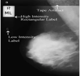
[image:2.612.237.375.575.706.2]The main aim of the pre-processing is to improve the image quality to make it ready to further processing by removing or reducing the unrelated and surplus parts in the background of the mammogram images Mammogram is nothing but a medical images that complicated to interpret. Hence pre-processing is used to improve the quality[2]. Before any image-processing algorithm can be applied on mammogram, preprocessing steps are very important in order to limit the search for abnormalities without undue influence from background of the mammogram.
Technology (IJRASET)
The noise and high frequency components removed by using filters. Filtering is a technique for modifying or enhancing an image. For that purpose median filter is used in this paper. Median filter preserves edges while removing noise[3]. The median filter is a sliding-window spatial filter. It replaces the value of the center pixel with the median of the intensity values in the neighborhood of that pixel. A median filter is more effective than convolution when the goal is to simultaneously reduce noise and preserve edges.
(s,t) Sxy
(x, y)
(s, t)
f
median g
Sxy represent the set of coordinates in a rectangular subimage window (neighborhood) of size m*n, centered at point (x,y). The input
image is g(s,t)[4]. Median filter works on a monochrome color image. It reduces “speckle” and “salt and paper” noise. It does not require convolution. Its edge preserving nature makes it useful in many cases[5]. The median value selected will be exactly equal to one of the existing brightness value so that no round-off error is involved when we work independently with integer brightness values comparing to the other filters.
Figure 2: Original Image Figure 3: Filtered Image
III. PECTORAL MUSCLE REMOVAL
This operation is important in medio-lateral oblique view (MLO), where the pectoral muscle, slightly brighter compared to the rest of the breast tissue, can appear in the mammogram. In properly imaged MLO mammograms, the pectoral muscle is visible as a triangular region of high-density at the upper posterior part of the image. Texture of the pectoral muscle may also be similar to some abnormalities and may cause false positives in the detection of suspicious masses. Pectoral muscles are the regions in mammograms that contain brightest pixels. These regions must be removed before detecting the tumor cells so that mass detection can be done efficiently. Artifact is nothing but some unwanted part which is present in the mammogram [6]. This mammogram images contains wedges and labels in some of the images, breast region is connected with tape artifact. This unnecessary part is removed by using connected component labeling. Because of that only breast part is present in the mammogram.
[image:3.612.140.476.224.362.2]In the 8-neighborhood connectivity in that pixels are connected if their edges or corners touch. This means that if two adjoining pixels are on, they are part of the same object, regardless of whether they are connected along the horizontal, vertical, or diagonal direction.
Figure 4: 8-neighborhood connectivity
Technology (IJRASET)
way connected with each other.
Figure 5: Pectoral Muscle Part Detected Figure 6: Image after Removing Pectoral Muscle
IV. SEGMENTATION
Image segmentation is typically used to locate objects and boundaries (lines, curves, etc.) in images. The result of image segmentation is a set of regions that collectively cover the entire image[12]. Each of the pixels in a region are similar with respect to some characteristic or computed property, such as color, intensity, or texture. Adjacent regions are significantly different with respect to the same characteristics. Several general-purpose algorithms and techniques have been developed for image segmentation. so that the entropy segmentation method is used for segmentation[10].
Steps-
A. Calculating Histogram of an image
B. Calculating the entropy of an image
C. Then choose the threshold value & comparing with entropy of an image.
D. Using this thresholding calculate the segmented portion of an image.
Histogram of an image-
Histograms show the distribution of data values across a data range[8]. They do this by dividing the data range into a certain number of intervals, tabulating the number of values that fall into each interval, and plotting the values in the bins using bars or wedges of varying height. The functions that create histograms are hist and rose.
Calculating Entropy-
Suppose
{ }
P
i in1 be the probability distribution for the image. At the threshold, t this distribution is divided into two sub distributions; one for the foreground (class f) and the other for the background (class b) given byP
f{ }
t 1i i
P
and1
P
b
{ }
P
i i tn respectively. Thus, the generalized Rànyi entropies for the two distributions as functions of t are given as1 2 1
1
(t)
log
1
t k f k t k kP
H
P
1 2 11
(t)
log
1
n kb k t
n k k t
P
H
P
The value of α is greater than zero and less than one so that is gives better result[11].
Thresholding-
Technology (IJRASET)
[image:5.612.247.383.171.300.2]identifying common properties[9]. Or, similarly, we identify contours by identifying differences between regions (edges). The simplest property that pixels in a region can share is intensity. So, a natural way to segment such regions is through thresholding, the separation of light and dark regions. Thresholding creates binary images from grey-level ones by turning all pixels below some threshold to zero and all pixels about that threshold to one. Thresholding method in image segmentation that yields all the pixels and assumes the algorithm in two cases i.e. darkness and brightness. The output might be two label “object” or “background” which due to its dichotomous nature, can be represented as a Boolean variable “1” or “0”. The output from the test condition could be based by some other properties, rather than simple brightness. Using this thresholding calculate the segmented portion of an image.
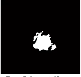
Figure 7: Segmented Image
V. CONCLUSION
Although In this study, system is used to detect the ROI in breast cancer by using entropy segmentation is presented. Also by using several preprocessing techniques the unwanted part is removed in the image. Using Entropy segmentation it shows the tumor area in the image. This system is very useful for diagnosing the tumor area.
REFERENCES
[1] M. Siddiqui, M. Anand, P. Mehrotra, R. Sarangi, N. Mathur, Biomonitoring of organochlorines in women with benign and malignant breast disease, Environmental Research 98 (2) (2005) 250–257.
[2] R. Ramani, Dr. N.Suthanthira Vanitha, S. Valarmathy. “The Pre-Processing Techniques for Breast Cancer Detection in Mammography Images”, I.J. Image, Graphics and Signal Processing, 2013, 5, pp- 47-54
[3] H.D. Cheng, JuanShan, WenJu, YanhuiGuo, LingZhang, “Automated breast cancer detection and classification using ultrasound images: A survey”, Pattern Recognition(20100),pp-299-317.
[4] R. C. Gonzalez. “Digital Image processing using Matlab” Pearson publication, 2005.
[5] Matthias Kirchner a and Jessica Fridrich, “On Detection of Median Filtering in Digital Images”.
[6] H. Abdellatif, t. E. Taha, o. F. Zahran, w. Al-nauimy, f. E. Abd el-samie, “k9. Automatic segmentation of digital mammograms to detect masses,” 30th national radio science conference, 2013, pp-557-565.
[7] R.J. Ferrari, R.M. Rangayyan, J.E. Desautels, R.A. Borges, A.F. Fr`ere,, “Automatic identification of the pectoral muscle in mammo-grams,” IEEE Trans on Medical Imaging, vol. 23,(2) 2004 pp. 232–245.
[8] G. Cao, Y. Zhao, and R. Ni, “Detection of image sharpening based on histogram aberration and ringing artifacts," in Proceedings of the 2009 IEEE International Conference on Multimedia and Expo (ICME 2009), pp. 1026-1029.
[9] Chengxin Yan a, Nong Sang a, Tianxu Zhang, “Local entropy-based transition region extraction and thresholding,” Pattern Recognition Letters 24 (2003), pp 2935–2941.
[10] Amar Partap Singh Pharwaha, Baljit Singh, “Shannon and Non-Shannon Measures of Entropy for Statistical Texture Feature Extraction in Digitized Mammograms,” Proceedings of the World Congress on Engineering and Computer Science 2009 Vol II., October 20-22, 2009, ISBN:978-988-18210-2-7 [11] Samy Sadek, Sayed Abdel-Khalek “ Generalized α-Entropy Based Medical Image Segmentation” Journal of Software Engineering and Applications, 2014, 7,
pp- 62-67