New horizons for studying human hepatotropic
infections
Ype P. de Jong, … , Charles M. Rice, Alexander Ploss
J Clin Invest.
2010;
120(3)
:650-653.
https://doi.org/10.1172/JCI42338
.
The liver serves as a target organ for several important pathogens, including hepatitis B and
C viruses (HBV and HCV, respectively) and the human malaria parasites, all of which
represent serious global health problems. Because these pathogens are restricted to human
hepatocytes, research in small animals has been compromised by the frailty of the current
mouse xenotransplantation models. In this issue of the
JCI
, Bissig et al. demonstrate robust
HBV and HCV infection in a novel xenotransplantation model in which large numbers of
immunodeficient mice with liver injury were engrafted with significant quantities of human
hepatocytes. This technical advance paves the way for more widespread use of human liver
chimeric mice and forms the basis for creating increasingly complex humanized mouse
models that could prove useful for studying immunopathogenesis and vaccine development
against hepatotropic pathogens.
Commentary
Find the latest version:
http://jci.me/42338/pdf
New horizons for studying
human hepatotropic infections
Ype P. de Jong,1,2 Charles M. Rice,1 and Alexander Ploss1
1Center for the Study of Hepatitis C, The Rockefeller University, New York, New York. 2Division of Gastroenterology, Mount Sinai School of Medicine, New York, New York.
The liver serves as a target organ for several important pathogens,
includ-ing hepatitis B and C viruses (HBV and HCV, respectively) and the human
malaria parasites, all of which represent serious global health problems.
Because these pathogens are restricted to human hepatocytes, research in
small animals has been compromised by the frailty of the current mouse
xenotransplantation models. In this issue of the
JCI
, Bissig et al.
demon-strate robust HBV and HCV infection in a novel xenotransplantation model
in which large numbers of immunodeficient mice with liver injury were
engrafted with significant quantities of human hepatocytes. This
techni-cal advance paves the way for more widespread use of human liver
chime-ric mice and forms the basis for creating increasingly complex humanized
mouse models that could prove useful for studying immunopathogenesis
and vaccine development against hepatotropic pathogens.
More than 800 million people are chroni-cally infected with hepatitis B and C viruses (HBV and HCV, respectively) and malaria, causing over 1.5 million deaths annually (1, 2). Persistent HBV and HCV infections can lead to cirrhosis and/or hepatocellular carcinoma (HCC). Basic research, as well as the development of drugs and vaccines targeting human hepatotropic patho-gens, has been handicapped by the lack of robust in vitro and in vivo platforms that mimic human liver biology and disease susceptibility. The narrow species tropism of HBV and HCV restricts preclinical stud- ies to chimpanzees or human liver–chime-ric mice. Large primate studies are often limited by financial and ethical concerns, and humanized mice are currently cum-bersome and low-throughput. In culture, HCC-derived cell lines and immortalized hepatocytes have been useful for study-ing various aspects of HBV and HCV life cycles, but these cells differ phenotypically and functionally from human hepatocytes in vivo. Primary hepatocytes, which can be coaxed to maintain liver-specific functions by culture with fibroblasts, hold promise as a more physiologically relevant in vitro substrate for HCV infection (3). Addressing
fundamental questions about hepatotropic pathogen biology in vivo, however, requires a suitable small animal model to comple-ment and guide more challenging and expensive studies, including clinical trials.
Human liver–chimeric mouse models for human hepatotropic infections
Currently, the most advanced in vivo sys-tems for modeling human hepatotropic infectious disease are human liver–chime-ric mice. A high degree of chimerism (up to 99%) can be achieved by transplanting human hepatocytes into immunodefi-cient mice in which liver injury has been induced to ablate the endogenous murine hepatocytes (Figure 1). The best-charac- terized model to date is the immunode- ficient urokinase-type plasminogen acti-vator (uPA) mouse, in which an albumin (Alb) promoter directs high-level toxic expression of uPA (Alb-uPA mice) (4, 5). The hepatotoxicity creates a permissive environment for the expansion of func-tional, transgene-free human hepatocytes. Human liver–chimeric immunodeficient Alb-uPA mice are susceptible to hepato-tropic human pathogens (4, 5) and have been used in studies ranging from viral evolution to preclinical testing of antivi-ral compounds (6, 7). Working with the Alb-uPA model, however, poses serious challenges (Table 1). The selective pressure needed to achieve and maintain a high level of human chimerism can only be achieved
in homozygous Alb-uPA immunodeficient recipients, which suffer from infertility. Therefore large, costly, hemizygous breed-er cohorts, or rescue of breeder pairs by transplantation with normal hepatocytes, is required to propagate these mice (8). The severe liver injury of homozygous Alb-uPA immunodeficient pups necessitates engraftment surgery in the first weeks of life — a procedure that is complicated by the susceptibility of these animals to fatal hemorrhaging (9). Because of these factors, along with the need for high-quality adult human hepatocytes, only modest numbers of experimental mice can be generated, and at great expense.
To overcome some of these challenges, alternative liver injury models have been developed, including a less toxic Alb-uPA line (10), mice characterized by induc-ible uPA transgene expression (11), or mice in which the major urinary protein (MUP) promoter is used to delay uPA expression (12). Recently, two groups have reported successful engraftment of human hepatocytes into fumaryl acetoac-etate hydrolase–deficient (FAH-deficient) mice bred with mice of a Rag2–/–Il2rgnull
immunodeficient background (FRG mice) (13, 14) (Table 1). FAH is the last enzyme in the tyrosine breakdown pathway (Figure 2), and its deficiency leads to lethal type 1 hypertyrosinemia in humans and liver fail- ure in mice. Treatment with 2-(2-nitro-4- trifluoromethylbenzyol)-cyclohexane-1,3-dione (NTBC) prevents the accumulation of toxic metabolites and hepatotoxicity (15), allowing liver injury to be induced at will by withdrawal of the drug.
Using mice of the FRG background, Bis-sig et al. report consistently high, on average 42%, human liver chimerism after transplan-tation of human adult hepatocytes (Table 1) (16). The trick to achieving this high chime-rism seems to be the use of 3–5 times more human adult hepatocytes than in previous studies (13, 14). The impressively large num-ber of successfully transplanted animals revealed a clear correlation between the level
Conflict of interest: C.M. Rice holds equity in Apath LLC, and AlphaVax Inc. and serves as an advisor for Genentech, GlaxoSmithKline, iTherX, Merck, Novartis Vaccines and Diagnostics, Pfizer, and Vertex.
commentaries
of chimerism and human serum albumin — providing a convenient, minimally inva-sive test to monitor engraftment (16). The authors then tested whether the engrafted animals were susceptible to HBV and HCV infection. Similar to previous studies with immunodeficient Alb-uPA mice (5, 17), FRG mice were shown to be susceptible to HBV infection, irrespective of the level of human chimerism. Hallmarks of viral replication, including the presence of serum HBV DNA, covalently closed circular HBV DNA, and HBV core antigen in liver tissue, were readily observed. In contrast, successful HCV infec-tion could only be achieved if more than 10% of hepatocytes were of human origin, again in line with previous observations in
immunodeficient Alb-uPA mice (4, 5). Inter- estingly, above this 10% threshold of chime- rism, no clear correlation between hepato-cyte engraftment levels and serum viremia was observed. One can only speculate as to why HCV requires substantially higher human chimerism — the short in vivo half-life of the virus may require rapid uptake by new permissive target cells, or successful propagation may necessitate densities of human hepatocytes sufficient for cell-to-cell spread. Importantly, HCV infection could be initiated using cell culture–grown or patient-derived virus, with the highest vire-mia level achieved using a patient-derived isolate. Furthermore, HCV antigens could be detected in the livers of infected animals, traditionally a highly technically difficult achievement (18).
Potential of human liver–chimeric mice in preclinical drug development
[image:3.585.48.544.87.388.2]The susceptibility of humanized FRG mice to hepatotropic infections holds promise for preclinical drug efficacy test-ing. The authors demonstrate a modest decrease in HBV viremia after short-term treatment with the reverse transcriptase inhibitor adefovir dipivoxil, similar to the slow kinetics of viral decline seen in humans treated with this drug (16). For HCV, the current treatment is pegylated IFN-a (peg-IFN) and ribavirin, a combi- nation therapy that is often poorly toler- ated and unsuccessful. Several drug can-didates targeting viral and host proteins are in preclinical or clinical development, and a major question is whether the virus
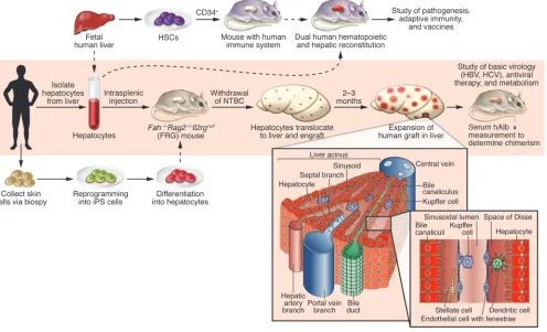
Figure 1
Creation and future improvements of human liver–chimeric FRG mice. In the model described in the current issue by Bissig et al. (16), enclosed in the shaded rectangle, hepatocytes are isolated from adult human liver tissue and injected into FRG mice that are cycled off the protective drug NTBC. Human hepatocyte engraftment levels are then monitored by serial human albumin (hAlb) measurements in the serum of transplanted mice. Over 2–3 months, the human hepatocytes expand and can repopulate up to 97% of the FRG mouse liver, with the remainder of liver-resident cells likely of murine origin. This robust system can be used to study HBV and HCV infections in vivo and can serve as a scaffold for more complex humanized mouse models. The engrafted FRG mice reported by Bissig et al. could potentially be combined with mice bred to posses a human immune system, which are generated by transplantation of human CD34+ stem cells (HSCs) (23). Use of donor hepatocytes and HSCs derived
can be eliminated using solely a cocktail of specific antivirals. Identification of the most effective drug combinations, and the ability to monitor resistance, is an area where model animals could have great utility. Bissig et al. show that a combination of peg-IFN and Debio 025 — an HCV inhibitor targeting the host factor cyclophilin A — is as effective as peg-IFN plus ribavirin over a short-term treatment period. Overall, the results of the Bissig et al. study recapitulate many aspects of HBV and HCV infection in humans, illustrating the promise of this new model for basic studies of human hepatitis viruses and preclinical drug efficacy assessments.
The next steps
The development of more robust human liver–chimeric mice is an important step forward, but further refinements are needed. Despite prolonged high vire-mia, none of the commonly observed sequelae associated with HBV or HCV infections in humans, namely fibrosis or
HCC, were observed in the current study (16). (Patho)physiological processes may require crosstalk between hepatocytes and other liver-resident cells, and, while human hepatocytes are abundant in highly engrafted chimeric mice, non-parenchymal cells are of murine-recipient origin. Kupffer cells and liver sinusoidal endothelial cells, for example, appear to be critical to the ability of Plasmodium sporozoites (the transmission form of
the malaria parasite) to infect the liver, as permissive hepatocytes are not directly accessible to the sporozoites (19). While infection of human liver–chimeric mice with virulent and live-attenuated Plasmo-dium species has been reported (20, 21), infection frequencies were low and may in the future possibly be improved by addi-tion of other human non-parenchymal liver cell subsets.
Another obvious caveat of the current human liver–chimeric models is their immunodeficient background. Chronic inflammation is thought to be a major contributor to fibrosis during persis-tent HBV and HCV infection. Thus, dual engraftment of these animals, resulting in a human liver and a human immune system, is not only critical to monitor immune responses to infection, but also to more faithfully mimic disease patho-genesis (22). Furthermore, since viral hepatitis has become the leading mor-bidity in HIV-infected individuals in the Western world, modeling such coinfec-tions in dually reconstituted mice would be another clinically important applica-tion (Figure 1).
[image:4.585.54.358.118.365.2]In summary, human liver–chimeric mice can serve as valuable tools in preclinical drug efficacy, toxicity, and pharmacokinet-ic applications but also facilitate the study of human hepatotropic pathogens. After more than a decade of working with frail immunodeficient Alb-uPA mice, the FRG model reported by Bissig and colleagues lends itself to become the cornerstone for a wide variety of investigations and will likely provide the platform for further improvements (16).
Table 1
Comparison between FRG mice and immunodeficient Alb-uPA human liver–chimeric mice
FRG mice Immunodeficient Alb-uPA mice
Liver injury
Mutation Fumaryl acetoacetate Urokinase plasminogen hydrolase deficiency activator overexpression Occurrence Inducible upon NTBC withdrawal In utero
Transplantation
Age Any (adult) 7–21 days after birth
Hepatocytes: adult + +
Hepatocytes: fetal Not tested – Hepatocytes: ES-derived Not tested Not tested Hepatocytes: iPS-derived Not yet available Not tested
Human chimerism 3%–97% (average, 42%) Up to 99%
Throughput Medium–high Low
Additional challenges Murine HCC after long-term Infertility of homozygous NTBC withdrawal transgenic mice; coagulopathy
Susceptibility to
Hepatitis B + +
Hepatitis C: HCVcc (genotype 2a) + + Hepatitis C: patient samples + +
Plasmodium spp. Not tested +
Human liver disease after HCV or HBV infection
Fibrosis – –
HCC – –
iPS, induced pluripotent stem; HCVcc, cell culture–derived HCV.
Figure 2
[image:4.585.223.533.542.739.2]commentaries
Acknowledgments
The authors thank Catherine Murray (The Rockefeller University) for editing the manuscript. This work was supported by grants from the Gates Foundation through the Grand Challenges in Global Health ini-tiative (to all authors) and funded in part by the Greenberg Medical Research Insti-tute, the Ellison Medical Foundation, the Starr Foundation, the Ronald A. Shellow Memorial Fund, the Richard Salomon Family Foundation (to C.M. Rice), and the NIH through the NIH Roadmap for Medi-cal Research, grant 1 R01 DK085713-01 (to C.M. Rice and A. Ploss). C.M. Rice is an Ellison Medical Foundation Senior Scholar in Global Infectious Diseases. Address correspondence to: Alexander Ploss or Charles M. Rice, Center for the Study of Hepatitis C, The Rockefeller University, 1230 York Avenue, Box 64, New York, NY 10065. Phone: 212.327.7066; Fax: 212.327.7048; E-mail: [email protected] (A. Ploss). Phone: 212.327.7046; Fax: 212.327.7048; E-mail: [email protected] (C.M. Rice). 1. Alter MJ. Epidemiology and prevention of hepatitis B. Semin Liver Dis. 2003;23(1):39–46.
2. Shepard CW, Finelli L, Alter MJ. Global epidemiol-ogy of hepatitis C virus infection. Lancet Infect Dis. 2005;5(9):558–567.
3. Ploss A, et al. Persistent hepatitis C virus infection in microscale primary human hepatocyte cultures.
Proc Natl Acad Sci U S A. In press.
4. Mercer DF, et al. Hepatitis C virus replication
in mice with chimeric human livers. Nat Med. 2001;7(8):927–933.
5. Meuleman P, et al. Morphological and biochemical characterization of a human liver in a uPA-SCID mouse chimera. Hepatology. 2005;41(4):847–856. 6. Kaul A, Woerz I, Meuleman P, Leroux-Roels G,
Bartenschlager R. Cell culture adaptation of hepati-tis C virus and in vivo viability of an adapted variant.
J Virol. 2007;81(23):13168–13179.
7. Meuleman P, Leroux-Roels G. The human liver-uPA-SCID mouse: a model for the evaluation of antiviral compounds against HBV and HCV. Anti-viral Res. 2008;80(3):231–238.
8. Brezillon NM, et al. Rescue of fertility in homozy- gous mice for the urokinase plasminogen activa-tor transgene by the transplantation of mouse hepatocytes. Cell Transplant. 2008;17(7):803–812. 9. Heckel JL, Sandgren EP, Degen JL, Palmiter RD, Brinster RL. Neonatal bleeding in transgenic mice expressing urokinase-type plasminogen activator. Cell. 1990;62(3):447–456. 10. Suemizu H, et al. Establishment of a humanized model of liver using NOD/Shi-scid IL2Rgnull mice.
Biochem Biophys Res Commun. 2008;377(1):248–252. 11. Song X, et al. A mouse model of inducible liver injury caused by tet-on regulated urokinase for studies of hepatocyte transplantation. Am J Pathol. 2009;175(5):1975–1983.
12. Weglarz TC, Degen JL, Sandgren EP. Hepatocyte transplantation into diseased mouse liver. Kinetics of parenchymal repopulation and identification of the proliferative capacity of tetraploid and octaploid hepatocytes. Am J Pathol. 2000;157(6):1963–1974. 13. Azuma H, et al. Robust expansion of human
hepatocytes in Fah–/–/Rag2–/–/Il2rg–/– mice. Nat Biotechnol. 2007;25(8):903–910.
14. Bissig KD, Le TT, Woods NB, Verma IM. Repopu-lation of adult and neonatal mice with human hepatocytes: a chimeric animal model. Proc Natl Acad Sci U S A. 2007;104(51):20507–20511. 15. Grompe M, et al. Pharmacological correction of
neonatal lethal hepatic dysfunction in a murine model of hereditary tyrosinaemia type I. Nat Genet. 1995;10(4):453–460.
16. Bissig K-D, et al. Human liver chimeric mice pro-vide a model for hepatitis B and C virus infection and treatment. J Clin Invest. 2010;120(3):924–930. 17. Dandri M, et al. Repopulation of mouse liver with
human hepatocytes and in vivo infection with hep-atitis B virus. Hepatology. 2001;33(4):981–988.
18. Liang Y, et al. Visualizing hepatitis C virus infec-tions in human liver by two-photon microscopy.
Gastroenterology. 2009;137(4):1448–1458. 19. Frevert U, Usynin I, Baer K, Klotz C. Plasmodium
sporozoite passage across the sinusoidal cell layer.
Subcell Biochem. 2008;47:182–197.
20. VanBuskirk KM, et al. Preerythrocytic, live-attenuated Plasmodium falciparum vaccine candidates by design. Proc Natl Acad Sci U S A. 2009;106(31):13004–13009.
21. Morosan S, et al. Liver-stage development of Plas-modium falciparum, in a humanized mouse model.
J Infect Dis. 2006;193(7):996–1004.
22. Legrand N, et al. Humanized mice for modeling human infectious disease: challenges, progress, and outlook. Cell Host Microbe. 2009;6(1):5–9. 23. Shultz LD, Ishikawa F, Greiner DL. Humanized
mice in translational biomedical research. Nat Rev Immunol. 2007;7(2):118–130.
24. Sullivan GJ, et al. Generation of functional human hepatic endoderm from human induced pluripo-tent stem cells. Hepatology. 2010;51(1):329–335. 25. Si-Tayeb K, et al. Highly efficient generation of
human hepatocyte-like cells from induced pluripo-tent stem cells. Hepatology. 2010;51(1):297–305. 26. Snykers S, De Kock J, Rogiers V, Vanhaecke T. In
vitro differentiation of embryonic and adult stem cells into hepatocytes: state of the art. Stem Cells. 2009;27(3):577–605.
27. Kneteman NM, Mercer DF. Mice with chimeric liv-ers: who says supermodels have to be tall? Hepatology. 2005;41(4):703–706.
28. Adams DH, Eksteen B. Aberrant homing of muco-sal T cells and extra-intestinal manifestations of inflammatory bowel disease. Nat Rev Immunol. 2006;6(3):244–251.
29. Ploss A, Rice CM. Towards a small animal model for hepatitis C. EMBO Rep. 2009;10(11):1220–1227.
Bidirectional homing of Tregs
between the skin and lymph nodes
Hironori Matsushima and Akira Takashima
Department of Medical Microbiology and Immunology, University of Toledo College of Medicine, Ohio.
Although several homing receptors are known to be differentially
expressed by Tregs in lymphoid tissues compared with those found in
peripheral tissues, it remains unclear whether these cells traffic between
the two locations. In this issue of the
JCI
, Tomura et al. report
steady-state Treg migration from the skin to draining LNs in mice.
Further-more, they report that not only does skin inflammation exacerbate
LN-directed Treg homing, it also triggers reverse circulation of Tregs
from LNs to skin, whereby these cells contribute to regulation of the
immune response. These results now form a new framework for our
understanding of Treg homing.
Conflict of interest: The authors have declared that no conflict of interest exists.