Skin exposure promotes a Th2-dependent
sensitization to peanut allergens
Leticia Tordesillas, … , Hugh A. Sampson, M. Cecilia Berin
J Clin Invest. 2014;124(11):4965-4975. https://doi.org/10.1172/JCI75660.
Sensitization to foods often occurs in infancy, without a known prior oral exposure,
suggesting that alternative exposure routes contribute to food allergy. Here, we tested the
hypothesis that peanut proteins activate innate immune pathways in the skin that promote
sensitization. We exposed mice to peanut protein extract on undamaged areas of skin and
observed that repeated topical exposure to peanut allergens led to sensitization and
anaphylaxis upon rechallenge. In mice, this epicutaneous peanut exposure induced
sensitization to the peanut components Ara h 1 and Ara h 2, which is also observed in
human peanut allergy. Both crude peanut extract and Ara h 2 alone served as adjuvants, as
both induced a bystander sensitization that was similar to that induced by the atopic
dermatitis-associated staphylococcal enterotoxin B. In cultured human keratinocytes and in
murine skin, peanut extract directly induced cytokine expression. Moreover, topical peanut
extract application induced an alteration dependent on the IL-33 receptor ST2 in
skin-draining DCs, resulting in Th2 cytokine production from T cells. Together, our data support
the hypothesis that peanuts are allergenic due to inherent adjuvant activity and suggest that
skin exposure to food allergens contributes to sensitization to foods in early life.
Research Article
Immunology
Find the latest version:
Introduction
IgE-mediated food allergy affects 6%–8% of children, and although allergies to foods such as milk or egg are frequently outgrown, allergies to peanut are commonly lifelong and affect 1%–2% of the population (1). Prevalence of peanut allergy is increasing (2), and reactions to peanut can be severe and occur at a very low dose. It is difficult to avoid peanut due to many fac-tors, including cross-contamination during food processing. What makes peanut such a potent allergen is not understood, although in human in vitro systems it was described that Ara h 1 from pea-nut could bind to DC-specific ICAM3-grabbing nonintegrin (DC-SIGN) in a glycosylation-dependent manner, resulting in a modi-fied DC phenotype and increased capacity for Th2 skewing (3, 4). Innate recognition of allergens has been described for other clini-cally relevant allergens, including dust mite allergens that activate epithelial cells and DCs through TLR4 and dectin-1 (5, 6).
The vast majority of first reactions to peanut and tree nuts occur on the first known ingestion (7), suggesting that expo-sure resulting in primary sensitization likely occurred through another route. There is mixed evidence about a potential role of exposure to peanut in utero or through breast milk (8, 9).
House-hold use of peanut, independent of maternal or infant ingestion of peanut, is a risk factor for development of peanut allergy (10). Furthermore, peanut antigen is readily detectable in household dust (11, 12), indicating that environmental exposure to peanut is likely. Studies on peripheral blood from patients with peanut allergy demonstrate that peanut-responsive T cells are found within the fraction bearing the skin-homing receptor CLA (13), providing support for the hypothesis that initial priming may have occurred through the skin. Ara h 1–MHC II tetramers iden-tify CD4+ T cells in peanut-allergic subjects that express high levels of CCR4 (14), another skin-homing receptor, suggesting that skin exposure may be a highly relevant route of exposure for peanut sensitization. It is not clear if this is unique to peanut or may be common to all foods.
Previous studies have shown that epicutaneous exposure of mice to allergens results in sensitization when skin has been lightly damaged through tape stripping and that this is downstream of an altered immune milieu in the skin, including elevated TSLP and IL-21 (15–17). Tape stripping is a model of excoriation of the skin analogous to that observed in atopic dermatitis, a condition that is a risk factor for the development of food allergy. Furthermore, filaggrin mutations that are associated with defective skin barrier also convey increased risk for peanut allergy (18). Although food allergy is a common feature in children with atopic skin inflamma-tion, children with healthy skin also become sensitized to peanut. We have shown previously that exposure of mice to milk allergens by the epicutaneous route can induce sensitization, but only in the Sensitization to foods often occurs in infancy, without a known prior oral exposure, suggesting that alternative
exposure routes contribute to food allergy. Here, we tested the hypothesis that peanut proteins activate innate immune pathways in the skin that promote sensitization. We exposed mice to peanut protein extract on undamaged areas of skin and observed that repeated topical exposure to peanut allergens led to sensitization and anaphylaxis upon rechallenge. In mice, this epicutaneous peanut exposure induced sensitization to the peanut components Ara h 1 and Ara h 2, which is also observed in human peanut allergy. Both crude peanut extract and Ara h 2 alone served as adjuvants, as both induced a bystander sensitization that was similar to that induced by the atopic dermatitis-associated staphylococcal enterotoxin B. In cultured human keratinocytes and in murine skin, peanut extract directly induced cytokine expression. Moreover, topical peanut extract application induced an alteration dependent on the IL-33 receptor ST2 in skin-draining DCs, resulting in Th2 cytokine production from T cells. Together, our data support the hypothesis that peanuts are allergenic due to inherent adjuvant activity and suggest that skin exposure to food allergens contributes to sensitization to foods in early life.
Skin exposure promotes a Th2-dependent sensitization
to peanut allergens
Leticia Tordesillas,1,2 Ritobrata Goswami,1,2 Sara Benedé,3 Galina Grishina,1 David Dunkin,1 Kirsi M. Järvinen,4
Soheila J. Maleki,5 Hugh A. Sampson,1,2,6 and M. Cecilia Berin1,2,6,7
1Department of Pediatrics and 2Immunology Institute, Icahn School of Medicine at Mount Sinai, New York, New York, USA. 3Institute of Food Science Research (CIAL),
Spanish National Research Council–Autonomous University of Madrid, Madrid, Spain. 4Department of Medicine and Center for Immunology and Microbial Diseases,
Albany Medical College, Albany, New York, USA. 5Department of Agriculture, Agricultural Research Service, Southern Regional Research Center, New Orleans, Louisiana, USA. 6Mindich Child Health and Development Institute and 7Tisch Cancer Institute, Icahn School of Medicine at Mount Sinai, New York, New York, USA.
Authorship note: Leticia Tordesillas and Ritobrata Goswami contributed equally to
this work.
Conflict of interest: The authors have declared that no conflict of interest exists. Submitted: February 18, 2014; Accepted: September 4, 2014.
iment, milk allergen α-lactalbumin (ALA) exposure was used as a negative control group, and mice exposed to ALA alone did not develop ALA-specific IgE and IgG1 responses (data not shown). To further explore the relationship between known allergenicity and responses in this mouse model, mice were epicutaneously exposed to extract from cashew, a tree nut with high allergenicity, and green bean, a legume with low allergenicity in humans (Sup-plemental Figure 2). Mice exposed to cashew without exogenous adjuvant developed cashew-specific IgE and IgG1 responses, while mice exposed to green bean extract developed IgE and IgG1 responses only in the presence of exogenous adjuvant (cholera toxin). These results suggest that the response of mice to epicuta-neous food allergen exposure is consistent with the allergenicity of these foods in humans.
To determine whether the process of hair removal contributed to the sensitization response to peanut, perhaps through emptying of the hair follicle that is densely surrounded by DCs (20), the experi-ment was repeated by applying peanut extract to the ear, without any pretreatment of the skin. Exposure of mice to peanut extract topically on the ear induced peanut-specific IgE and IgG1 responses, basophil activation after in vitro allergen stimulation, and anaphylaxis in mice when rechallenged (Supplemental Figure 3). Altogether, these results show that peanut, similar to cashew and unlike weak allergens such as soy, green bean, or milk ALA, is capable of inducing adjuvant-inde-pendent sensitization by topical exposure on healthy skin.
We next examined which allergens were recognized in mice sensitized by the epicutaneous route (Supplemental Figure 4B) and compared this to the sensitization profile of peanut-allergic subjects (Supplemental Figure 4A). Immunoblotting of peanut extract performed with human sera identified 3 bands in the 14- to 20-kDa range, consistent with Ara h 2 (approximately 17 kDa) and Ara h 6 (approximately 14 kDa). These 3 bands were also detect-able in mice epicutaneously exposed to crude peanut extract (CPE). A higher molecular weight band was also detectable in both humans and mice, consistent with Ara h 1 (63–68 kDa) or Ara h 3 (57 kDa). Using purified Ara h 1 and Ara h 2, we examined the contribution of these allergens in mice sensitized to CPE. Mice exposed to peanut developed detectable IgE antibodies against both Ara h 1 and Ara h 2 (Supplemental Figure 4C), and basophil activation tests performed with whole blood from peanut-sensi-tized mice showed a similar threshold of reactivity to the 2 dom-presence of adjuvant, which was needed to drive dermal DCs to
the draining lymph node (19). Our previous studies indicate that skin exposure is not inherently sensitizing and that some form of adjuvant activity is needed to drive sensitization through the skin. In this study, we show that peanut can induce sensitization when applied to healthy intact skin and that it does so by activating innate immunity in the skin, altering the DC phenotype through mechanisms mediated by the IL-33 receptor ST2, and driving a Th2-polarized T cell response. Our results with peanut allergen support the hypothesis that environmental exposure to peanut is an important risk factor for sensitization.
Results
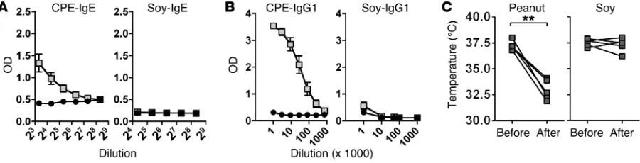
[image:3.585.58.508.57.172.2]Adjuvant-independent sensitization to peanut through epicutaneous exposure. We demonstrated previously that mice can be sensitized to cow’s milk allergens through the epicutaneous route, but only in the presence of exogenous adjuvant (19). To determine whether this adjuvant dependence also applies to the potent food allergen peanut, we exposed mice to crude protein extracts from peanut. Soy extract was used as a comparison extract. The major peanut allergens, Ara h 1, Ara h 2, and Ara h 3, are highly homologous to proteins found within soy, yet soy allergy is rarely persistent or severe. Extracts were applied epicutaneously once per week for 6 weeks. Briefly, hair was removed with depilatory cream, but no tape stripping was performed and antigen was added topically in PBS without occlusive bandages. Histology of the skin after hair removal confirmed that the stratum corneum was intact (data not shown). Mice were then rechallenged and monitored for a drop in body temperature indicative of anaphylaxis. Mice exposed to peanut extract developed robust peanut-specific IgE and IgG1 responses and, when challenged by the intraperitoneal route with 100 μg peanut extract, developed severe anaphylaxis (Figure 1). In contrast, mice exposed to soy extract generated no soy-specific antibodies and did not develop anaphylaxis on soy challenge. However, when mice were exposed to soy in the presence of the superantigen staphylococcal enterotoxin B (SEB), they developed IgE and IgG1 antibody responses to soy (Supple-mental Figure 1; supple(Supple-mental material available online with this article; doi:10.1172/JCI75660DS1). SEB did not further amplify the IgE and IgG1 response to peanut but increased the low IgG2a response observed with peanut alone. In the same
exper-Figure 1. Adjuvant-independent sensitization to peanut through epicutaneous exposure. Mice were topically exposed to 1 mg CPE or soy extract weekly
for 6 weeks. Antigen-specific (A) IgE and (B) IgG1 were measured prior to allergen challenge. Black circles represent values in naive mice, and gray
did not have detectable levels of ALA-specific IgE and had low levels of ALA-specific IgG1 (Figure 2, A and B). Mice exposed to ALA in the presence of either SEB as adjuvant or peanut developed a sig-nificant ALA-specific antibody response, including IgE and IgG1 antibodies (Figure 2, A and B), and to a lesser extent IgG2a (data not shown). To test the functional consequence of bystander sensi-tization, mice were challenged with ALA by oral gavage (Figure 2C). Mice exposed to ALA alone did not undergo anaphylaxis when they were orally rechallenged with ALA, but when mice exposed to ALA together with either SEB or peanut were orally rechallenged with ALA alone, they developed anaphylaxis, as measured by a signifi-cant drop in body temperature. We then tested whether Ara h 2, the dominant allergen in peanut, could induce bystander sensitization. Similar to the results with CPE, topical exposure to Ara h 2 resulted in bystander sensitization to ALA (Figure 2, D and E) and anaphy-laxis in mice orally challenged with ALA alone (Figure 2F). These data conclusively demonstrate that peanut, and its major allergen Ara h 2, have adjuvant activity when applied to the skin.
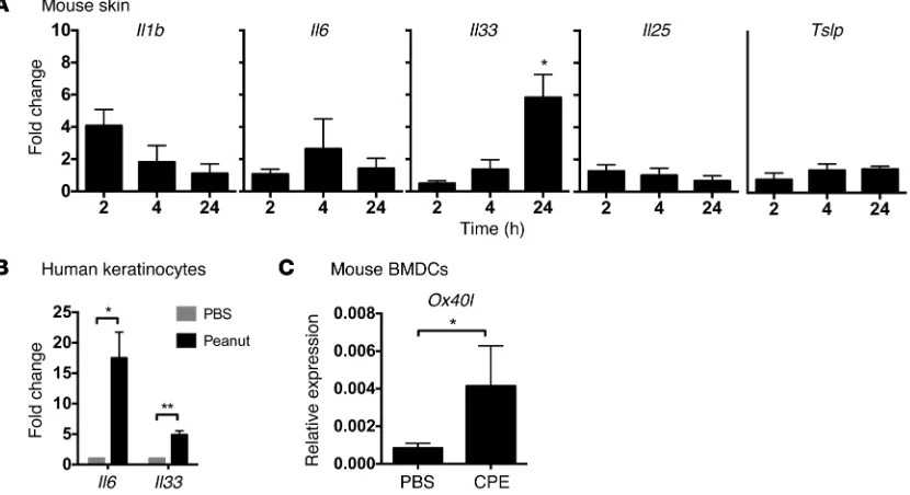
Peanut induces an innate cytokine response. To determine whether peanut can induce innate responses in vivo, peanut extract was applied on the ear, followed by harvesting of tissue RNA over time (2–24 hours) and assessment of cytokine expres-sion by RT-PCR (Figure 3A). We examined expresexpres-sion of the cytokines Il1b, Il6, Il33, Il25, and Tslp. These cytokines were cho-sen since they can alter the phenotype of resident DCs and drive the adaptive immune response away from one of tolerance. We observed significant upregulation of Il33 expression and a trend of increased expression of Il1b but did not observe upregulation of other Th2-promoting cytokines (Il25, Tslp) or proinflammatory cytokines. To test whether keratinocytes could directly recog-nize and respond to peanut exposure, we used primary neonatal human keratinocytes (Figure 3B). We examined expression of the cytokines IL1A, IL1B, TNFA, IL6, GMCSF, TSLP, IL25, and IL33 after stimulation with CPE that had been cleaned of endotoxin. Of these cytokines, IL6 and IL33 were significantly upregulated by exposure of keratinocytes to peanut extract. IL-6 was confirmed at the protein level to be released in response to peanut in a dose- dependent manner (data not shown). Peanut has been shown pre-viously to have direct effects on mouse and human DCs (3, 24). We stimulated bone marrow–derived DCs with peanut extract and assessed phenotype by flow cytometry and real-time PCR. Stimu-lation of cells with peanut extract did not alter the expression of MHC II, Cd80, or Cd86 or significantly change the expression of cytokines, including Il1b, Il6, or Il10. However, stimulation with peanut extract did induce a significant upregulation of Ox40l (Fig-ure 3C), a costimulatory molecule that we and others have previ-ously shown is involved in the induction of Th2 responses after oral sensitization using cholera toxin adjuvant (25, 26).
inant allergens (Supplemental Figure 4D). A titrated intraperito-neal challenge of peanut-sensitized mice demonstrated that both Ara h 1 and Ara h 2 could trigger anaphylaxis, beginning at a dose of 10 μg, but Ara h 2 induced a stronger anaphylactic reaction than Ara h 1 (Supplemental Figure 4E).
To determine whether sensitization could also be established using purified allergens on exposure, we exposed mice to peanut extract or purified Ara h 1 or Ara h 2 epicutaneously without adju-vant (Supplemental Figure 5). Mice developed marked antibody responses to the peanut components (Supplemental Figure 5A), indicating that other components in crude extract are not neces-sary for sensitization. Despite the induction of antibody responses to Ara h 1 and Ara h 2, only mice exposed to Ara h 2 experienced significant anaphylactic reactions following intraperitoneal chal-lenge (Supplemental Figure 5B). Ara h 2 is the component of pea-nut linked most closely to clinical reactivity in humans (21).
Peanut has adjuvant activity on the skin. Sensitization to anti-gens after epicutaneous exposure has been shown previously to be dependent on additional factors, including skin damage (17), pres-ence of inflammation (22), or use of exogenous adjuvants such as SEB (23) or cholera toxin (19). We hypothesized that peanut could be inducing “adjuvant-free” sensitization by providing inherent adjuvant activity. To test this, we examined the capacity of peanut to induce bystander sensitization to the milk allergen ALA that we have previously shown is adjuvant dependent (19). Mice were exposed to ALA alone or in the presence of peanut or SEB as pos-itive control once a week for 6 weeks. Mice exposed to ALA alone
Figure 2. Peanut and Ara h 2 induce bystander sensitization, demonstrat-ing adjuvant activity. (A–C) Mice were exposed to 0.1 mg ALA on the ear,
alone, or in the presence of 10 μg SEB or 1 mg CPE once a week for 6 weeks. ALA-specific (A) IgE and (B) IgG1 were measured in serum prior to oral
challenge with ALA. (C) Body temperature was measured at baseline and
30 minutes after challenge. n = 5 mice per group. (D–F) The experiment was
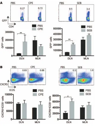
affinity maturation. We hypothesized that peanut or SEB promote bystander sensitization or self-sensitization by inducing Th2 and/ or Tfh responses. To test whether there was an induction of Th2 immunity after sensitization, 4get mice (which have GFP expres-sion in IL-4–expressing cells) (28) were epicutaneously exposed to PBS, SEB, or peanut. One week after exposure, the draining lymph nodes were harvested and assessed for GFP. We observed a sig-nificant increase of GFP-positive CD4+ T cells in the skin-drain-ing lymph nodes (Figure 5A) in response to either peanut or SEB. Induction of reporter activity was restricted to the draining lymph nodes and was not observed at distal lymph nodes (the mesen-teric lymph nodes). GFP reporter activity was observed primar-ily within the memory T cell population (CD44hiCD4+ cells, data not shown). We verified the peanut-dependent induction of Th2 responses by using IL-13 reporter mice (IL-13eGFP mice) (29). An increase in IL-13 reporter activity was also observed in the skin-draining lymph nodes after epicutaneous exposure to pea-nut in total CD4+ and CD44hiCD4+ cells (Supplemental Figure 6 and data not shown). Induction of memory Th2 cells is therefore a common response to exposure to peanut and SEB.
We next wanted to determine whether there was a local induction of Tfh cells after epicutaneous exposure to peanut or SEB. CXCR5+ICOS+ cells in the CD4+ population were defined as the Tfh cell population. Mice exposed to SEB via skin showed an increased population of CXCR5+ICOS+ cells in skin-draining lymph nodes within both total CD4+ and CD44hi memory popu-lations (Figure 5B and data not shown). In contrast, mice epicu-taneously exposed to peanut did not have an expansion of local Tfh cells, indicating that SEB induces a broader immune response than that generated by peanut. IgE class switching has been reported to occur outside of germinal centers (30); however, we do not have direct evidence to suggest non-germinal center local-ization of the peanut-specific IgE response.
DCs from peanut-exposed mice are sufficient to drive Th2 priming. To determine whether epicutaneous peanut exposure modifies the phenotype of DCs in vivo, resulting in functional consequences on T cell priming, we purified DCs from the skin-draining lymph nodes of mice 24 hours after epicutaneous exposure to ovalbumin (OVA) in the presence or absence of peanut. Purified DCs were cocultured with OVA-specific CD4+ T cells from DO11.10 mice at a 1:5 ratio for 3 days, followed by restimulation with anti-CD3/ CD28 to maximize cytokine output. Cytokine secretion was mea-sured by ELISA (Figure 4). DCs harvested from mice epicutane-ously exposed to OVA in the presence of peanut induced signifi-cantly greater Th2 (IL-4 and IL-5) responses compared with those from mice exposed to OVA alone, while IL-10 was unchanged. IL-13, IFN-γ, and IL-17 secretion was elevated but did not reach the level of statistical significance. CPE alone was used as a negative control (without OVA to activate the OVA-specific T cells), with-out significant differences with respect to PBS or OVA alone (data not shown). These results demonstrate that topical peanut expo-sure alters the phenotype of skin-draining DCs in vivo, resulting in an amplification of adaptive immunity, including an elevated Th2 immune response. Interestingly, the DC-driven increase in Th2 priming was not observed when we used SEB rather than CPE as an adjuvant in this model (data not shown), indicating that the mechanism mediating the induction of Th2 responses is different depending on the nature of the adjuvant factor.
[image:5.585.77.492.60.286.2]Induction of Th2 immunity can be driven by peanut or adjuvant, but only adjuvant induces T follicular helper cells. The generation of IgE antibodies resulting in clinical food allergy could potentially be driven by 2 T helper subsets: Th2 cells producing IL-4 and IL-13 and T follicular helper (Tfh) cells. Tfh cells are a subset of CD4+ T cells, which induce immunoglobulin class switching and pro-mote germinal center reactions (27). These cells also secrete low levels of IL-4, which is required for somatic hypermutation and
Figure 3. Innate response to epicutaneous peanut exposure. (A) Mice were exposed to peanut or PBS as control on the ear, followed by RNA isolation and
RT-PCR for cytokines. (B) Human keratinocytes or (C) mouse bone marrow DCs were exposed in vitro to CPE or PBS as control. Fold change is shown with respect
ing lymph nodes but not distal lymph nodes (Figure 7A). ST2 is expressed on a number of different cell types, including DCs, T cells, and innate lymphoid cells. We hypothesized that IL-33 from keratinocytes was modulating the function of DCs. Mice were treated with isotype or anti-ST2 antibody prior to epicutaneous exposure to OVA with peanut as adjuvant. DCs were then purified IL-1, IL-6, and IL-33 link innate and adaptive immunity in the
skin. We observed that peanut induces an innate cytokine response from keratinocytes and alters the phenotype of DCs, contributing to altered T cell priming. We sought to establish a link between the innate immune response and the adaptive immune response in vivo. We first blocked cytokines, specifically IL-6 and IL-1, that we had observed were upregulated in keratinocytes
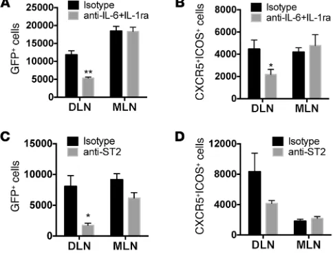
or skin by peanut exposure. IL-6 was blocked with neutralizing anti–IL-6 antibody, while IL-1 sig-naling was blocked with IL-1 receptor antagonist (anakinra). Blockade of IL-6 and IL-1 signaling prior to SEB exposure resulted in a significant sup-pression of IL-4 reporter activity in the skin-drain-ing lymph nodes but did not influence constitutive IL-4 reporter activity in a distal lymph node (Fig-ure 6A). We also observed attenuated numbers of CXCR5+ICOS+ Tfh cells in response to SEB when IL-1 and IL-6 signaling was blocked (Figure 6B). Blockade of ST2, the receptor for IL-33, also led to a significant reduction in IL-4 reporter activity in vivo (Figure 6C) and a trend toward a reduced number of Tfh cells (Figure 6D).
In contrast to the findings with SEB, blocking IL-6 and IL-1 had no effect on peanut-induced IL-4 reporter activity in draining lymph nodes (Supplemental Figure 7). However, similar to that with SEB-induced IL-4 production, neu-tralizing anti-ST2 antibody administered before epicutaneous exposure to peanut significantly suppressed IL-4 reporter activity in
skin-drain-Figure 4. Effect of in vivo exposure to CPE on priming by DCs.
DCs were purified from the skin-draining lymph nodes of BALB/c mice epicutaneously exposed to PBS, OVA, or OVA and CPE and cocultured with DO11.10 CD4+ T cells for 3 days, followed
[image:6.585.42.324.56.229.2]by restimulation with CD3/CD28. Data are mean ± SEM of 7 independent experiments, with each experiment consisting of 3 pooled mice per group. *P < 0.05, **P < 0.01.
Figure 5. Effect of CPE or SEB on Th2 and Tfh cells.
4get mice were epicutaneously exposed to PBS, peanut (CPE), or SEB. One week later, skin-draining lymph nodes (DLN) and mesenteric lymph nodes (MLN) were assessed for (A) IL-4 reporter expression
or (B) Tfh markers CXCR5 and ICOS. Representative
plots of IL-4+ or CXCR5+ICOS+ cells of total CD4+ cells in
skin-draining lymph nodes are shown above summary plots of the mean ± SEM number of cells normalized per million CD4+ T cells for 3 mice per condition.
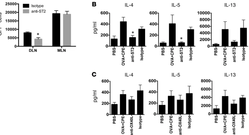
[image:6.585.240.551.336.749.2]from the skin-draining lymph nodes and cocultured with DO11.10 T cells. DCs from peanut-exposed mice induced Th2 priming that was significantly reduced in mice treated with anti-ST2 antibody (Figure 7B), indicating that ST2 was acting at or above the level of the DC, resulting in an altered DC phenotype. We and others have shown that cholera toxin–driven sensitization by gastrointestinal DCs is dependent on OX40L expression by DCs (25, 26); however, blockade of OX40L in the skin DC–T cell cocultures resulted only in a minor reduction in Th2 cytokine production (Figure 7C). The lack of significant effect of OX40L neutralization in response to CPE was also observed using 4get mice in vivo (data not shown). Together, these data suggest that a diverse set of sensitizing stimuli at the level of the skin can induce Th2-skewed immune responses through a ST2-dependent modulation of skin DCs.
Discussion
Peanut is one of the most common foods causing allergic reac-tions, and peanut allergy is increasing in prevalence in the US (2) and the UK (31). Peanut allergy, unlike milk or egg allergy, is com-monly lifelong and is one of the causes of potentially life-threat-ening anaphylactic reactions (32, 33). In this report, we identify novel innate responses to epicutaneous peanut exposure in vivo that contribute to the generation of Th2-biased adaptive responses to peanut. Our results point to inherent adjuvant factors in peanut combined with a nonoral exposure route as being critical to the development of peanut allergy.
Environmental exposure to peanut is a risk factor for the devel-opment of peanut allergy while early dietary exposure of infants to peanut is associated with low prevalence of peanut allergy (10, 34). The skin is of particular interest as a route of allergen exposure, since infant exposure to peanut oil–containing ointments was found to be a risk factor for peanut allergy in the UK (35). Exposure to wheat hydrolysates in facial soap has also been reported to be associated with self-reported wheat allergy (36). We have previously demon-strated that sensitization to milk allergens can be induced via mul-tiple physiological routes in mice, including the epicutaneous route, but only in the presence of an exogenous adjuvant (19). These pre-vious results suggest that skin exposure is not inherently allergenic but, given the appropriate immune environment, can result in
sen-sitization. For example, the presence of filaggrin mutations, which confer susceptibility to atopic dermatitis, is a risk factor for peanut allergy independent of atopic dermatitis (18, 37). This is supported by preclinical data showing increased susceptibility to sensitiza-tion in mice with filaggrin mutasensitiza-tions (38). In addisensitiza-tion, models of skin sensitization to food allergens commonly use tape stripping that causes mechanical injury to promote skin inflammation and the induction of TSLP and Th2 cytokines that lead to sensitization (16, 17, 39, 40). Vitamin D analogs, which upregulate TSLP expres-sion in the skin, can also induce sensitization to food allergens via the skin (22). However, here we show evidence that mice can be sensitized to peanut or cashew via intact skin in an adjuvant-inde-pendent fashion, while soy or green bean extracts, used as control legume extracts, do not sensitize. This suggests that while the skin is not inherently sensitizing upon antigen exposure, it is permissive to sensitization to peanut in the absence of adjuvants, unlike oral exposure, which leads to tolerance. This finding supports the clin-ical observations that exposure to peanut through nonoral routes promotes sensitization (10), while early oral exposure is associated with lower risk of peanut sensitization (34). Our findings also indi-cate that this paradigm may not fit all allergens, as it does not apply to milk (19). The mechanism by which skin, but not gut, exposure to peanut alone can induce sensitization is not clear but may relate to features of the resident DCs within each site, such as differential expression of innate receptors.
Using a series of in vivo approaches, we demonstrated that peanut elicits an innate response after topical application to intact skin, followed by an induction of the Th2-priming potential of DCs in the draining lymph node, leading to the induction of IL-4– and IL-13–expressing CD4+ T cells. Although there was an induction of IL-1β and IL-6 from skin and keratinocytes, respectively, and these cytokines were required for the induction of Th2 and Tfh responses using the adjuvant SEB, we did not find evidence for a role of IL-1β
[image:7.585.37.279.60.248.2]and IL-6 signaling in the induction of Th2 responses to peanut. In both intact mouse skin and human keratinocytes, there was an upregulation of IL-33 in response to peanut exposure. In keratino-cytes this was a late response (24 hours), occurring after the early peak of proinflammatory cytokines (2 hours), suggesting that IL-33 could be upregulated in response to autocrine signals. Blockade of ST2, the IL-33 receptor, led to a significant reduction in Th2 cells in response to epicutaneous peanut exposure. There is growing evidence for a central role of the IL-1 family member IL-33 or ST2 in the induction of sensitization at multiple tissue sites. Sensitiza-tion to dust mite through airway exposure is dependent on ST2 (26, 41), downstream of the innate recognition of dust mite through a TLR4-dependent mechanism (41). Oral sensitization to peanut (requiring cholera toxin adjuvant) is also ST2 dependent (26). ST2-expressing resident eosinophils in the gastrointestinal tract were Figure 6. Contribution of IL-1, IL-6, and IL-33 to Th2 and Tfh induction by SEB. 4get mice were pretreated with either isotype control or (A and B) neutralizing IL-6 antibodies plus IL-1 receptor antagonist or (C and D)
neutralizing anti-ST2 antibody before epicutaneous exposure to SEB. Draining lymph nodes and mesenteric lymph nodes were harvested 1 week later. Summary plots of (A and C) IL-4+ cells or (B and D) CXCR5+ICOS+ cells
per million CD4+ T cells are shown. Data are mean ± SEM of 3 mice per
recently reported to be essential for this oral sensitization to pea-nut by driving CD103+ DCs expressing elevated levels of CD86 and OX40L from the lamina propria to the mesenteric lymph node (42). Kobayashi et al. recently showed that airway administration of exogenous IL-1β or IL-33 together with OVA or ragweed extract could induce IgE production against the allergens (43). Interest-ingly, IL-1βinduced IL-17 production in addition to Th2 cytokines, while IL-33 induced a more restricted Th2 cytokine induction. Sim-ilarly, we observed a broader immunologic impact when mice were exposed to SEB, which induced Th2 and Tfh cells via IL-1–, IL-6–, and ST2-dependent pathways, compared with that when mice were exposed to peanut that induced Th2 cells solely through an ST2-dependent pathway. ST2 is found on a number of different cell types, including innate lymphoid cells (44), allergic effector cells (45, 46), and DCs (47). We did not observe any increase in IL-4 or IL-13 reporter activity in non-CD4+ T cells after peanut exposure in the draining lymph nodes. Furthermore, purified DCs from pea-nut-exposed mice were able to induce Th2 responses from naive T cells in the absence of any other accessory cells, suggesting that DCs are sufficient for the induction of Th2-skewed immune responses to epicutaneous peanut exposure, although the finding does not necessarily rule out contribution from innate sources of Th2 cytokines in vivo. Although TSLP can promote sensitization to allergens via the skin (17, 22, 39), neither mouse skin nor human keratinocytes upregulated TSLP upon peanut exposure, and sen-sitization of mice to peanut through the skin was normal in TSLP receptor–deficient mice (data not shown). This is consistent with our previous findings and those of other groups that primary sensi-tization and generation of IgE in response to oral sensisensi-tization can occur through TSLP-independent pathways (26, 48).
[image:8.585.92.491.55.272.2]Keratinocytes are presumed to be a major source of Th2- inducing cytokines, including IL-33, IL-25, and TSLP, and, con-sistent with a keratinocyte source of IL-33 in our model, we observed that IL33 mRNA expression was regulated by peanut exposure in human keratinocytes in vitro. The receptor for IL-33 is expressed by DCs, among many other cell types, and treatment of DCs with IL-33 has been shown to upregulate OX40L expres-sion (49). ST2 may modulate DC function upstream through an intermediate cell. As an example, gastrointestinal eosinophils express ST2 and are required for oral sensitization to peanut through eosinophil peroxidase-dependent modulation of DC phenotype (42). Recently, two reports identified a skin-draining DC subset as being critical for the induction of Th2 immunity (50, 51). The markers CD301b and PD-L2 were identified on these Th2-inducing DCs, and this DC subset expressed high lev-els of the IL-33 receptor ST2 (50). Furthermore, these DCs were required for induction of Th2 responses but not Tfh responses. In addition to acting on DCs, IL-33 can also be produced by DCs and act directly on T cells. IL-33 was recently shown to be regu-lated by IRF-4 in DCs, and DC-derived IL-33 was necessary for the induction of Th2 immunity (52). In a model of bee venom– induced Th2 immunity, IL-33 was shown to be central to the induction of Th2 immunity but via direct actions on T cells rather than through actions on a DC subset (53). Our data suggest that ST2 is acting at the level of the DC or higher, rather than directly on the T cells or through innate lymphoid cells, since neutral-ization of ST2 in vivo suppressed the ability of DCs to directly induce Th2 priming after peanut exposure. ST2 also negatively regulates TLR2 signaling (54), suggesting that ST2 blockade may have effects beyond neutralization of IL-33 signaling.
Figure 7. Peanut induces Th2 polarization through ST2-mediated effects on skin-draining DCs. 4get mice were pretreated with either isotype control or
neutralizing ST2 antibodies before epicutaneous exposure to CPE. One week after exposure, draining lymph nodes and mesenteric lymph nodes were har-vested, and (A) IL-4 reporter activity was quantified. Summary data (mean ± SEM) showing number of cells per million CD4+ T cells are shown. n = 3 mice
per condition. *P < 0.05. (B) BALB/c mice were pretreated with isotype control or anti-ST2 antibody prior to exposure to PBS or OVA and CPE. DCs from
skin-draining lymph nodes were cocultured with DO11.10 T cells, and cytokine output was measured. Data are mean ± SEM of 3 independent experiments. *P < 0.05, compared with isotype control. (C) DCs from skin-draining lymph nodes were isolated after exposure to OVA and CPE and cocultured with
extracts were prepared as described above. ALA and OVA (grade V) were purchased from Sigma-Aldrich. Purified Ara h 1 and Ara h 2 for in vivo studies and basophil activation tests were prepared as previously described (63, 64). Extracts used in cell culture were purified from endotoxin using Detoxi-Gel columns (Pierce). SEB was from Toxin Technologies. Cholera toxin was from List Biolog-ical Laboratories.
Mice. C3H/HeJ and BALB/c mice were obtained from National
Cancer Institute, and C3H/HeOuJ mice were obtained from Jackson Laboratories. 4get (28) and DO11.10 mice (Jackson Laboratories) were maintained as breeding colonies at Mount Sinai. IL-13eGFP (29) mice were originally developed by Andrew McKenzie (MRC Laboratory of Molecular Biology, Cambridge, United Kingdom) and provided by Paul Bryce (Northwestern University, Chicago, Illinois, USA).
Epicutaneous sensitization of mice. Six- to eight-week-old female
mice were anesthetized, and abdominal fur was removed with depil-atory cream (Veet; Reckitt Benckiser), immediately followed by expo-sure to antigens (1 mg crude extract or 0.1 mg purified antigens) in 50 to 100 μl PBS spread thinly on the abdominal skin to dry. SEB or cholera toxin were applied at a dose of 10 μg. In some experiments, antigens were applied to the ear pinnae. Mice were exposed weekly for a total of 6 exposures and challenged a minimum of 1 week after the last exposure.
Allergen challenge and assessment of anaphylaxis. Mice underwent
an oral challenge to allergen (50 mg ALA or OVA) and were observed for 30 minutes following challenge. Body temperature was measured before and 30 minutes after challenge. For extracts, mice were chal-lenged by intraperitoneal route with 100 μg antigen and followed for a further 30 minutes prior to measuring temperature with a rectal ther-mometer (WPI Instruments).
Cytokine neutralization. During neutralization experiments, mice
were pretreated with neutralizing anti–IL-6 (0.5 mg) or isotype anti-body (BioXCell) together with the IL-1 receptor antagonist anakinra (0.1 mg per mouse, provided by Raphaela Goldbach-Mansky, NIH, Bethesda, Maryland, USA). IL-33 receptor (ST2) was blocked with neutralizing anti-ST2 (20 μg) or isotype control antibody (R&D Sys-tems). Mice were then epicutaneously exposed to OVA and SEB or CPE. Mice undergoing IL-6/IL-1 or ST2 neutralization received half of the initial antibody amount administered every other day for 7 days. OX40L was blocked with anti-OX40L (0.2 mg per mouse) or isotype control antibody (R&D Systems) twice in 1 week.
Flow cytometry. One week after epicutaneous exposure mice were
sacrificed, and cells from skin-draining lymph nodes and mesenteric lymph nodes were harvested, washed, and stained with anti-mouse CXCR5 (BD Biosciences), mouse CD44 (Biolegend), anti-mouse CD4 (eBioscience), and anti-anti-mouse ICOS (eBioscience), in the presence of FcBlock (eBioscience). Cells were acquired on a BD LSR Fortessa cytometer (BD Biosciences). Data were analyzed using FlowJo software (TreeStar Inc.).
Skin gene expression. CPE (1 mg) applied on the left ears and right
ears of mice was used as negative control. Ears were harvested 2, 4, and 24 hours after the CPE application. Total RNA was isolated with Trizol (Invitrogen, Life Technologies), followed by RNA clean up with the RNeasy Mini Kit (Qiagen).
Human keratinocytes. Human epidermal keratinocytes
(neona-tal cells, Invitrogen, Life Technologies) were cultured using EpiLife medium with growth supplements (all from Gibco, Life Technologies). We and others have shown that OX40L on DCs is responsible
for DC-mediated Th2 skewing and sensitization in the gastroin-testinal tract (25, 26, 55). In the current studies, OX40L neutraliza-tion had only a minor inhibitory effect on DC-driven Th2 cytokine production in vitro and in vivo, indicating that other mechanisms for DC-driven Th2 priming must predominate in the skin.
Our data contributes to the growing body of literature showing that allergens activate innate immunity. Allergy to house dust mite is one of the most common allergic responses worldwide (56). In the case of house dust mite, there are several allergens that have pro-tease activity (Der p 1, Der p 3, Der p 6, Der p 9) and a capacity to bind to lipids (Der p 2) (6, 57, 58). Der p 2 activates the TLR4 signal-ing pathway by actsignal-ing as a MD2 mimetic and induces sensitization in a TLR4-dependent manner that is dependent on presence of LPS within the preparation (5). In pollens, the presence of pollen-associ-ated lipid mediators that promote Th2 responses has been described (59). Lectins derived from legumes are able to activate basophils to release IL-4 and IL-13 (60). Peanut has innate activity on human DCs, in part through Ara h 1 binding to DC-SIGN (3). Peanut can also activate complement that may influence adaptive immune responses (61). We found that purified Ara h 2 was sufficient to induce IgE pro-duction in mice, leading to anaphylaxis upon challenge, and could induce bystander sensitization, demonstrating inherent adjuvant activity. Ara h 2 is the dominant allergen in peanut, and component testing has indicated that IgE against Ara h 2 is predictive of clinical reactivity in patients (21). Interestingly, we did not observe innate effects of Ara h 2 on human keratinocytes (data not shown), and, unlike Ara h 1, Ara h 2 is not significantly glycosylated. Innate effects of peanut extract on human DCs were previously shown to be glyco-sylation dependent, and purified Ara h 2 did not induce a Th2-skew-ing phenotype in human DCs (3). Thus, the molecular mechanism by which Ara h 2 may induce bystander sensitization is unclear. It was shown, using timothy grass extract, that Th2-inducing activity could be discriminated from IgE binding, and several novel proteins were identified that were capable of Th2-inducing activity but were not recognized by IgE (62). Similar approaches are needed to iden-tify all potential T cell modulatory fractions of peanut that may be distinct from the dominant allergens.
In summary, we have shown that the skin is permissive to aller-gic sensitization on exposure to peanut without additional adjuvants or tissue damage and that peanut induces Th2 skewing through a DC-autonomous pathway that is ST2 dependent. We contrast this with sensitization induced by the superantigen SEB, which induces both Th2 skewing and Tfh induction through IL-1– and IL-6–depen-dent as well as ST2-depenIL-6–depen-dent pathways. Our data provide a mech-anistic explanation for the epidemiologic observations that nonoral household exposure to peanut is a risk factor for peanut allergy. Fur-thermore, this model of adjuvant-independent sensitization to pea-nut provides a clinically relevant model of peapea-nut allergy that can be used to dissect the molecular basis of peanut allergenicity.
Methods
Antigens and adjuvants. Defatted CPE was prepared as previously
tralization, DCs were cocultured with T cells in the presence of anti-OX40L (10 μg/ml) or isotype control antibody (R&D Systems).
Immunoblotting. CPE (15 μg/cm) was separated on SDS-PAGE
(NuPage MES, 4% to 12% ZOOM gels, Invitrogen). The protein was transferred onto nitrocellulose membranes (Bio-Rad). After blocking, membranes were incubated with diluted human sera (1:50), followed by detection with mouse Anti-Human IgE-Alkaline Phosphatase (Sigma-Aldrich), alkaline phosphatase chemiluminescent substrate (CDP-Star, Sigma-Aldrich), and exposure to film (30–90 minutes). Alternatively, membranes were incubated with diluted mouse sera (1:20), followed by detection with sheep anti-mouse IgE (Binding Site), donkey anti-sheep HRP (Binding Site), and HRP substrate (Mil-lipore). The blot was exposed for 1 to 5 minutes.
Basophil activation tests. Basophil activation tests were
per-formed as previously described (66). Blood was diluted in RPMI and incubated at 37°C for 90 minutes with extracts or purified allergen at 50 μg/ml except where indicated. Red blood cells were lysed, and cells were stained with CD49b and IgE to detect the population of basophils, after gating out T and B cells with CD3/CD19 staining. CD200R was used as basophil activation marker (all antibodies were from eBioscience).
Statistics. Statistical analyses were performed using GraphPad Prism
software, version 6.0e (GraphPad). Two-tailed Student’s t test or 2-way ANOVA followed by Bonferroni analysis were used for determining statistical significance (P < 0.05). Results are expressed as mean ± SEM.
Study approval. All animal procedures were approved by the
Insti-tutional Animal Care and Use Committee of the Icahn School of Med-icine at Mount Sinai. Clinical specimens were provided by the Jaffe Food Allergy Resource Initiative. Inform consent was obtained for all the studied subjects. All the procedures were approved by the Institu-tional Review Board at Mount Sinai School of Medicine.
Acknowledgments
The work was supported by NIH grants AI044236 (to H.A. Samp-son and M.C. Berin), AI093577 (to M.C. Berin), and AI091655 (to K.M. Järvinen) and Environmental Protection Agency grant R834064 (to M.C. Berin). Clinical specimens were provided by the Jaffe Food Allergy Resource Initiative, funded by Food Allergy Research and Education. We thank Wei Wang for technical contri-butions to the project.
Address correspondence to: Cecilia Berin, Pediatric Allergy and Immunology, Box 1198, Icahn School of Medicine at Mount Sinai, One Gustave L. Levy Place, New York, New York 10029, USA. Phone: 212.824.8442; E-mail: cecilia.berin@mssm.edu.
Cells were seeded in 24-well plates and stimulated with CPE or soy extract. Cells were recovered at several time points for RNA isolation with the RNeasy Mini Kit (Qiagen).
Bone marrow–derived DCs. Bone marrow cells were collected
from femurs and cultured for 10 days in RPMI 1640 with l-glutamine (Gibco, Life Technologies), supplemented with 10% fetal bovine serum (Gemini Bio-products), penicillin/streptomycin (Gibco, Life Tech-nologies), 50 μM 2-Mercaptoethanol (Sigma-Aldrich), and 20 ng/ml GM-CSF (Peprotech). At day 10, bone marrow–derived DCs were col-lected and resuspended in complete RPMI at a concentration of 5 × 10⁵ cells per ml. Cells were stimulated for 3 hours with CPE (50 μg/ml), followed by RNA isolation with the RNeasy Mini Kit (Qiagen).
Real-time PCR. RT-PCR was performed, starting from 1 μg total
RNA, using SuperScript II Reverse Transcriptase (Invitrogen, Life Technologies). cDNA was amplified using the Power SYBR Green PCR Master Mix (Applied Biosystems, Life Technologies) and run on an Applied Biosystems 7300 Real-Time Detection System, using the following primers: mouse Il6, F, TTCCATCCAGTTGCCTTCTTG; R, GGGAGTGGTATCCTCTGTGAAGTC; mouse Il1b (65), F, TTGACGGACCCCAAAAGAT; R, GAGCGCTCACGAACAGTTG; mouse Il10 (25), F, TGCTATGCTGCCTGCTCTTA; R, TCATTTC-CGATAAGGCTTGG; mouse Ox40l (25), F, CCCTCCAATCCAAA-GACTCA; R, ATCCTTCGACCATCGTTCAG; mouse Il33, F, ATC-GGGTACCAAGCATGAAG; R, GACTTGCAGGACAGGGAGAC; mouse Tslp (48), F, GAGAGAAATGACGGTACTCA; R, CTACAGT-TAGGTTTGCCCTA; mouse Il25, F, TGTTGCATTCTTGGCAAT-GATC; R, GACTGCAGCCCTCCTGGAT; human IL6, F, AAA-GAGGCACTGGCAGAAAA; R, CAGGGGTGGTTATTGCATCT; human IL33, F, GAGCTAAGGCCACTGAGGAA; R, TGGGCCTTT-GAAGTTCCATA.
GADPH and β-actin were used as the housekeeping genes.
Rel-ative quantification was performed using the comparRel-ative threshold cycle method (2–ΔΔCt). The changes in gene expression were calculated with respect to the untreated cells. All amplifications were carried out in duplicates.
Antigen presentation assay. Antigen presentation assay was
per-formed as previously described (19). BALB/c mice were exposed to 10 mg OVA in the presence or absence of 1 mg CPE. After 24 hours, DCs were purified from inguinal lymph nodes by using CD11c microbeads (Mil-tenyi Biotec). DCs were cultured at a ratio of 1:5 with DO11.10 CD4+ T cells. After 72 hours, cells were restimulated with anti-CD3/CD28, supernatants were harvested, and cytokines were measured by ELISA according to manufacturer’s instructions (all from eBioscience).
In neutralization experiments, anti-ST2 was injected prior to exposure with OVA and CPE, as described above. For the OX40L
1. Sicherer SH, Sampson HA. Food allergy: epidemi-ology, pathogenesis, diagnosis, and treatment. J Allergy Clin Immunol. 2014;133(2):291–307. 2. Sicherer SH, Munoz-Furlong A, Godbold JH,
Samp-son HA. US prevalence of self-reported peanut, tree nut, and sesame allergy: 11-year follow-up. J Allergy Clin Immunol. 2010;125(6):1322–1326. 3. Shreffler WG, et al. The major glycoprotein
aller-gen from Arachis hypogaea, Ara h 1, is a ligand of dendritic cell-specific ICAM-grabbing noninteg-rin and acts as a Th2 adjuvant in vitro. J Immunol. 2006;177(6):3677–3685.
4. Hsu SC, et al. Functional interaction of common allergens and a C-type lectin receptor, dendritic cell-specific ICAM3-grabbing non-integrin (DC-SIGN), on human dendritic cells. J Biol Chem. 2010;285(11):7903–7910.
5. Trompette A, et al. Allergenicity resulting from functional mimicry of a Toll-like receptor com-plex protein. Nature. 2009;457(7229):585–588. 6. Barrett NA, et al. Dectin-2 mediates Th2
immu-nity through the generation of cysteinyl leu-kotrienes. J Exp Med. 2011;208(3):593–604. 7. Sicherer SH, Burks AW, Sampson HA. Clinical
features of acute allergic reactions to peanut and tree nuts in children. Pediatrics. 1998;102(1):e6. 8. Sicherer SH, et al. Maternal consumption of
peanut during pregnancy is associated with pea-nut sensitization in atopic infants. J Allergy Clin Immunol. 2010;126(6):1191–1197.
10. Fox AT, Sasieni P, du Toit G, Syed H, Lack G. Household peanut consumption as a risk factor for the development of peanut allergy. J Allergy Clin Immunol. 2009;123(2):417–423. 11. Trendelenburg V, Ahrens B, Wehrmann AK,
Kalb B, Niggemann B, Beyer K. Peanut aller-gen in house dust of eating area and bed — a risk factor for peanut sensitization? Allergy. 2013;68(11):1460–1462.
12. Brough HA, et al. Peanut protein in household dust is related to household peanut consumption and is biologically active. J Allergy Clin Immunol. 2013;132(3):630–638.
13. Chan SM, Turcanu V, Stephens AC, Fox AT, Grieve AP, Lack G. Cutaneous lymphocyte anti-gen and alpha4beta7 T-lymphocyte responses are associated with peanut allergy and tolerance in children. Allergy. 2012;67(3):336–342. 14. DeLong JH, Simpson KH, Wambre E, James EA,
Robinson D, Kwok WW. Ara h 1-reactive T cells in individuals with peanut allergy. J Allergy Clin Immunol. 2011;127(5):1211–1218.e3.
15. Spergel JM, Mizoguchi E, Brewer JP, Martin TR, Bhan AK, Geha RS. Epicutaneous sensitization with protein antigen induces localized allergic dermatitis and hyperresponsiveness to metha-choline after single exposure to aerosolized anti-gen in mice. J Clin Invest. 1998;101(8):1614–1622. 16. Jin H, et al. IL-21R is essential for epicutaneous sen-sitization and allergic skin inflammation in humans and mice. J Clin Invest. 2009;119(1):47–60. 17. Oyoshi MK, Larson RP, Ziegler SF, Geha RS.
Mechanical injury polarizes skin dendritic cells to elicit a T(H)2 response by inducing cutaneous thymic stromal lymphopoietin expression. J Allergy Clin Immunol. 2010;126(5):976–984. 18. Brown SJ, et al. Loss-of-function variants in
the filaggrin gene are a significant risk factor for peanut allergy. J Allergy Clin Immunol. 2011;127(3):661–667.
19. Dunkin D, Berin MC, Mayer L. Allergic sensiti-zation can be induced via multiple physiologic routes in an adjuvant-dependent manner. J Allergy Clin Immunol. 2011;128(6):1251–1258.e2. 20. Nagao K, et al. Stress-induced production of
chemokines by hair follicles regulates the traf-ficking of dendritic cells in skin. Nat Immunol. 2012;13(8):744–752.
21. Lieberman JA, Glaumann S, Batelson S, Borres MP, Sampson HA, Nilsson C. The utility of pea-nut components in the diagnosis of IgE-mediated peanut allergy among distinct populations. J Allergy Clin Immunol Pract. 2013;1(1):75–82. 22. Noti M, et al. Thymic stromal
lymphopoiet-in-elicited basophil responses promote eosino-philic esophagitis. Nat Med. 2013;19(8):1005–1013. 23. Forbes-Blom E, Camberis M, Prout M, Tang SC,
Le Gros G. Staphylococcal-derived superantigen enhances peanut induced Th2 responses in the skin. Clin Exp Allergy. 2012;42(2):305–314. 24. Pochard P, et al. Targeting Toll-like receptors on
dendritic cells modifies the T(H)2 response to peanut allergens in vitro. J Allergy Clin Immunol. 2010;126(1):92–97.e5.
25. Blazquez AB, Berin MC. Gastrointestinal den-dritic cells promote Th2 skewing via OX40L. J Immunol. 2008;180(7):4441–4450. 26. Chu DK, et al. IL-33, but not thymic stromal
lymphopoietin or IL-25, is central to mite peanut allergic sensitization. J Allergy Clin Immunol. 2013;131(1):187–200.e1–e8.
27. Crotty S. Follicular helper CD4 T cells (TFH). Annu Rev Immunol. 2011;29:621–663. 28. Mohrs M, Shinkai K, Mohrs K, Locksley RM.
Anal-ysis of type 2 immunity in vivo with a bicistronic IL-4 reporter. Immunity. 2001;15(2):303–311. 29. Neill DR, et al. Nuocytes represent a new innate
effector leukocyte that mediates type-2 immu-nity. Nature. 2010;464(7293):1367–1370. 30. Wu LC, Zarrin AA. The production and
regula-tion of IgE by the immune system. Nat Rev Immu-nol. 2014;14(4):247–259.
31. Kotz D, Simpson CR, Sheikh A. Incidence, prevalence, and trends of general practi-tioner-recorded diagnosis of peanut allergy in England, 2001 to 2005. J Allergy Clin Immunol. 2011;127(3):623–630.e1.
32. Bock SA, Munoz-Furlong A, Sampson HA. Fatalities due to anaphylactic reactions to foods. J Allergy Clin Immunol. 2001;107(1):191–193. 33. Moneret-Vautrin DA, Morisset M, Flabbee
J, Beaudouin E, Kanny G. Epidemiology of life-threatening and lethal anaphylaxis: a review. Allergy. 2005;60(4):443–451.
34. Du Toit G, et al. Early consumption of peanuts in infancy is associated with a low prevalence of peanut allergy. J Allergy Clin Immunol. 2008;122(5):984–991.
35. Lack G, Fox D, Northstone K, Golding J. Factors associated with the development of peanut allergy in childhood. N Engl J Med. 2003;348(11):977–985. 36. Fukutomi Y, Taniguchi M, Nakamura H, Akiyama K. Epidemiological link between wheat allergy exposure to hydrolyzed wheat protein in facial soap. Allergy. 2014;69(10):1405–1411. 37. Rogers AJ, Celedon JC, Lasky-Su JA, Weiss ST,
Raby BA. Filaggrin mutations confer susceptibil-ity to atopic dermatitis but not to asthma. J Allergy Clin Immunol. 2007;120(6):1332–1337.
38. Oyoshi MK, Murphy GF, Geha RS. Filaggrin-defi-cient mice exhibit TH17-dominated skin inflam-mation and permissiveness to epicutaneous sensitization with protein antigen. J Allergy Clin Immunol. 2009;124(3):485–493.
39. He R, Oyoshi MK, Garibyan L, Kumar L, Ziegler SF, Geha RS. TSLP acts on infiltrating effector T cells to drive allergic skin inflammation. Proc Natl Acad Sci U S A. 2008;105(33):11875–11880. 40. Laouini D, et al. Epicutaneous sensitization with
superantigen induces allergic skin inflammation. J Allergy Clin Immunol. 2003;112(5):981–987. 41. Willart MA, et al. Interleukin-1alpha controls
allergic sensitization to inhaled house dust mite via the epithelial release of GM-CSF and IL-33. J Exp Med. 2012;209(8):1505–1517.
42. Chu DK, et al. Indigenous enteric eosinophils con-trol DCs to initiate a primary Th2 immune response in vivo. J Exp Med. 2014;211(8):1657–1672. 43. Kobayashi T, Iijima K, Checkel JL, Kita H. IL-1
family cytokines drive Th2 and Th17 cells to innocuous airborne antigens. Am J Respir Cell Mol Biol. 2013;49(6):989–998.
44. Mjosberg JM, et al. Human IL-25- and IL-33-re-sponsive type 2 innate lymphoid cells are defined by expression of CRTH2 and CD161. Nat Immu-nol. 2011;12(11):1055–1062.
45. Allakhverdi Z, Smith DE, Comeau MR, Delespesse G. Cutting edge: The ST2 ligand IL-33 potently activates and drives maturation of human mast cells. J Immunol. 2007;179(4):2051–2054. 46. Junttila IS, et al. Efficient cytokine-induced
IL-13 production by mast cells requires both IL-33 and IL-3. J Allergy Clin Immunol. 2013;132(3):704–712.e10.
47. Rank MA, Kobayashi T, Kozaki H, Bartemes KR, Squillace DL, Kita H. IL-33-activated ædendritic cells induce an atypical TH2-type response. J Allergy Clin Immunol. 2009;123(5):1047–1054. 48. Blazquez AB, Mayer L, Berin MC. Thymic
stro-mal lymphopoietin is required for gastrointesti-nal allergy but not oral tolerance. Gastroenterol-ogy. 2010;139(4):1301–1309.
49. Besnard AG, Togbe D, Guillou N, Erard F, Quesniaux V, Ryffel B. IL-33-activated dendritic cells are critical for allergic airway inflammation. Eur J Immunol. 2011;41(6):1675–1686.
50. Kumamoto Y, Linehan M, Weinstein JS, Laidlaw BJ, Craft JE, Iwasaki A. CD301b(+) dermal den-dritic cells drive T helper 2 cell-mediated immu-nity. Immuimmu-nity. 2013;39(4):733–743.
51. Gao Y, et al. Control of T helper 2 responses by transcription factor IRF4-dependent dendritic cells. Immunity. 2013;39(4):722–732. 52. Williams JW, et al. Transcription factor IRF4
drives dendritic cells to promote Th2 differentia-tion. Nat Commun. 2013;4:2990.
53. Palm NW, Rosenstein RK, Yu S, Schenten DD, Florsheim E, Medzhitov R. Bee venom phospholipase A2 induces a primary type 2 response that is dependent on the receptor ST2 and confers protective immunity. Immunity. 2013;39(5):976–985.
54. Liu J, Buckley JM, Redmond HP, Wang JH. ST2 negatively regulates TLR2 signaling, but is not required for bacterial lipoprotein-induced toler-ance. J Immunol. 2010;184(10):5802–5808. 55. Chu DK, et al. T helper cell IL-4 drives intestinal
Th2 priming to oral peanut antigen, under the con-trol of OX40L independent of innate-like lympho-cytes [published online ahead of print April 30, 2014]. Mucosal Immunol. doi:10.1038/mi.2014.29. 56. Gregory LG, Lloyd CM. Orchestrating house dust mite-associated allergy in the lung. Trends Immu-nol. 2011;32(9):402–411.
57. Ruiter B, Shreffler WG. Innate immuno-stimulatory properties of allergens and their relevance to food allergy. Semin Immunopathol. 2012;34(5):617–632.
58. Ryu JH, et al. Distinct TLR-mediated pathways regulate house dust mite-induced allergic dis-ease in the upper and lower airways. J Allergy Clin Immunol. 2013;131(2):549–561.
59. Traidl-Hoffmann C, et al. Pollen-associated phy-toprostanes inhibit dendritic cell interleukin-12 production and augment T helper type 2 cell polarization. J Exp Med. 2005;201(4):627–636. 60. Haas H, et al. Dietary lectins can induce in vitro
release of IL-4 and IL-13 from human basophils. Eur J Immunol. 1999;29(3):918–927.
61. Khodoun M, et al. Peanuts can contribute to ana-phylactic shock by activating complement. J Allergy Clin Immunol. 2009;123(2):342–351. 62. Schulten V, et al. Previously undescribed
of T helper 2 cytokine-producing T cells in allergic individuals. Proc Natl Acad Sci U S A. 2013;110(9):3459–3464.
63. Maleki SJ, et al. Structure of the major pea-nut allergen Ara h 1 may protect IgE-bind-ing epitopes from degradation. J Immunol. 2000;164(11):5844–5849.
64. Sen M, Kopper R, Pons L, Abraham EC, Burks AW, Bannon GA. Protein structure plays a critical role in peanut allergen stability and may deter-mine immunodominant IgE-binding epitopes. J Immunol. 2002;169(2):882–887.
65. Strid J, Sobolev O, Zafirova B, Polic B, Hayday A. The intraepithelial T cell response to NKG2D-
ligands links lymphoid stress surveillance to atopy. Science. 2011;334(6060):1293–1297. 66. Leonard SA, Martos G, Wang W,