to the pathogenesis of Shiga toxin–associated
hemolytic uremic syndrome in humans and
mice
Tania N. Petruzziello-Pellegrini, … , James L. Brunton, Philip
A. Marsden
J Clin Invest.
2012;
122(2)
:759-776.
https://doi.org/10.1172/JCI57313
.
Hemolytic uremic syndrome (HUS) is a potentially life-threatening condition. It often occurs
after gastrointestinal infection with
E. coli
O157:H7, which produces Shiga toxins (Stx) that
cause hemolytic anemia, thrombocytopenia, and renal injury. Stx-mediated changes in
endothelial phenotype have been linked to the pathogenesis of HUS. Here we report our
studies investigating Stx-induced changes in gene expression and their contribution to the
pathogenesis of HUS. Stx function by inactivating host ribosomes but can also alter gene
expression at concentrations that minimally affect global protein synthesis. Gene expression
profiling of human microvascular endothelium treated with Stx implicated a role for
activation of CXCR4 and CXCR7 by their shared cognate chemokine ligand (stromal cell–
derived factor-1 [SDF-1]) in Stx-mediated pathophysiology. The changes in gene
expression required a catalytically active Stx A subunit and were mediated by enhanced
transcription and mRNA stability. Stx also enhanced the association of
CXCR4
,
CXCR7
,
and
SDF1
mRNAs with ribosomes. In a mouse model of Stx-mediated pathology, we noted
changes in plasma and tissue content of CXCR4, CXCR7, and SDF-1 after Stx exposure.
Furthermore, inhibition of the CXCR4/SDF-1 interaction decreased endothelial activation
and organ injury and improved animal survival. Finally, in children infected with
E. coli
O157:H7, plasma SDF-1 levels were elevated in individuals who progressed to HUS.
Collectively, these data implicate the CXCR4/CXCR7/SDF-1 pathway in Stx-mediated
pathogenesis and suggest […]
Research Article
Nephrology
Find the latest version:
The CXCR4/CXCR7/SDF-1 pathway
contributes to the pathogenesis
of Shiga toxin–associated hemolytic uremic
syndrome in humans and mice
Tania N. Petruzziello-Pellegrini,1,2 Darren A. Yuen,2,3 Andrea V. Page,4 Sajedabanu Patel,5
Anna M. Soltyk,6 Charles C. Matouk,3 Dennis K. Wong,3 Paul J. Turgeon,1,2 Jason E. Fish,3
J.J. David Ho,2 Brent M. Steer,2 Vahid Khajoee,7 Jayesh Tigdi,7 Warren L. Lee,2,7 David G. Motto,8
Andrew Advani,2 Richard E. Gilbert,2 S. Ananth Karumanchi,9 Lisa A. Robinson,5 Phillip I. Tarr,10
W. Conrad Liles,4,6 James L. Brunton,1,6 and Philip A. Marsden1,2,3
1Department of Laboratory Medicine and Pathobiology, University of Toronto, Toronto, Ontario, Canada. 2Keenan Research Centre and
Li Ka Shing Knowledge Institute, St. Michael’s Hospital, Department of Medicine, University of Toronto, Toronto, Ontario, Canada.
3Renal Division and Department of Medicine, St. Michael’s Hospital, Toronto, Ontario, Canada. 4Department of Medicine, Toronto General Research Institute,
McLaughlin-Rotman Centre for Global Health, McLaughlin Centre for Molecular Medicine, University Health Network, University of Toronto, Toronto, Ontario, Canada. 5Program in Cell Biology, Hospital for Sick Children Research Institute, Toronto, Ontario, Canada. 6Division of Infectious Diseases,
University Health Network Department of Medicine, University of Toronto, Toronto, Ontario, Canada. 7Respirology and Critical Care Medicine Divisions,
Department of Medicine, St. Michael’s Hospital, Toronto, Ontario, Canada. 8Departments of Internal Medicine and Pediatrics,
University of Iowa College of Medicine, Iowa City, Iowa, USA. 9Center for Vascular Biology Research, Division of Molecular and Vascular Medicine,
and Department of Medicine, Beth Israel Deaconess Medical Center, Boston, Massachusetts, USA. 10Department of Pediatrics,
Washington University School of Medicine, St. Louis, Missouri, USA.
Hemolytic uremic syndrome (HUS) is a potentially life-threatening condition. It often occurs after
gastro-intestinal infection with
E. coli
O157:H7, which produces Shiga toxins (Stx) that cause hemolytic anemia,
thrombocytopenia, and renal injury. Stx-mediated changes in endothelial phenotype have been linked to
the pathogenesis of HUS. Here we report our studies investigating Stx-induced changes in gene expression
and their contribution to the pathogenesis of HUS. Stx function by inactivating host ribosomes but can
also alter gene expression at concentrations that minimally affect global protein synthesis. Gene
expres-sion profiling of human microvascular endothelium treated with Stx implicated a role for activation of
CXCR4 and CXCR7 by their shared cognate chemokine ligand (stromal cell–derived factor-1 [SDF-1]) in
Stx-mediated pathophysiology. The changes in gene expression required a catalytically active Stx A subunit
and were mediated by enhanced transcription and mRNA stability. Stx also enhanced the association of
CXCR4
,
CXCR7
, and
SDF1
mRNAs with ribosomes. In a mouse model of Stx-mediated pathology, we noted
changes in plasma and tissue content of CXCR4, CXCR7, and SDF-1 after Stx exposure. Furthermore,
inhi-bition of the CXCR4/SDF-1 interaction decreased endothelial activation and organ injury and improved
animal survival. Finally, in children infected with
E. coli
O157:H7, plasma SDF-1 levels were elevated in
individuals who progressed to HUS. Collectively, these data implicate the CXCR4/CXCR7/SDF-1 pathway
in Stx-mediated pathogenesis and suggest novel therapeutic strategies for prevention and/or treatment of
complications associated with
E. coli
O157:H7 infection.
Introduction
Hemolytic uremic syndrome (HUS) is a dramatic illness charac-terized by nonimmune hemolytic anemia, thrombocytopenia, and acute renal failure (1). A major breakthrough in our understand- ing of this disease occurred 3 decades ago, with the epidemiologi-cal association of HUS, hemorrhagic colitis, and stool isolates of
E. coli, specifically serotype O157:H7. Since then, whenever there are reports of food- or water-source epidemics of virulent E. coli, the public and governments immediately become engaged, even more so since children are especially at risk of diarrhea-associat-ed HUS, a leading cause of acquired pediatric acute renal failure.
E. coli
O157:H7 produce Shiga toxins (Stx), also known as Shiga-like toxins or verotoxins. These protein exotoxins are strongly implicated in the pathogenesis of this microangiopathy.
Beginning in May 2011, a large outbreak of diarrhea-associated HUS was documented in deaths in Northern Germany (2, 3). Rather than the typical enterohemorrhagic (EHEC) E. coli O157:H7, the serotype for this epidemic was an enteroaggregative (EAEC) E. coli O104:H4 that had acquired the stx2 gene, presumably via horizon- tal gene transfer (4, 5). This especially virulent strain was associ-ated with a high incidence of HUS that resulted in over 40 deaths (2, 3). Sadly, this new epidemic underscores the seminal role that Stx play in the pathophysiology of diarrhea-associated HUS.
The family of Stx comprises various forms, including Stx1, Stx1c, Stx2, Stx2c, Stx2d, Stx2e, and Stx2f (6). Stx2 is the variant associated with more severe human disease (7, 8). It has long been appreciated that Stx are enzymatic ribosome-inactivating proteins
Conflict of interest: The authors have declared that no conflict of interest exists.
that enter cells via binding to globotriaosylceramide (Gb3) (9) and
catalyze the N-glycosidase–mediated removal of a single adenine residue within the 28S ribosomal RNA of the large 60S ribosome subunit (10). However, it is becoming increasingly apparent that Stx can have profound effects on endothelial gene expression and phenotype at concentrations that have only minimal effects on new protein synthesis (11). The extent to which Stx-induced phe- notypic changes are caused by inhibition of global protein synthe-sis instead of activation of specific cellular pathways is currently unclear. We have previously shown that low concentrations of Stx selectively increase specific mRNA levels in endothelial cells (11). In these studies, Stx was found to potently increase preproendo- thelin-1 mRNA, the precursor of the endothelial vasoconstric-tor endothelin-1 (ET-1). Moreover, the observed upregulation of ET-1 was attributed to Stx-induced stabilization of the transcript. Thus, low levels of Stx may act to modulate gene expression and endothelial phenotype in HUS.
To gain a greater understanding of Stx biology, we developed an in vitro model of extremely sensitive human microvascular endothelial cells (HMVECs) and performed gene expression pro- filing after Stx treatment. This agnosic approach identified per-turbation of the CXCR4/CXCR7/ stromal cell–derived factor-1 (CXCR4/CXCR7/SDF-1) pathway, which consists of 2 G protein– coupled receptors (CXCR4, CXCR7) and their shared cognate che-mokine ligand (SDF-1/CXCL12), as a key target of Stx-induced changes in mRNA expression. An emerging theme indicates that decreased and increased activity of the CXCR4/SDF-1 axis is rel-evant to diseases of the vasculature. For instance, CXCR4 and/or SDF-1 deficiency have been shown to cause perinatal lethality in mice, due, in part, to a defective vasculature (12–14). Cxcr4–/– and
Sdf1–/–
mice exhibit defective vascular remodeling in the gastroin-testinal tract (12, 13) and disorganized glomerular blood vessels in the kidneys (14). In the latter study, podocyte-derived SDF-1 was shown to be required for proper patterning of CXCR4+ endothelial
cells. This axis has also been shown to be fundamental in postnatal angiogenesis and vascular repair (15–18). In contrast, an overactive CXCR4/SDF-1 pathway has been associated with various forms of kidney pathology, including glomerulopathies (19–21). For exam-ple, podocyte-specific expression of CXCR4 was shown to disrupt glomerular function (19). Taken together, these findings suggest that tight regulation of this axis is essential for homeostasis.
Given its central roles in kidney and endothelial biology, the CXCR4/CXCR7/SDF-1 pathway was of particular interest in our studies on Stx-mediated pathophysiology. In the studies reported here, we have defined a mechanism by which Stx affects gene expression, especially engagement of select target RNAs on ribo-somes in Stx-treated cells. In addition, we have developed and characterized a murine model of pathogenesis and discovered a key role for this important biological pathway in mediating experi-mental Stx-induced disease. Finally, the clinical relevance of the work was addressed. We found elevated levels of SDF-1 in plasma samples from children who were destined to develop HUS as a complication of E. coli O157:H7 infection. Taken together, our data implicate an important role for the CXCR4/CXCR7/SDF-1 pathway in the pathogenesis of HUS.
Results
Low concentrations of Stx affect global metabolism in the microvascu-lar endothelium. To study Stx-mediated pathogenesis of HUS, we established an in vitro model using primary HMVECs. The
Stx sensitivity of HMVECs isolated from multiple donors was assessed after 24 hours of exposure to Stx1 or Stx2 (Figure 1A and Supplemental Figure 1A; supplemental material available online with this article; doi:10.1172/JCI57313DS1). HMVECs showed a concentration-dependent inhibition of leucine incor-poration into nascent proteins, with decreased levels of protein synthesis observed at concentrations as low as 10 fM Stx2. The IC50 for leucine incorporation in HMVECs was 170.9 ± 50.3 fM
Stx2, more than 3 × 10⁵ times less than the IC
50
observed for aor-tic endothelial cells used in previous studies (11). Similar results were obtained using [3H]methionine incorporation as an alternate
measure of nascent protein synthesis (Supplemental Figure 1B). Stx2 also inhibited DNA and RNA synthesis, although this inhibi- tion required higher concentrations of toxin (Supplemental Fig-ure 1, C and D). Protein synthesis was also measured in HUVECs, a classic in vitro endothelial model. Consistent with previous reports (22, 23), protein synthesis inhibition in this macrovascular endothelial cell type was shown to be approximately 105 times less
sensitive to Stx (Figure 1A), highlighting important phenotypic differences, especially sensitivity to Stx, among endothelial cells cultured from different vascular beds. Global protein synthesis was unaffected in cells treated with the receptor-binding B sub-unit alone or a holotoxin containing 2 amino acid substitutions in the enzymatic A subunit (Figure 1B). Lactate dehydrogenase (LDH) release by cells treated with Stx2 demonstrated minimal or no cytotoxicity with 1,000 fM or 10 fM, respectively (Supple-mental Figure 1E). Annexin V staining also indicated only minor apoptosis (1,000 fM, 24 hours), which could be blocked by the pan-caspase inhibitor, ZVAD (Supplemental Figure 1F).
Stx potently affect microvascular endothelial gene expression . To bet-ter understand the role of Stx in pathogenesis, we conducted gene expression profiling studies on HMVECs treated with Stx2 (10 fM, 24 hours; n = 6). The approach in this study was to focus on a concentration of Stx2 that had relatively minor effects on overall protein synthesis. The reasoning behind this approach is twofold. Because free Stx has never been detected in human blood, it has been speculated that endothelial cells are exposed to very small amounts of Stx at any given time. More importantly, at low con- centrations of Stx, the majority of the cellular translation machin-ery is still functional, so changes in mRNA levels are more likely to correspond to changes in protein levels.
The endothelial gene expression pattern observed was highly gene specific, as Stx altered expression of only 369 (2.5%) of the 14,500 genes represented on the microarray (Supplemental Table 1). Of the 369 unique, differentially expressed genes, 318 (86.2%) were upregulated (Figure 1C), compared with only 51 (13.8%) that were downregulated. A variety of preprocessing steps yielded com-parable findings. Quantitative RT-PCR (qRT-PCR) was used to independently verify the microarray results for 11 genes, includ-ing those that were upregulated and downregulated. In addition to validating genes that displayed some of the highest absolute fold changes, we also confirmed increased expression for genes that were at the user-defined 2-fold threshold used in the statisti-cal analysis of the arrays (Supplemental Figure 2). For example,
ICAM1, predicted to be induced approximately 2 fold by our microarray analysis, was also confirmed by qRT-PCR.
and 7.1 fold, respectively. In addition, we discovered increased expression (2.0 fold) of the common cognate ligand for both of these receptors, SDF-1. Taken together, these data highlighted a CXCR4/CXCR7/SDF-1 pathway that was perturbed by Stx in the microvascular endothelium.
Stx alter the CXCR4/CXCR7/SDF-1 axis . In addition to its criti-cal roles in blood vessel development, CXCR4/SDF-1 signaling is important in revascularization of ischemic tissues and mainte-nance of the vasculature (16, 18, 24, 25). A disrupted CXCR4/SDF-1 axis has also been described in a variety of thrombotic and renal
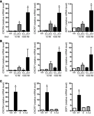
disorders (19–21, 26). Less is currently known about the biology of CXCR7, although it has been shown to be impor-tant in cardiac development and biology (27, 28). Since HUS is a microangiopathic disorder, at least in humans, we were interested in further examining the involvement of the CXCR4/CXCR7/SDF-1 pathway in Stx pathogenesis. We independently confirmed the microarray results using qRT-PCR and found time- and concentration-dependent increases in all 3 genes (Figure 2A). These findings were reca-pitulated in HMVECs treated with another Stx variant, Stx1 (Figure 2B), which shares 58% identity in amino acid sequence with Stx2 (29). SDF-1 is the sole known ligand for CXCR4 to date, while CXCR7 binds to SDF-1 as well as to interferon-inducible T cell α chemoattractant (I-TAC) (30). Although I-TAC was repre-sented by 2 independent probe sets on the HG-U133A arrays, its expression was not signifi-cantly altered by Stx2 treat-ment (10 fM, 24 hours), and these findings were confirmed by qRT-PCR (data not shown).
Changes in gene expression require a catalytically active Stx A subunit . Stx consist of pen-tameric receptor-binding B subunits covalently linked to an enzymatic A subunit, which depurinates eukaryotic 28S rRNA and inhibits pro-tein synthesis (Figure 1, A and B, and ref. 10). To determine the basis for differential gene expression, we tested whether the B subunit alone was suf-ficient to upregulate CXCR4,
CXCR7, and SDF1. Whereas 24-hour treatment of HMVECs with 1,000 fM wild-type Stx2 substantially increased expression of these genes, equimolar concentrations of the Stx1 B subunit, as expected, had no effect on protein synthesis (Figure 1B) and did not change mRNA levels (Figure 2C). This indicated that bind-ing of the toxin to its receptor, Gb3, was not sufficient to induce
[image:4.585.44.412.82.448.2]expression of these genes. Similarly, when cells were treated with an inactive Stx2 holotoxin mutant containing 2 amino acid sub-stitutions within the A subunit (E166Q, R169L) (31, 32), mRNA levels were not changed (Figure 2C). Taken together, these studies
Figure 1
Stx have potent effects on endothelial cell metabolism and phenotype. HMVECs or HUVECs were treated with vehicle (Veh) or the indicated concentrations of (A) Stx2, (B) Stx1 B subunit alone (B) (1,000 fM), or holotoxin with a double mutation in the A subunit (A mut) (1,000 fM) for 24 hours. One hour before harvest, 1 μCi [3H]leucine was added to assess de novo protein synthesis. Radioactivity incorporated into
TCA-precipitable material was quantitated, and data were normalized to vehicle-treated cells. The mean ± SEM of 14 experiments (HMVECs with Stx2), 5 experiments (HUVECs), or 3 experiments (mutants), triplicate determinations, is shown. (C) HMVECs (n = 6 replicates) were treated with Stx2 (10 fM, 24 hours) or vehicle and subjected to Affymetrix GeneChip gene expression profiling. By combining probe sets identified as dif-ferentially expressed by all preprocessing algorithms and removing redundant probe sets, i.e., probe sets mapping to the same gene, it was determined that 86.2% of differentially expressed genes (318 out of 369 unique genes) were upregulated by treatment with Stx. (D) The top 30 differentially expressed probe sets (based on magnitude of differential expression) were summarized in a heat map with hierarchical clustering. Processed signal intensities (computed using the mmgMOS preprocessing algorithm) are depicted on a log2
indicate that the A subunit must be present and catalytically active to elicit changes in gene expression.
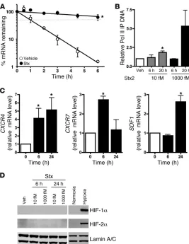
Stx increases steady-state CXCR4 mRNA by transcriptional as well as posttranscriptional mechanisms. We and others have shown that Stx increases levels of mRNAs via transcript stabilization, without tran-scriptional activation (11, 33–35). These observations compel study of the role of ribosome and mRNA interactions, especially because related ribosome-inactivating proteins, such as ricin, have compa-rable effects on gene expression (11). Like ET1, CXCR4 is a short-lived mRNA with a half-life of 1.18 ± 0.09 hours (Figure 3A). Upon Stx treatment (1,000 fM, 20 hours), its mRNA half-life increased to 19.2 ± 7.8 hours (Figure 3A). Although we observed a dramatic posttranscriptional effect, a role for transcription could not be excluded. We therefore assessed the levels of RNA polymerase II associated with the CXCR4 promoter in Stx-treated HMVECs by ChIP. A concentration- and time-dependent increase in polymerase II recruited to the CXCR4 locus was observed (Figure 3B). Hence, even though transcriptional effects were modest relative to those of posttranscriptional mechanisms, a combination of both nucle-ar events and posttranscriptional mechanisms contributed to the marked mRNA induction of CXCR4 by Stx. This could explain why
CXCR4 was among the most highly differentially expressed mRNA transcripts in our microarray studies (Figure 1D).
The CXCR4/CXCR7/SDF-1 pathway is regulated specifically by Stx through mechanisms distinct from those of other classic endothelial stimuli. TNF-α is an important proinflammatory cytokine and potent endothelial cell activator, which has been suggested to medi-ate many of the in vivo effects of Stx. To determine whether the CXCR4/CXCR7/SDF-1 pathway responds nonspecifically when the endothelium is activated, or whether this response is specific to Stx, HMVECs were treated with TNF-α (10 ng/ml), and mRNA lev- els were measured using qRT-PCR (Supplemental Figure 3). Simi-lar to that with Stx treatment, TNF-α increased CXCR7 expression, but, in contrast to that with Stx treatment, CXCR4 and SDF1 lev-els were dramatically reduced by TNF-α. These data indicate that Stx-mediated stimulation of the CXCR4/CXCR7/SDF-1 pathway in microvascular endothelium clearly differs from expression pat-terns produced by another classic endothelial activator.
[image:5.585.43.355.84.471.2]An additional, biologically important vascular stimulus is hypoxia. Similar to the Stx-induced response, hypoxia increased expression of CXCR4, CXCR7, and SDF1 mRNAs (Figure 3C). How-ever, Stx induced upregulation of all 3 components of the pathway by 24 hours, while hypoxia increased these receptor messages by 6 hours, followed by a delayed increase in their ligand at 24 hours. CXCR4 and SDF-1 expression is increased in a variety of hypoxic tissues and cells, and this pair is a target of HIF (16, 36, 37). CXCR7
Figure 2
Stx activates the CXCR4/CXCR7/SDF-1 pathway in microvascular endothelium by a mechanism requiring enzymatic activity of the Stx A subunit. HMVECs were treated with the indicated concen-trations of (A) Stx2 or (B) Stx1 for 6 or 24 hours, and mRNA levels were quantitated by qRT-PCR. (C) HMVECs were treated with 1,000 fM wild-type Stx2 (WT), Stx1 B subunit alone, or Stx2 holotox-in consistholotox-ing of a mutant A subunit and wild-type B subunit (A mut). mRNA levels were measured by qRT-PCR. In all cases, data are normal-ized for GAPDH and 18S and are shown rela-tive to vehicle-treated cells. Similar trends were observed whether or not data were normalized to housekeeping genes. Basal levels of CXCR4,
CXCR7, and SDF1 were 1.53 ± 0.43 × 10⁴,
7.40 ± 2.20 × 10⁴, and 2.21 ± 0.69 × 10⁴ copies/
has been shown to increase in lung, but not other tissues, during hypoxia (38). The similarity of the effects of Stx and hypoxia on the CXCR4/CXCR7/SDF-1 pathway, and our observation that CXCR4 is partly induced by transcription by Stx, prompted us to examine the potential involvement of HIF in Stx biology. Neither HIF-1α nor HIF-2α accumulated in Stx-treated cells as they did in hypoxic HMVECs (1 % O2, 24 hours) (Figure 3D). Hence, HIF
appears not to have a direct transcriptional effect on this pathway in the context of Stx, and this discordance suggests that Stx and hypoxia target the pathway via distinct mechanisms.
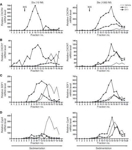
Stx enhances association of CXCR4, CXCR7, and SDF-1 transcripts with cellular polyribosomes . Because Stx in sufficient concentrations inac-tivates protein synthesis, it was important to address translation of CXCR4, CXCR7, and SDF-1 in addition to steady-state mRNA levels. We assessed whether the individual transcripts were being loaded onto ribosomes for translation by fractionating cellular ribosomes and their associated RNAs from Stx- and vehicle-treat-ed HMVECs. We also used a synthetic exogenous mRNA transcript to control for mRNA recovery and first-strand cDNA synthesis efficiency for rigorous quantification of total cellular RNA and ribosome fraction RNA content. CXCR4 and CXCR7 transcripts accumulated in polyribosome fractions (heavier fractions) after 24-hour treatment with 10 fM and 1,000 fM Stx, compared with those in vehicle-treated cells (Figure 4, A and B). The most dra-matic effect was the increase in CXCR4 mRNA associated with polyribosomes after addition of 10 fM Stx for 24 hours. This effect was also evident for both CXCR4 and SDF1. Importantly, there was
increased abundance of these transcripts in the polyribosome frac- tions after treatment with only 10 fM Stx for 24 hours, a concen-tration at which there is only minimal inhibition of global protein synthesis (Figure 1A). A key finding is that, even in the absence of a significant increase in total levels of CXCR4 (1.00-fold change vs. vehicle) and CXCR7 (2.06-fold change vs. vehicle) mRNA after 6-hour treatment with 1,000 fM Stx (Supplemental Figure 4, B and 4C), there was increased association of these transcripts with the polyribosome fractions (Figure 4, A and B), suggesting increased translational efficiency for these particular transcripts. Increased endothelial cell surface expression of CXCR4 was also observed by flow cytometry (Supplemental Figure 4A). The observed trans-lational effects of Stx on SDF1 were less pronounced than those on the SDF-1 receptors (Figure 4C) but were consistent with a previous report of enhanced SDF-1 release from microvascular endothelial cells exposed to Stx (39). The translational changes observed for these Stx target genes are specific and clearly different from other genes, such as cyclophilin A, which appears to fall off the ribosomes in the presence of only 10 fM Stx (Figure 4D). These findings indicate that Stx, a bacterial derived exotoxin that catalyt-ically modifies the 28S ribosomal RNA of the large 60S ribosome subunit, has transcript-specific effects on the functional interac-tion between mRNA transcripts and the translating ribosome.
[image:6.585.44.311.81.426.2]Functional role of the CXCR4/CXCR7/SDF-1 pathway in Stx-medi-ated endothelial injury. To explore the involvement of an activated CXCR4/CXCR7/SDF-1 pathway in Stx-mediated endothelial injury, we examined endothelial monolayer integrity using in
Figure 3
Stx enhances CXCR4 expression by both transcriptional and posttranscriptional mechanisms. (A) HMVECs treated with vehicle or 1,000 fM Stx2 for 20 hours were subjected to transcription arrest with 10 μg/ml actinomycin D, and RNA was harvested at various times thereafter. Remaining
CXCR4 mRNA was determined using qRT-PCR and nor-malized to 18S. The mean ± SEM of 3 independent experi-ments is shown. Half-lives were calculated using exponen-tial regression to be 1.18 ± 0.09 hours in vehicle-treated cells and 19.2 ± 7.8 hours in Stx-treated cells. (B) HMVECs were exposed to 1,000 fM Stx2 for 20 hours, followed by ChIP using anti-RNA polymerase II (Pol II) antibodies. qRT-PCR for CXCR4 was then used to determine the amount of immunoprecipitated DNA (IP DNA). The mean ± SEM of at least 4 independent experiments is shown. (C) HMVECs were maintained under normoxic (21% O2) or hypoxic (<1%
O2) conditions for the indicated times. qRT-PCR was used to
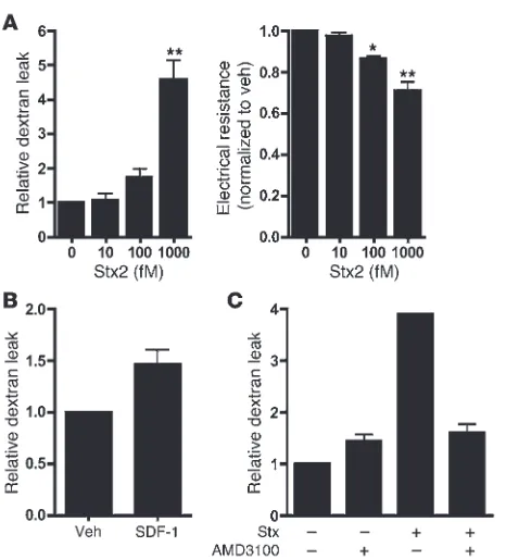
vitro permeability assays. Dextran leak and reduced electrical resis-tance were evident across monolayers treated with Stx (Figure 5A). As previously shown (40), SDF-1α induces modest increases in endothelial permeability (Figure 5B). Inhibition of SDF-1/CXCR4 signaling using the CXCR4 antagonist AMD3100/plerixafor reduced permeability of Stx-treated HMVECs (1,000 fM Stx, 24 hours, n = 3; Figure 5C), thus implicating a role for this pathway in Stx-mediated endothelial damage.
Stx causes kidney dysfunction in an animal model of pathogenesis. Based on our in vitro findings, we hypothesized that the CXCR4/CXCR7/ SDF-1 pathway plays a role in Stx-mediated pathogenesis in vivo. To test this hypothesis, we established and characterized a mouse model of Stx exposure. Importantly, murine in vivo Stx models imperfectly recapitulate human HUS. Motto et al. have previously developed mice deficient in Adamts13, the vWF protease (41). When
[image:7.585.81.509.81.566.2]Adamts13 was ablated via gene targeting in the widely used inbred
Figure 4
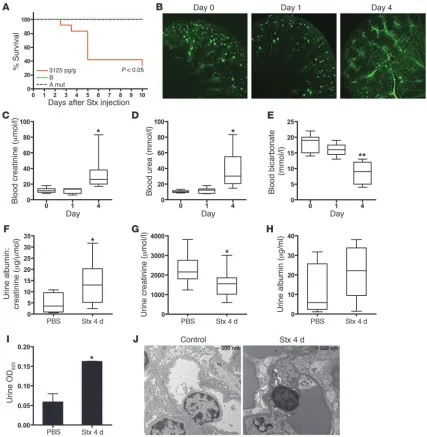
lab strain, C57BL/6, no phenotype was observed; however, knock-out of this gene in the wild-derived Mus musculus castaneus (CAST/ Ei) strain rendered these mice extremely susceptible to Stx-induced thrombotic microangiopathy. It was demonstrated that C57BL/6 mice express a truncated form of ADAMTS13 due to an insertional element within the genetic locus and are already hypomorphic for this protease (42). Interestingly, however, these mice do not devel-op overt thrombotic microangiopathy, and genetic modifiers that protect these inbred strains from such diseases have been mapped (41, 43). Consistent with this, castaneus mice are naturally more susceptible to thrombotic diseases. For these reasons, we opted to use CAST/Ei mice for our in vivo studies. Male and female CAST/ Ei mice received a single i.v. injection of various concentrations of purified Stx2, and survival was monitored for a period of 10 days. While both Stx1 and Stx2 affected the CXCR4/CXCR7/SDF-1 pathway, our studies focused specifically on Stx2, as this is the isoform associated with the most severe forms of human disease (7, 8). As expected, increasing concentrations of Stx resulted in increased mortality, with a LD50
approximating 2,400 pg/g. In con-trast, we noted that neither the Stx2 A subunit mutant (10 times the molar LD50) nor the Stx1 B subunit alone (50times the LD50)
were lethal over a 10-day observation interval (Figure 6A). Of note, animals treated with wild-type Stx2 exhibited significant weight
loss (86.1% ± 1.9% of original weight at day 0; mean ± SEM; P < 0.05 vs. weight at day 0) within 4 days of challenge.
Numerous measures used to evaluate kidney function were indicative of renal impairment. Fluorescence microangiography (fluorescent microbead size averaging 0.02 m) showed normal microvascular architecture within the renal cortex and medulla in control animals and those treated with Stx for 24 hours (Figure 6B). However, 4 days after Stx injection, cortical perfusion of the functional vasculature was severely affected, with some mice dem-onstrating marked changes in glomerular microperfusion (Figure 6B). At the same time, blood creatinine and urea were increased in Stx-treated animals (Figure 6, C and D). Decreased bicarbonate lev- els were also observed (Figure 6E). Urinalysis provided further evi-dence of kidney dysfunction. Urine albumin/creatinine ratios were significantly higher in Stx-treated animals (Figure 6F) as a result of significantly lower urine creatinine (Figure 6G) and elevated albumin leak (Figure 6H). Additionally, mice injected i.v. with Evans Blue dye, which binds to albumin and serves as a marker of albumin leak from the vasculature (44), showed release of Evans Blue/albumin complexes into the urine (Figure 6I). Consistent with these parameters of disruption of the glomerular permse- lectivity barrier by Stx, transmission electron microscopic exami-nation of the glomerular microvasculature demonstrated partial effacement of podocyte foot processes and glomerular endothelial cell ultrastructural alterations, specifically swelling and vacuoliza-tion of the endothelial cells (Figure 6J). The fall in urine creatinine concentrations, together with the observed elevated plasma creati-nine levels, are indicative of Stx-induced acute renal failure in this mouse model. Animals injected with either mutant form of the toxin showed blood creatinine and urea levels within the normal range for this strain (data not shown).
Effects of Stx on hematology and gastrointestinal biology . Throm- bocytopenia and hemolytic anemia are prominent pathophysi-ological features of HUS in patients. The majority of previously described mouse models do not report anemia, and development of thrombocytopenia in one model was attributed to cotreatment with endotoxin (45). Hematologic analysis in our model showed a modest but significant decrease in hematocrit levels by 24 hours after Stx injection, which then increased to basal levels by day 4 (Figure 7A). As we previously noted (41) in the outbred CAST/Ei strain, fragmented red blood cells were not pronounced in periph- eral blood smears (data not shown). Platelet levels were not signifi-cantly altered by 24 hours after toxin injection; however, they were substantially increased above basal levels by day 4 (Figure 7B), as in mice that do not develop thrombocytopenia following Stx (41).
E. coli O157:H7 cause bloody diarrhea and gut injury that nor- mally precedes HUS. Previous studies have demonstrated expres-sion of Gb3 on human gut epithelial cells (46). Additionally, Stx
has previously been shown to induce cecal or colonic hemorrhagic lesions in rabbits (47), and both purified Stx and culture filtrate from O157:H7 produced hemorrhagic lesions in a rat model (48). Purified Stx damages the stomach and intestinal microvasculature, causing microinfarctions and both intramucosal and lumenal hemorrhagic lesions, including frank intraluminal blood content with upper gastrointestinal hemorrhage, in approximately 60% of treated mice (Figure 7, C–E).
[image:8.585.48.281.79.336.2]Stx regulation of the CXCR4/CXCR7/SDF-1 pathway in vivo . To deter-mine whether Stx alters the CXCR4/CXCR7/SDF-1 axis in vivo, mice were injected with Stx, and expression of these messages was surveyed across tissues by in situ cRNA hybridization (Figure 8A
Figure 5
and Supplemental Figure 6). Consistent with previous studies, CXCR4 and SDF-1 were highly expressed in the bone marrow (Supplemental Figure 5), and CXCR7 was expressed in the hearts of control animals (Supplemental Figure 6). Perturbation of the pathway at the mRNA level was evident as early as 24 hours after Stx injection. In particular, CXCR4 expression was increased in the thymus (Supplemental Figure 6). In contrast, CXCR7 levels decreased in cardiac tissue by 24 hours after Stx and remained
[image:9.585.79.506.83.520.2]low at day 4 (Supplemental Figure 6 and data not shown). In situ hybridization revealed increases in SDF1 mRNA levels in the kidney and liver by 1 day after toxin treatment (Figure 8A and Supplemen-tal Figure 6), and immunohistochemistry demonstrated increased SDF-1 protein in the kidney of toxin-treated animals (Figure 8, A and B). A significant increase in the secreted SDF-1 peptide was observed in the blood of treated animals by day 4 (median, 2.1 ng/ ml; interquartile range [IQR], 1.6–2.7 ng/ml) compared with that
Figure 6
Stx challenge causes impaired renal function and mortality in vivo. (A) CAST/Ei mice were injected i.v. with wild-type Stx2 (3,125 pg/g), Stx2 A subunit mutant (10 times LD50), or Stx1 B subunit (50 times LD50), and survival was monitored for 10 days. (B) Mice were injected with Stx2
at day 0 (median, 1.1 ng/ml; IQR, 0.6–1.7 ng/ml) (Figure 8C). It is possible that decreased expression of CXCR7, which is a scaveng-ing receptor (49, 50), contributed to increased blood levels. It is of great interest that our in vitro mechanistic studies demonstrated enhanced translation of the mRNA transcripts for CXCR4, CXCR7,
SDF1 after Stx treatment.
CXCR4 inhibition improves animal survival. Given the multifaceted nature of the CXCR4/CXCR7/SDF-1 pathway, from proangiogen-ic functions for tissue repair (12, 13, 51) to its pathological roles in renal disease and inflammation (19–21), we examined its role in Stx pathogenesis in our in vivo model. We determined the effects of the CXCR4 antagonist, AMD3100/plerixafor, which is currently approved for stem cell mobilization in non-Hodgkin’s lymphoma and multiple myeloma and represents an attractive option avenue for patients (52). In conjunction with AMD3100/plerixafor admin-istration, animals received 200–300 μl saline. We chose to replace fluids, because animals treated with Stx lost weight compared with untreated animals, and their hematocrit levels increased signifi- cantly between days 1 and 4 after Stx injection (Figure 7A). In addi-tion, we observed albumin leak from the microvasculature in the kidney (Figure 6, G and H). Administration of AMD3100/plerixa-for beginning 1 day after Stx challenge improved animal survival over the course of 10 days (Figure 9A). Survival between days 0 and 5 was identical in both the AMD3100/plerixafor and PBS groups, with 57.7% survival (15 out of 26 mice in each group). Beyond day 5,
animal survival in the group receiving AMD3100/plerixafor improved significantly compared with that in the group receiving PBS (P < 0.05). 42.3% of mice receiving AMD3100/plerixafor sur-vived to day 10, compared with only 23.1% of mice receiving PBS control injections. These data suggest a key role for the CXCR4/ CXCR7/SDF-1 pathway in Stx pathogenesis and demonstrate that AMD3100/plerixafor treatment after Stx exposure enhances sur-vival of Stx-treated animals.
[image:10.585.45.268.80.326.2]Kidney function was also restored in part after AMD3100/ plerixafor treatment, as demonstrated by reductions in both blood creatinine and urea levels (Figure 9, B and C). Because common inbred lab mouse strains (45) and the wild-derived castaneus strain described here do not naturally develop remarkable anemia or thrombocytopenia when treated with purified Stx, we evaluated the effects of AMD3100/plerixafor on hematologic parameters in an Adamts13-deficient castaneus mouse model of thrombotic microangiopathy described elsewhere (41). Nolasco et al. have shown that high concentrations of Stx can induce ultra-large vWF multimer secretion from endothelial cells in culture and delayed multimer cleavage by ADAMTS13, which, the authors propose,
Figure 7
[image:10.585.301.532.445.740.2]Effects of Stx on animal hematology and gastrointestinal biology. (A) CAST/Ei mice were injected i.v. with Stx2 (2,400 pg/g) on day 0. Blood samples were taken via saphenous vein at the indicated times. (A) Hematocrit (HCT) and (B) platelet levels are shown (n = 13 per time point). (C–E) Approximately 60% of mice develop gastrointestinal hemorrhage. (C) Gross images of stomach and small intestine from PBS- and Stx-treated mice. (D) H&E staining shows intraluminal hem-orrhage (original magnification, ×50) and (E) intramucosal hemorrhage (arrows; original magnification, ×100). **P < 0.001.
Figure 8
leads to enhanced platelet aggregation (53). As previously reported (41), Adamts13–/–
mice treated with Stx developed features con-sistent with thrombotic microangiopathy, including hemolytic anemia, with red blood cell fragments and marked reticulocytosis evident in a peripheral blood smear (Figure 10A), and thrombocy-topenia (Figure 10B). Importantly, platelet counts were restored to normal levels with AMD3100/plerixafor treatment, while saline administration alone had no effect (Figure 10B). Despite the effect on platelets, no beneficial effect of the drug was observed with respect to hematocrit levels (data not shown). To further inves-tigate the mechanism by which this may occur, platelet adhesion to endothelial cells under fluid flow conditions was measured in vitro after Stx treatment in the presence and absence of AMD3100/ plerixafor. Toxin-treated endothelial cells exhibited enhanced platelet adhesion, and inhibition of CXCR4/SDF-1 interaction significantly reduced platelet strings attached to the endothelial monolayer (Figure 10, C and D). Other thrombotic abnormalities observed in HMVECs include increased tissue factor (Tf ) (Supple-mental Figure 7, A and B) and decreased tissue factor pathway inhibitor (TFPI) mRNA, protein levels, and biochemical activity (Supplemental Figure 7, C and D).
SDF-1 levels in patients with gastrointestinal E. coli O157:H7 infection. To examine whether the CXCR4/CXCR7/SDF-1 axis is altered in patients with Stx-induced disease, SDF-1 levels were measured in plasma collected from children younger than 10 years of age with documented gastrointestinal E. coli O157:H7 infection (54, 55). The samples were obtained after a similar numbers of days of illness in 2 groups of children with infection: those with uncomplicated infection and those in whom HUS subsequently developed. The hematocrit, platelet count, and serum creatinine concentration were normal and similar in these 2 groups, as described previously (55). Thus, hemolysis, quantitative platelet abnormalities, and renal insufficiency had yet to evolve in the children in whom HUS subsequently developed. Circulating SDF-1 levels were compared in children whose infection resolved without further complication
and in those children who subsequently developed HUS (Figure 11A). Despite the absence of clinically evident microangiopathic changes or renal insufficiency, the children in whom HUS subse-quently developed had a 4-fold increase in median plasma levels of SDF-1 in the days preceding onset of HUS (median, 94.2 pg/ml, IQR, 79.3–181.6 pg/ml vs. median, 23.0 pg/ml, IQR, 7.3–69.46 pg/ ml in individuals who were later diagnosed with the disease com-pared with children with uncomplicated infection, respectively). Median SDF-1 levels during HUS (median, 62.1 pg/ml; IQR, 33.69–242.2 pg/ml) remained significantly higher compared with those in individuals with uncomplicated infection. Importantly, the observed increases in blood SDF-1 levels preceded development of HUS, suggesting that aberrant regulation of the pathway, either as a cause or a consequence of endothelial injury, contributes to progression of the disease. Serial measurements were available for some patients and showed that SDF-1 levels peaked prior to devel-opment of HUS and subsequently decreased (Figure 11B).
Discussion
Stx-mediated HUS is a clinically important microangiopathy that may follow infection with enterohemorrhagic E. coli, particularly serotype O157:H7 and the newer O104:H4 enteroaggregative strain, which has acquired Stx expression. We have established an in vitro system to study Stx-mediated injury to cells that are relevant to Stx-mediated HUS and demonstrated the importance of the CXCR4/CXCR7/SDF-1 axis in eukaryotic injury. An under-standing of the effects of Stx on its target vascular cells is critical to development of efficient intervention strategies. To this end, we established an in vitro model using primary microvascular endothelial cells. In many studies to date, HUVECs have been the endothelial model of choice to study effects of Stx, despite the fact that they express quantitatively less of the toxin receptor (Gb3) on
[image:11.585.49.278.80.337.2]their surface and, therefore, exhibit less sensitivity to the toxin (22, 23). Moreover, published reports have shown fundamental differ-ences in the gene expression patterns of endothelial cells isolated
Figure 9
from different vascular beds, in particular macrovascular- and microvascular-derived endothelium (56, 57). The microvascular localization of Stx pathology therefore emphasizes the relevance of our microvascular cell model. In addition, the exquisite sensitivity of microvascular endothelium allowed us to focus specifically on the effects of Stx, whereas other cells, such as HUVECs, have usu-ally required sensitization with cytokine pretreatment (58, 59).
In the current study, we expanded our previous work, demon-strating that Stx upregulated ET1 mRNA that occurred at Stx concentrations that had minimal effects on global cellular protein synthesis (11). Specifically, we now more comprehensively charac-terized the gene expression profile of HMVECs after treatment with 10 fM Stx2, which produces only approximately 10% inhibition of protein synthesis. It is interesting that this low concentration of Stx had such potent and reproducible effects on endothelial phenotype. Previous expression profiling studies have been performed using Stx in HUVECs and in vivo (45, 60). We identified many of the same
genes in our work; however, modulation of the CXCR4/CXCR7/ SDF-1 pathway was not described in either previous study. This might be attributable to the cell model used in the former study, or source of tissue studied, or LPS cotreatment in the latter study. Interestingly, Guessous et al. observed elevated SDF-1α protein lev-els in conditioned media from HMVECs treated with Stx (39).
Our in vitro and in vivo data indicate that A subunit activity is indispensable for disruption of the CXCR4/CXCR7/SDF-1 path-way and for Stx-mediated mortality. First, studies with the mutant holotoxin and the receptor-binding B subunit demonstrated that the toxin must possess a functional A subunit in order to regulate expression of the CXCR4/CXCR7/SDF-1 pathway both in vitro and in vivo. Second, though our studies do not exclude important biological roles of the B subunit (61), our in vivo studies using the B subunit demonstrated 100% animal survival.
[image:12.585.85.498.81.426.2]Most of the genes whose expression was affected were upreg-ulated. We and others have previously shown that Stx increases
Figure 10
Blockade of CXCR4/SDF-1 interaction by AMD3100 restores platelet levels in vivo and inhibits platelet adhesion to endothelial cells under flow conditions in vitro. (A) Blood smears from Adamts13–/– mice injected with Stx2 (625 pg/g) show evidence of red blood cell fragments 4 days after
Stx exposure, with high numbers of reticulocytes (original magnification, ×150). (B) Adamts13–/– mice were injected with Stx2 (625 pg/g) on day 0,
followed by daily injections of AMD3100 (10 μg/g) or PBS. Blood was analyzed for platelet levels prior to Stx injection and again at day 4 after toxin injection in both groups (n = 9–21 animals per group). *P < 0.05 vs. control animals that received neither Stx nor AMD3100. (C) HMVECs were grown to confluence in microfluidic channels and were either left untreated or were treated with 10 fM Stx2 (24 hours), after which calcein-labeled platelets were perfused at constant shear rates of 2.5 dyn/cm2 over the endothelial monolayer. Preincubation of HMVECs with AMD3100 (10 μM)
particular mRNAs by increasing the half-life of the transcript (11, 33–35). We have also observed Stx-induced ET1 and ICAM1 mRNA expression via transcript stabilization but observed no change in nuclear run-off signal (ref. 11 and P.A. Marsden, unpublished observations). CXCR4, SDF-1, CXCR7, ICAM-1, and ET-1 share commonalities of basal mRNA lability, enhanced association with polyribosomes after Stx addition, and stabilization of mRNA tran- scripts after Stx treatment (ref. 11 and our unpublished observa-tions). It is of great interest, therefore, that CXCR4 transcription is also enhanced by Stx, explaining the marked increases in CXCR4 mRNA steady-state levels in Stx-treated HMVECs. However, nucle-ar events play a smaller part in the overall increase in steady-state mRNA, as the slight induction in transcription of CXCR4 with 10 fM Stx cannot explain the substantial increase in transcript levels at that concentration.
The stark differences between the regulation of the CXCR4/ CXCR7/SDF-1 pathway by Stx and TNF-α suggest that the effect of Stx on endothelial gene regulation is a function of this particular stimulus. CXCR4 and SDF-1 are well-known transcriptional targets of HIF, and as we showed, CXCR7 expression is also augmented in endothelium during hypoxia. Therefore, we considered the possibil-ity that Stx is a hypoxia mimetic, possibly via stabilization of HIF-α subunits. However, HIF-1α and HIF-2α levels were not affected by Stx in HMVECs. Notably, while a subset of hypoxia-regulated genes were upregulated in our studies (e.g., VEGF, CXCL1), most did not change (e.g., GLUT1, GLUT3, VEGFR2, TIE2, ANGPTL4). This dichotomy provides further evidence that Stx acts via a mechanism distinct from the cellular response to hypoxic stress.
It is key mechanistically that, even without changes in mRNA levels, we observed substantial increases in polyribosome loading of specific target transcripts only 6 hours after Stx addition and with very low concentrations of toxin. Moreover, the magnitude of change in polyribosome association induced by Stx far outweighed the changes in mRNA steady-state levels (up to 700 fold vs. 40 fold,
respectively). Comparison analysis demonstrates that only select mRNAs are abundantly associated with polyri-bosomes during Stx exposure. For instance, cyclophilin A mRNA transcripts exhibited decreased polyribosome association in Stx-treated endothelial cells. These find-ings highlight that changes in mRNA levels alone do not explain the effects and that specific translational regula-tion must be taken into consideration, especially in the case of a bacterial toxin that modifies ribosome activity. Together, these findings suggest the intriguing possibil- ity that low levels of ribosome damage by small quanti-ties of Stx may modulate selected cell signaling pathways that change ribosome function. Future mechanistic studies will be needed to gain greater understanding of how these putative signaling pathways modify the cell’s expression profile and phenotype and whether activa-tion of the CXCR4/CXCR7/SDF-1 pathway is common to other ribosome inhibiting proteins.
To assess the biological relevance of our in vitro find-ings, we established an in vivo model of Stx toxicity. Kidney impairment is evident in this model, but hemo-concentration and gastrointestinal vascular damage also figure prominently. Our studies also implicated the CXCR4/CXCR7/SDF-1 pathway as a mediator of Stx pathology in a murine model. The pattern of tissue dam-age in our mouse model coincided with tissues in which the CXCR4/CXCR7/SDF-1 pathway has previously been demon- strated to play critical roles, including the gastrointestinal vascu- lature (12, 13) and kidneys (14, 19, 20). Interestingly, overexpres-sion of CXCR4 or SDF-1 in the glomerulus causes kidney disease and induces proteinuria (19, 20). In our studies, the Stx-induced albuminuria and hemoconcentration were consistent with the observed in vitro endothelial permeability caused by Stx and SDF-1, which was reduced in our in vitro studies by inhibition of the path-way using AMD3100/plerixafor.
[image:13.585.42.316.84.226.2]The aberrant regulation of the complex and biologically impor- tant CXCR4/CXCR7/SDF-1 axis offers new insight into the patho-genesis of HUS. We acknowledge the limitation of our studies, in that the murine model is not a robust clinical surrogate for Stx- induced HUS. However, advances in understanding the complexi-ties of studying procoagulant pathways in murine models has led to the utilization of the outbred CAST/Ei strain, especially when deficient in ADAMTS13. We found that CXCR4 was expressed in lymphoid organs, and its expression was increased in thymus after Stx exposure. CXCR4 is expressed on a variety of cells, including T- and B-thymocytes, and its altered expression here might indi-cate an immune response to the toxin. CXCR7 has recently been described as a scavenger receptor that effectively creates and main-tains a SDF-1 gradient to support CXCR4 signaling functions (49) by binding SDF-1 and targeting the chemokine for degradation (50). Stx decreases the amount of CXCR7 in the heart and, there-fore, may increase the amount of free SDF-1 available to signal through CXCR4. Interestingly, while CXCR4/SDF-1 is essential in a variety of organ systems, CXCR7 is particularly important in proper development and function of the heart (27, 28), and CXCR7 present in cardiac tissue has previously been shown to sequester SDF-1 (50). To date, transgenic overexpression of SDF-1 in the heart has not been studied. However, adenovirus-mediated overexpression of CXCR4 in cardiac tissue increased SDF-1 lev-els and has been reported to enhance ischemia/reperfusion injury
Figure 11
Circulating SDF-1 levels in HUS patients. SDF-1 levels were measured in plas-ma samples from individuals infected with E. coli O157:H7. (A) Infected sam-ples obtained from patients with uncomplicated O157:H7 infection within 4 days after the onset of diarrhea (n = 36). Horizontal bars indicate the median values, boxes indicate 25th to 75th percentiles, and whiskers indicate the minimum and maximum values. Pre-HUS, samples collected prior to HUS diagnosis in those individuals who later progressed to HUS (n = 7); HUS, samples collected on the day of HUS diagnosis (n = 22). *P < 0.05. (B) SDF-1 levels over the course of
(62). The CXCR4/SDF-1 axis is emerging as a key pathway in vas- cular and other tissue biology, and, thus, a tight balance is neces-sary to maintain homeostasis. The increase in free SDF-1 might tip the balance toward CXCR4 signaling on cells expressing the receptor and contribute to the endothelial dysfunction character-istic of Stx-mediated disease. It is important to note that our in vivo studies specifically addressed mRNA levels, but as we have shown in vitro, Stx pathobiology can involve profound changes in translational efficiency of CXCR4, CXCR7, and SDF1 transcripts, and this mechanism may also be important in vivo in tissues in which changes in mRNA levels are not observed.
We addressed the in vivo role of the CXCR4/CXCR7/SDF-1 pathway using the CXCR4 inhibitor AMD3100/plerixafor. This bicylam compound is a specific antagonist of CXCR4 and does not appear to inhibit CXCR7 function (63). AMD3100/plerixafor increases CXCR7 affinity for SDF-1 and functions as a CXCR7 agonist (64). When AMD3100/plerixafor therapy was initiated in a clinically relevant setting, namely 1 day after Stx challenge, we observed a beneficial effect on overall survival (Figure 9A). Impor-tantly, CXCR4 inhibition is beneficial to mice already exposed to the toxin. This has important clinical implications for patients who visit the hospital only after becoming ill from enteric infec-tion with E. coli O157:H7.
Our data also demonstrate involvement of the CXCR4/CXCR7/ SDF-1 axis in pediatric HUS cases, using plasma from a pro-spective cohort study of patients with E. coli O157:H7infection (54, 55). Children in this phase of infection have prothrombotic abnormalities, which appear before, and plausibly contribute to, HUS. We demonstrated marked elevation in blood SDF-1 before the onset of HUS and azotemia, which could contribute to the pathogenesis of HUS. SDF-1 has been shown to enhance platelet activation by low levels of ADP (65) or thrombin (66), leading to increased aggregation (67) through CXCR4 platelet surface expres-sion. Platelet thrombi are a characteristic feature of HUS. Our data indicate that Stx augment platelet adhesion in an in vitro flow model and that AMD3100/plerixafor could inhibit this. More-over, we found increased and decreased activity of Tf and TFPI, respectively. Therefore, increased thrombin generation observed in HUS (55), in combination with increased SDF-1 levels, may be an important mechanism involved in Stx pathogenesis. The relationship between increased tissue factor, decreased tissue fac-tor pathway inhibitor, and the effects of thrombin and SDF-1 on platelet aggregation in the in vivo setting warrants further study. SDF-1 is also stored in and released by activated platelets (68) and could exacerbate thrombosis in later stages of disease. Our in vitro studies demonstrate that AMD3100/plerixafor inhibited forma-tion of platelet strings on the endothelium and may explain why
Adamts13–/–
mice receiving the drug displayed normal platelet lev-els after Stx challenge compared with those of animals that did not receive the drug.
Taken together, these studies identify Stx-induced activation of the CXCR4/CXCR7/SDF-1 pathway in vitro and in vivo, demon-strate that this pathway contributes to Stx-mediated pathogenesis, and present targets which we believe to be novel for therapeutic intervention. We have found that the median SDF-1 level decreases in overt HUS. SDF-1 binding to CXCR4 induces permanent inter-nalization of the receptor from the cell surface, so that by the time HUS ensues, SDF-1 might be cleared from the circulation by CXCR4 or its other scavenger receptor, CXCR7. Alternatively, platelet decreases during HUS may account for the fall in median
SDF-1 levels. Little overlap exists between the IQRs of SDF-1 mea-surements in children with uncomplicated infection and those who later developed HUS. Further studies are therefore warranted to determine the potential value of plasma SDF-1 level as a predictive indicator of complications and progression to HUS after infection with E. coli O157:H7.
In summary, aberrant activation of the CXCR4/CXCR7/SDF-1 pathway in Stx-mediated pathogenesis could provide novel insight into the molecular mechanisms underlying HUS. Specifically, CXCR4 inhibition improves renal function and, ultimately, surviv-al in a murine model, and circulating SDF-1 in O157:H7-infected patients provides complementary data in support of an important role for this axis in the development of HUS.
Methods Reagents
[methyl-3H]thymidine (70–90 Ci/mmol), [5,6-3H]uridine (30–50 Ci/mmol), and l-[3,4,5-3 H(N)]leucine (>140 Ci/mmol) were purchased from NEN-Dupont. l-[methyl-3H]methionine (70–85 Ci/mmol) was purchased from Amersham Biosciences. TNF-α was purchased from Sigma-Aldrich. cDNA I.M.A.G.E. clones were obtained from ATCC or Invitrogen.
Toxin purification
Wild-type Stx2 holotoxin and Stx2 holotoxin mutant (E167Q and R170L substitutions in the A subunit) were extracted from recombinant E. coli strains using a combination of 10 μg/ml lysozyme and 0.1 mg/ml colimycin in PBS in the presence of 5 mM EDTA and 0.1 g/l PMSF (Sigma-Aldrich). The extract was cleared by centrifugation, dialyzed against 50 mM acetate buffer (pH 5), resolved on DEAE Sepharose Fast Flow (Amersham Biosci-ences), and eluted using a NaCl gradient 0 M to 0.5 M in 50 mM acetate buffer. Fractions containing Stx were precipitated with 80% ammonium sulfate, backwashed with 0.8 M ammonium sulfate in 50 mM acetate buf- fer (pH 5), and diluted to 0.6 M ammonium sulfate in 50 mM acetate buf-fer (pH 5). Fractions were then loaded on Phenyl Sepharose HP (Amersham Biosciences), eluted with a decreasing salt gradient, dialyzed against 50 mM ammonium bicarbonate, lyophilized for storage, and resuspended in PBS. Stx1 B subunit was purified as previously described (69). Contaminating lipopolysaccharide was removed by incubating with Affi-Prep polymyxin B resin overnight (Bio-Rad) at 4°C. Endotoxin content was tested by Limulus Amebocyte Lysate assay (Associates of Cape Cod Inc.) and determined to be less than 1 pg endotoxin/μg Stx.
Cell culture
Human dermal neonatal microvascular endothelial cells (HMVECs) were purchased from Lonza, cultured on 0.2% gelatin, and maintained in EGM-2MV growth medium (Lonza). Studies were performed on HMVECs at passages 5 and 6. HUVECs were maintained in M199 (Invitrogen) con-taining 20% fetal bovine serum (Hyclone), 17 units/ml heparin, and 0.05 mg/ml endothelial mitogen (Biomedical Technologies Inc.), as previously described (70), and used at passages 3 and 4. Confluent monolayers were treated with the specified concentrations of Stx or TNF-α (10 ng/ml) for the indicated times before harvesting. For hypoxia studies, HMVECs were treated as previously described (71). Total cellular RNA was purified by CsCl ultracentrifugation, as previously described (72).
Metabolic assays