R E S E A R C H A R T I C L E
Open Access
An
in vitro
study of anti-inflammatory activity of
standardised
Andrographis paniculata
extracts
and pure andrographolide
Mitchell Low
1, Cheang S Khoo
1, Gerald Münch
1,2, Suresh Govindaraghavan
3and Nikolaus J Sucher
4*Abstract
Background:The anti-inflammatory activity ofAndrographis paniculata(Acanthaceae), a traditional medicine widely used in Asia, is commonly attributed to andrographolide, its main secondary metabolite. CommercialA. paniculata
extracts are standardised to andrographolide content. We undertook the present study to investigate 1) how selective enrichment of andrographolide in commercialA. paniculata extracts affects the variability of non-standardised phytochemical components and 2) if variability in the non-standardised components of the extract affects the pharmacological activity of andrographolide itself.
Methods: We characterized 12 commercial, standardised (≥30% andrographolide) batches ofA. paniculataextracts from India by HPLC profiling. We determined the antioxidant capacity of the extracts using 2,2-diphenyl-1-picrylhydrazyl (DPPH) free radical scavenging, oxygen radical antioxidant capacity (ORAC) and a Folin-Ciocalteu (FC) antioxidant assays. Their anti-inflammatory activity was assessed by assaying their inhibitory effect on the release of tumor necrosis factor alpha (TNF-α) in the human monocytic cell line THP-1.
Results:The andrographolide content in the samples was close to the claimed value (32.2 ± 2.1%, range 27.5 to 35.9%). Twenty-one non-standardised constituents exhibited more than 2-fold variation in HPLC peak intensities in the tested batches. The chlorogenic acid content of the batches varied more than 30-fold. The DPPH free radical scavenging activity varied ~3-fold, the ORAC and FC antioxidant capacity varied ~1.5 fold among batches. In contrast, the TNF-αinhibitory activity of the extracts exhibited little variation and comparison with pure andrographolide indicated that it was mostly due to their andrographolide content.
Conclusions:StandardisedA. paniculataextracts contained the claimed amount of andrographolide but exhibited considerable phytochemical background variation. DPPH radical scavenging activity of the extracts was mostly due to the flavonoid/phenlycarboxylic acid compounds in the extracts. The inhibitory effect of andrographolide on the release of TNF-αwas little affected by the quantitative variation of the non-standardised constituents.
Keywords:Andrographolide, Andrographis paniculata, Anti-inflammatory, Antioxidant, TNF-α, Phytochemistry
Background
Chronic inflammation is thought to be a contributing factor to many prevalent ageing-related diseases, such as acute and chronic neurodegenerative diseases, degen-erative musculoskeletal diseases, cardiovascular dis-eases, diabetes, and cancer [1-5]. Other common chronic inflammatory conditions include asthma, rheumatoid
arthritis, and inflammatory bowel disease. To date, pharmacotherapy of inflammatory conditions is based mainly on the use of non-steroidal anti-inflammatory drugs (NSAIDs), steroids and more recently tumor ne-crosis factor alpha (TNF-α) inhibitors [6-8]. However, the prolonged use of NSAIDs can cause serious gastrointes-tinal toxicity [9]. Some NSAIDs have also been linked to increased blood pressure, greatly increased risk of con-gestive heart failure and occurrence of thrombosis [10]. These findings illustrate the need to develop novel and safe anti-inflammatory medicines [11].
* Correspondence:[email protected] 4
Science Department, Roxbury Community College, 1234 Columbus Avenue, Roxbury Crossing, MA, 02120, USA
Full list of author information is available at the end of the article
Andrographis paniculata (Acanthaceae), which is en-dogenous to South India and South East Asia, is used as an herbal medicine in both traditional Indian and Chin-ese medicine (where it is known as kalmegh and chuan-xinlian, respectively) as well as in Malaysia and Thailand [12,13]. A. paniculata extracts exhibit anti-inflammatory activity [13] that is commonly attributed to the ent-labdane diterpenoid andrographolide, its charac-teristic and main secondary metabolite [14-30]. Andrographolide appears to be rapidly absorbed [31] and found to be non-toxic even at very high doses in animals [32] and is well tolerated by humans with no serious ad-verse effects at doses in the range of 1 to 2 mg/kg/day [33,34]. Intriguingly, andrographolide has been reported to exhibit gastro-protective and ulcer preventive ef-fects, which combined with its well-documented anti-inflammatory effects could make it a safe alternative to traditional NSAIDs [35]. A proprietary A. paniculata extract (HMPL-004, Hutchison MediPharma) is under development for the treatment of inflammatory bowel disease [36,37] and is currently being tested in a global phase III clinical trial (http://clinicaltrials.gov/show/ NCT01805791).
CommercialA. paniculatatablets standardised to requis-ite concentrations of andrographolide (5% or 30% w/w) are used in clinical studies with an assumption of consistency [38,39]. However with reported innate phytochemical vari-ation influenced by phytogeographical and spatiotemporal factors [40-44], it is not known how selective enrichment (standardization) of andrographolide in commercial prepa-rations affects the variability of non-standardised phyto-chemical components. It is also not known if variations in the non-standardised components affect the anti-inflammatory activity of the extracts. We undertook the present study to investigate 1) how selective enrichment of andrographolide in commercial A. paniculataextracts affects the variability of non-standardised phytochemical components and 2) if variability in the non-standardised components affects the pharmacological activity of the ex-tracts. To this end, we profiled the phytochemical com-position and antioxidant capacity of standardised A. paniculata extracts and compared the activity of the ex-tracts and purified andrographolide in an assay relevant to their anti-inflammatory activity.
Anti-inflammatory activity of andrographolide has been studied using a number ofin vivo andin vitro ex-perimental paradigms including human whole genome DNA microarrays [20]. The most commonly implicated molecular mechanism underpinning the anti-inflammatory and immunomodulatoy effects of andrographolide is inhib-ition of the mitogen-activated protein kinase/extracellular signal-regulated kinase (MAPK/ERK) signalling (specifically p38 MAPK/ERK1/2) pathway and downstream transcrip-tion factors such as nuclear factor kappa B (NF-κB) and
nuclear factor of activated T cells (NFAT) [24,45-50]. An exemplary experimentalin vitromodel where this mechan-ism has been implicated is the inhibition by andro-grapholide of the release of TNF-α from LPS stimulated macrophages [22,51-54]. Therefore, we chose TNF-α release from LPS stimulated monocytic leukaemia cells (THP-1) as a model to study anti-inflammatory activity of the extracts and purified andrographolide.
Methods
Extract provenance and preparation
Twelve commercial batch samples of A. paniculata extracts (extract ratio 14:1) standardised to≥30% andro-grapholide were kindly provided to us by LIPA Pharma-ceuticals Ltd (NSW, Australia). The extracts conform to the TGA guidelines for incorporation in herbal medicines manufactured in Australia. The whole plant starting ma-terial for each batch was sourced (during 2004 – 2008) and the extracts manufactured in India. A systematic bot-anist authenticated each batch’s starting material and the manufacturer provided traceability documents for each extract. All the samples were re-analysed by high-performance liquid chromatography (HPLC) with photo-diode array detection (PDA) for andrographolide content to reconfirm the manufacturer’s certificate of analysis.
Phytochemical analysis
We used HPLC to profile the phytochemical compos-ition of the A. paniculata extracts. Andrographolide (14.3 mg in 10 ml) and extract samples (125 mg in 50 ml) were dissolved by sonication in methanol. HPLC analysis of andrographolide and the extract samples was performed using a Varian Inc. (USA) HPLC system equipped with ProStar 335 photodiode array detector (PDA) and 1200 L quadrupole tandem mass spectrom-etry (MS/MS) detector. An Alltech Alltima (Alltech Australia) reverse phase C18 column (46 × 150 mm I.D., 5 μm) with a Phenomenex (California, USA) Security C18 guard column (20 mm × 4 mm, 5μm) were used in these experiments.
We generated HPLC-PDA and HPLC-MS/MS profiles using a 5 μl injection of extract samples. The mobile phase consisted of 0.1% (v/v) aqueous formic acid (mobile phase A) and 0.1% (v/v) formic acid in acetonitrile (mobile phase B). The mobile phase gradient was 10% B for 10 min with a linear increase; to 50% B at 63 min, 70% B at 72 min and then 100% B (wash) for 8 min before equilibrating at the starting composition for 5 min. Mobile phase flow rate was maintained at 1 ml/min. The post col-umn flow was split to send 80% to the PDA (200–500 nm) and 20% to the MS.
with the needle voltage 5000 V at 13 μA; nebulization gas (nitrogen) temperature of 350°C at 20 psi; shield voltage 175 V; capillary voltage 53 V and the detector voltage was 1600 V.
We quantified the andrographolide (0.14 - 1.4 mg/ml) content of the extracts at a detection wavelength of 240 nm and the chlorogenic acid (6.6 – 132.4 μg/ml) content at 330 nm, using five-point linear calibration curves. We generated chromatograms at 227 nm for the detection of diterpenes and at 261 nm and 330 nm for the detection of flavonoids and phenyl carboxylic acids, respectively.
While the PDA was used to quantify andrographolide and chlorogenic acid, the MS detector was used to con-firm the identity of these peaks by comparing the MS/ MS obtained for the sample and reference standard peaks. MS/MS data was also used to tentatively identify other diterpenes by comparison to published MS data. The UV spectra were used to assign tentatively some of the observed peaks as flavonoids, phenylcarboxylic acids or diterpenes as shown in Table 1.
We used the package ‘msProcess’ [56] (R Project) for Statistical Computing [57] to remove instrumental noise and baseline drift from the chromatograms as described in detail previously [58].
Antioxidant assays
We performed the DPPH (2,2-di(4-tert-octylphenyl)-1-picrylhydrazyl) radical scavenging capacity, oxygen rad-ical absorbance capacity (ORAC) and the total phenol assay (Folin-Ciocalteu assay; FC) on the extracts as re-ported in detail previously [58,59].
Cell culture and tumor necrosis factorα(TNF-α) assay We cultured human monocytic leukaemia cells (THP-1; American Type Culture Collection, Manassas, VA, USA) in RPMI (Roswell Park Memorial Institute) media con-taining 4.5 g/l D-Glucose and supplemented with 2 mM GlutaMax, 100 U/ml penicillin, 100μg/ml streptomycin, and 10% foetal bovine serum at 37°C, 5% CO2 in 95%
air. Cells (passage number between 10 and 25) were seeded at a density of 1 × 10⁵ cells/well and incubated for 48 h in phorbol-12-myristate-13-acetate (PMA; 100 nM). Non-adherent cells were removed by washing with fresh medium. The remaining cells were pre-incubated with different concentrations of extracts or andrographolide for 1 h and stimulated by lipopolysac-charide (LPS; 50 ng/ml) and interferon gamma (50 units) for 24 h. Andrographolide and extracts were prepared in DMSO and added at a final concentration of 0.1% DMSO. We determined the concentration of TNF-α in the THP-1 culture supernatant by a commercial sand-wich ELISA following the manufacturer’s instructions (PeproTech Inc., Rocky Hill, NJ, USA).
Potential cytotoxicity of the extracts and andrographolide was investigated using the MTT (3-(4,5-dimethylthiazol-2-yl)-2,5-diphenyltetrazolium bromide) assay. No toxic ef-fects of the extracts or andrographolide were observed at the tested concentrations (data not shown).
Statistical data analysis
All data is reported as mean ± standard deviation of the average of three replicates in experiments performed on three separate days. The TNF-αdose response data were fitted with a log (inhibitor) vs. normalized response with variable slope model using Prism 5 for Mac OSX (GraphPad Software, La Jolla, CA). IC50 values were
calculated from the fitted curves. F-test was used to compare if the best-fit values differed between the data sets. Differences were considered statistically signifi-cant ifp< 0.05.
Results and discussion
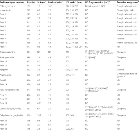
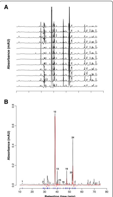
The phytochemical composition of 12 A.paniculata ex-tracts was characterized by HPLC-PDA (Figure 1A). Consistent with the intended standardization, to contain 30% andrographolide, the content was on average close to this value (32.2 ± 2.1%, range 27.5 to 35.9%; n= 12; peak #12 in Figure 1B). Correlation analysis of the andrographolide concentration versus storage time re-vealed that there was no observable loss in andrographo-lide concentration due to storage of the dry extracts (r2= 0.03; not illustrated). Non-standardised constitu-ents other than diterpenes (e. g. peaks 1–11), varied to a greater degree between the batches (Table 1). For ex-ample, the chlorogenic acid content exhibited >30 fold variation between batches (0.1 to 3.5 mg per g of extract; mean 1.4 ± 1.1 mg/g). The maximum variation observed between the chromatograms is illustrated by the red curve in Figure 1B.
scavenging was mainly due to peaks 1–11 (flavonoids and phenylcarboxylic acids) while the diterpenes (peaks #12, 13, 20, 24 and 25) were virtually devoid of DPPH radical scavenging capacity under the conditions of our experiments (Figure 2). The average ORAC of the extracts was 1.05 ± 0.16 mmol/g gallic acid equivalent and varied 1.6 fold. The average antioxidant activity of the extracts in the FC assay was 0.40 ± 0.05 mmol/g gallic acid equivalent and varied 1.5 fold.
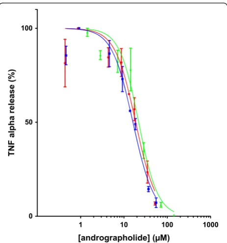
[image:4.595.58.538.110.562.2]Next, we assayed the activity of the extracts and puri-fied andrographolide on the inhibition of TNF-αrelease by LPS stimulated THP-1 cells. For this experiment, we obtained dose response curves of purified andrographo-lide and the extracts #3 and #9, the extracts with highest and lowest DPPH free radical scavenging activity. The results revealed that the dose response curves obtained upon application of pure andrographolide or the extracts were very similar (Figure 3), when normalized to
Table 1 Tentative assignment of HPLC chromatogram peaks as flavonoids, phenylcarboxylic acids or diterpenes based on UV absorbance and MS fragmentation patterns
Peakidentitycor number Rt (min) % Area3 Fold variation2 UV peaks1(nm) MS fragmentation (m/z)5 Tentative assignment4
Chlorogenic acid 12.2 1.0 34.3 327, 218, 235 Not determined (ND) Phenyl carboxylic acid
Isoquercetin 28.7 0.9 5.1 204, 255, 353 ND Flavonol glycoside
Peak 3 29.2 0.6 9.8 323, 219, 234 ND Phenyl carboxylic acid
Peak 4 29.7 3.5 3.8 325,219,235 ND Phenyl carboxylic acid
Peak 5 31 1.4 3.4 326, 219, 271 ND Phenyl carboxylic acid
Peak 6 32.2 4.1 6.1 324, 219, 234 ND Phenyl carboxylic acid
Peak 7 32.4 3.3 4.5 325, 220 ND Phenyl carboxylic acid
Peak 8 32.8 3.3 4.7 346, 224, 256 ND Phenyl carboxylic acid
Peak 9 33.7 2.1 4.5 204, 336, 266 ND Flavone
Peak 10 36.8 1.3 6.6 327, 219, 235 ND Phenyl carboxylic acid
Peak 11 37.7 0.8 4.4 327, 271, 223, 204 ND ND
Andrographolide 38.4 ND ND 227 351 [M+H]
+, 333 [M-H2O]+,
315 [M-2H2O]+, 297 [M-3H2O]+ Diterpene
Andropanoside 39.8 1.4 4.8 207 535 [M+K]+ Diterpene
Peak 14 40.2 8.0 7.1 225 ND ND
Peak 15 40.7 4.3 3.7 202 ND ND
Apigenin 42.2 2.6 6.7 211, 337, 267 ND Flavone
Wogonoside 45.2 0.7 2.3 265, 212 ND O-methylated flavone
glycoside
Peak18 46.6 0.7 4.6 ND ND ND
Peak 19 46.8 0.8 2.5 ND ND ND
Neoandrographolide 47.7 9.3 2.7 201 503 [M+Na]
+,519 [M+K]+,
319[M+H-Glu]+ Diterpene
Peak 21 48.6 0.5 3.5 ND ND ND
Peak 22 49.7 1.5 5.3 ND ND ND
Peak 23 50.2 0.76 7.7 ND ND ND
Deoxyandrographolide 52.1 6.7 3.7 200 357 [M+Na]
+
, 317 [M+H-H2O]+,
299 [M+H-2H2O]+ Diterpene
Dehydroandrographolide 52.6 35.1 2.1 200, 249 355 [M+Na]
+, 315 [M+H-H2O]+,
297 [M+H-2H2O]+ Diterpene
Peak 26 53.6 0.8 2.8 229 ND ND
Peak 27 54.3 2.0 6.1 229 ND ND
Peak 28 54.6 2.8 3.0 200, 263 ND ND
1
UV peaks are listed in order of intensity. 2
Fold variation = (Max. peak .area) / (Min. peak area). 3
Percent (%) area = average peak area from the 12 batches / total combined area × 100. Andrographolide was excluded from the % area calculations. 4
Assignments based on UV spectrum, MS fragmentation and/or comparison to reference standard RT. 5
andrographolide concentration. The half maximal inhibi-tory concentration (IC50) of pure andrographolide was
21.9μM (n= 3; 95% confidence interval: 18.1 - 26.5μM) compared to 16.4 μM (n= 3; 95% confidence interval: 13.8 - 18.8 μM) and 18.7 μM (n= 3; 95% confidence interval: 14.9 - 23.4 μM) for extracts #3 and #9,
respectively. Thus, the phytochemical background vari-ation of the extracts appeared not to influence significantly the activity of andrographolide in thisin vitroassay.
We characterized the phytochemical composition of 12 batches of commercial A. paniculata extracts stan-dardised for andrographolide content (30% w/w). We confirmed that the extracts contained the specified amount of andrographolide but observed substantial variation in the non-standardised components of the ex-tracts. Chlorogenic acid exhibited maximal (~30 fold) variation. This acid is one of the most abundant phen-olic compounds in the human diet and is present in sig-nificant amounts in coffee [63]. Various pharmacological effects of chlorogenic acid have been described and re-cent interest has focused on its effects on glucose and lipid metabolism [63]. Hypoglycaemic and hypocholes-terolemic effects of water and ethanolA. paniculata ex-tracts have been reported but the role of chlorogenic acid and its other phytochemical constituents and the molecular mechanisms underpinning these effects re-main to be established [64]. Patients with diabetes taking A. paniculata extracts may therefore need extra moni-toring and dietary counselling upon commencing or changing A. paniculata extract containing medications due to batch-to-batch variability of chlorogenic acid and its potential effects on blood glucose levels.
[image:5.595.302.539.87.287.2]Our results revealed that of the commonly used in vitro antioxidant assays the DPPH assay was a more
Figure 2Online DPPH assay.Representative chromatograms of A. paniculataextract #9. The chromatogram at 227 nm (black line) is contrasted with the DPPH absorbance at 529 nm (red line). Compounds that have DPPH antioxidant activity are observed as a negative peak at 529 nm. Numbered peaks were identified as chlorogenic acid (#1), andrographolide (#12), andropanoside (#13), neoandrogrpapholide (#19), deoxyandrographolide (#23), and 14-deoxy-11,12-didehydroandrographolide (#24).
A
B
[image:5.595.58.291.88.494.2]sensitive indicator of the phytochemical background variation in the standardised extracts than the ORAC and FC assays. The absolute increase in the activity of the extracts and reduced inter-extract variation observed in the ORAC and FC assays may be due to the activity of andrographolide and other diterpenes (and possibly additional unidentified compounds), which did not ex-hibit activity in the DPPH assay. The results suggest that at least under the conditions of our experiments the di-terpenes mainly functioned via hydrogen atom transfer (HAT) but not electron transfer (ET) in contrast to the flavonoids and phenyl carboxylic acids which exhibited activity in both the HAT and ET mechanisms [58]. The DPPH reactivity was correlated with the variation in chlorogenic acid (r2= 0.8), illustrating that chlorogenic acid contributed significantly to the ET antioxidant ac-tivity of the extracts. The chlorogenic acid quantity showed no correlation with the ORAC or FC results, despite chlorogenic acid being a HAT antioxidant; this is likely due to andrographolide masking chlorogenic acids contribution to the total HAT activity as andrographo-lide is in much greater abundance (~100 times).
There are many anti-inflammatory compounds re-ported in A. paniculata [52,65,66] but andrographolide is the most abundant [55]. In this study the most dis-similar extracts in terms of DPPH activity and chemical profile (#3 and #9) were compared to pure andrographo-lide to assess their inhibition of TNFαrelease from LPS stimulated macrophages. Our comparison between the purified andrographolide and extracts containing parallel amounts of andrographolide in this assay found very similar dose response curves. Thus, at this level of ana-lysis, there was no evidence for antagonistic, additive or synergistic effects between andrographolide and other phytochemical constituents of the extracts. The extract activity was almost entirely accounted for by the andro-grapholide content, thus the contribution of the other anti-inflammatory compounds present was minor, likely due to their lower abundance.
The IC50 of andrographolide (21.9 μM) was
com-parable to values obtained in a similar assay using mouse peritoneal macrophages [51]. A number of additional in vitro studies using various experimental paradigms have all reported effective concentrations of pure andrographolide in the range of 7 to 35 μM [20,24,45,47,48,66-68]. The concentration reported in ours and other in vitro studies are nominal concentra-tions and the “actual” concentration at the site of interaction between andrographolide and its potential (extra- and/or intracellular) target proteins (receptor(s) or enzymes) has not been determined. Nonetheless, it is noteworthy that the IC50 of andrographolide in our
in vitro assay was ~50 times higher than the maximal plasma concentration (0.5 μM) achieved following oral administration of 50 mg andrographolide in healthy hu-man volunteers [69], although significantly higher steady state blood concentrations (1.9 μM) have been reported in humans taking ~1 mg andrographolide per kg body weight per day [31]. In a prospective clinical study for the relief of rheumatoid arthritis symptoms, Burgos and col-leagues administered 3 times per day 100 mg ofA. pani-culataextract standardised to 30% andrographolide [38]. Although these authors observed some positive effects, thein vitrodata suggest that the administered dose might have been at the low end of the effective dose range and future clinical studies should consider testing higher doses. It will be interesting to investigate whether or not the absorption, distribution, metabolism and excretion of andrographolide alone or andrographolide administered as A. paniculata extract (with variable phytochemical background) differ or not.
Conclusion
[image:6.595.58.290.87.336.2]Supported by a considerable body of published evidence, standardisation of A. paniculata extracts for andro-grapholide content is based on the notion that this Figure 3Dose response curves for batches #9 (blue, squares),
#3 (red, circles) and andrographolide (green, triangles).IC50of
pure andrographolide: 21.9μM (n= 3; 95% confidence interval: 18.1 - 26.5μM), extract #3: 16.4μM (n= 3; 95% confidence interval: 13.8 - 18.8μM), extract #9: 18.7μM (n= 3; 95% confidence interval: 14.9 - 23.4μM). The concentrations have been normalized to concentration of andrographolide in the batches. The data were fitted with a log (inhibitor) vs. normalized response with variable slope model. The IC50was calculated from the fitted curve. F-test
compound accounts for the pharmacological effects of the complex extracts. To the best of our knowledge, however, our data represent the only quantitative direct comparison of the efficacy of pure andrographolide andA. paniculata extracts. Thus, our results support the development of andrographolide or andrographolide-derived compounds as anti-inflammatory drugs. Interestingly, A. paniculata related drug development efforts presently include both herbal medicine based approaches in the form of stan-dardised complex extracts as well as orthodox, synthetic drug based approaches [40,70-79]. We believe that well characterized standardised A. paniculataextracts present an excellent opportunity to further investigate the advan-tages and disadvanadvan-tages of the herbal vs. synthetic ap-proach in the treatment of inflammatory conditions.
Competing interests
The authors declare that they have no competing interests.
Authors’contributions
ML carried out the cell culture experiments and phytochemical analysis, participated in data analysis and manuscript writing. CK participated in the phytochemical and data analysis. GM, SG and NJS conceived the study, and collaborated in its design and coordination. NJS wrote the initial and final draft the manuscript. All authors read and approved the final manuscript.
Acknowledgements
We thank Mr. Dusko Pejnovic, Chief Executive Officer of LIPA Pharmaceuticals Ltd. for his support, and LIPA Pharmaceuticals for providing samples of A. paniculataextracts. This study was partially supported by a Research Partnership Grant from the University of Western Sydney and LIPA Pharmaceuticals (NJS) and a UWS RGS grant (NJS and GM).
Author details
1National Institute of Complementary Medicine, School of Science and Health,
University of Western Sydney, Locked Bag 1797, Penrith, N.S.W. 2751, Campbelltown, Australia.2Department of Pharmacology and Molecular Medicine Research Group, School of Medicine, University of Western Sydney, Campbelltown, Australia.3Network Nutrition-IMCD Australia, Unit 9, 7 Meridian Place, Bella Vista, NSW 2153, Australia.4Science Department, Roxbury Community College, 1234 Columbus Avenue, Roxbury Crossing, MA, 02120, USA.
Received: 7 April 2014 Accepted: 15 January 2015
References
1. Wyss-Coray T. Inflammation in Alzheimer disease: driving force, bystander or beneficial response? Nat Med. 2006;12(9):1005–15.
2. Hansson GK. Inflammation, atherosclerosis, and coronary artery disease. N Engl J Med. 2005;352(16):1685–95.
3. Balkwill F, Charles KA, Mantovani A. Smoldering and polarized inflammation in the initiation and promotion of malignant disease. Cancer Cell. 2005;7(3):211–7.
4. Wellen KE, Hotamisligil GS. Inflammation, stress, and diabetes. J Clin Invest. 2005;115(5):1111–9.
5. Tabas I, Glass CK. Anti-inflammatory therapy in chronic disease: challenges and opportunities. Science. 2013;339(6116):166–72.
6. Brooks PM, Day RO. Nonsteroidal antiinflammatory drugs–differences and similarities. N Engl J Med. 1991;324(24):1716–25.
7. St Clair EW. Tides of inflammation: impact of biologics. J Rheumatol Suppl. 2002;65:22–6.
8. Canvin JM, El-Gabalawy HS. Anti-inflammatory therapy. Phys Med Rehabil Clin N Am. 1999;10(2):301–17.
9. Wolfe MM, Lichtenstein DR, Singh G. Gastrointestinal toxicity of nonsteroidal antiinflammatory drugs. N Engl J Med. 1999;340(24):1888–99.
10. Pirmohamed M, James S, Meakin S, Green C, Scott AK, Walley TJ, et al. Adverse drug reactions as cause of admission to hospital: prospective analysis of 18 820 patients. BMJ. 2004;329(7456):15–9.
11. McCarberg B, Gibofsky A. Need to develop new nonsteroidal anti-inflammatory drug formulations. Clin Ther. 2012;34(9):1954–63.
12. Chao W-W, Lin B-F. Isolation and identification of bioactive compounds in Andrographis paniculata (Chuanxinlian). Chin Med. 2010;5:17.
13. Akbar S. Andrographis paniculata: a review of pharmacological activities and clinical effects. Altern Med Rev: J Clin Ther. 2011;16(1):66–77.
14. Cava MP, Chan WR, Haynes LJ, Johnson LF, Weinstein B. The structure of andrographolide. Tetrahedron. 1962;18(4):397–403.
15. Jarukamjorn K, Nemoto N. Pharmacological aspects of Andrographis paniculata on health and its major diterpenoid constituent andrographolide. J Health Sci. 2008;54(4):370–81.
16. Xia YF, Ye BQ, Li YD, Wang JG, He XJ, Lin X, et al. Andrographolide attenuates inflammation by inhibition of NF-kappa B activation through covalent modification of reduced cysteine 62 of p50. J Immunol. 2004;173(6):4207–17.
17. Chen HW, Lin AH, Chu HC, Li CC, Tsai CW, Chao CY, et al. Inhibition of TNF-alpha-Induced Inflammation by andrographolide via down-regulation of the PI3K/Akt signalling pathway. J Nat Prod. 2011;74(11):2408–13.
18. Chandrasekaran CV, Murali B, Deepak M, Agarwal A.In vitrocomparative evaluation of non-leaves and leaves extracts of Andrographis paniculata on modulation of inflammatory mediators. Anti-Inflamm Anti-Allergy Agents Med Chem. 2012;11(2):191–7.
19. Chandrasekaran CV, Thiyagarajan P, Deepak HB, Agarwal A.In vitro modulation of LPS/calcimycin induced inflammatory and allergic mediators by pure compounds of Andrographis paniculata (King of bitters) extract. Int Immunopharmacol. 2011;11(1):79–84.
20. Parichatikanond W, Suthisisang C, Dhepakson P, Herunsalee A. Study of anti-inflammatory activities of the pure compounds from Andrographis paniculata (burm.f.) Nees and their effects on gene expression. Int Immunopharmacol. 2010;10(11):1361–73.
21. Chandrasekaran CV, Gupta A, Agarwal A. Effect of an extract of Andrographis paniculata leaves on inflammatory and allergic mediators in vitro. J Ethnopharmacol. 2010;129(2):203–7.
22. Chao WW, Kuo YH, Lin BF. Anti-inflammatory activity of new compounds from Andrographis paniculata by NF-kappaB transactivation inhibition. J Agric Food Chem. 2010;58(4):2505–12.
23. Sheeja K, Shihab PK, Kuttan G. Antioxidant and anti-inflammatory activities of the plant Andrographis paniculata Nees. Immunopharmacol Immunotoxicol. 2006;28(1):129–40.
24. Shen T, Yang WS, Yi YS, Sung GH, Rhee MH, Poo H, et al. AP-1/IRF-3 Targeted Anti-Inflammatory Activity of Andrographolide Isolated from Andrographis paniculata. Evidence-Based Complementary Altern Med. 2013;2013:210736. 25. Shen YC, Chen CF, Chiou WF. Suppression of rat neutrophil reactive oxygen
species production and adhesion by the diterpenoid lactone andrographolide. Planta Med. 2000;66(4):314–7.
26. Shen YC, Chen CF, Chiou WF. Andrographolide prevents oxygen radical production by human neutrophils: possible mechanism(s) involved in its anti-inflammatory effect. Br J Pharmacol. 2002;135(2):399–406. 27. Jayakumar T, Hsieh C-Y, Lee J-J, Sheu J-R. Experimental and Clinical
Pharmacology of Andrographis paniculata and Its Major Bioactive Phytoconstituent Andrographolide. Evidence-Based ComplementaryAltern Med. 2013;2013:846740.
28. Abu-Ghefreh AA, Canatan H, Ezeamuzie CI.In vitroandin vivo anti-inflammatory effects of andrographolide. Int Immunopharmacol. 2009;9(3):313–8.
29. Levita J, Nawawi A, Mutholib A, Ibrahim S. Andrographolide inhibits COX-2 expression in human fibroblast cells Due to its interaction with arginine and histidine in cyclooxygenase site. J Appl Sci. 2010;10:1481–4.
30. Levita J, Nawawi A, Mutholib A, Ibrahim S. Andrographolide: a review of its anti-inflammatory activity via inhibition of NF-kappaB activation from computational chemistry aspects. Int J Pharmacol. 2010;6:569–76. 31. Panossian A, Hovhannisyan A, Mamikonyan G, Abrahamian H,
Hambardzumyan E, Gabrielian E, et al. Pharmacokinetic and oral bioavailability of andrographolide from Andrographis paniculata fixed combination Kan Jang in rats and human. Phytomedicine. 2000;7(5):351–64.
33. Gabrielian ES, Shukarian AK, Goukasova GI, Chandanian GL, Panossian AG, Wikman G, et al. A double blind, placebo-controlled study of Andrographis paniculata fixed combination Kan Jang in the treatment of acute upper respiratory tract infections including sinusitis. Phytomedicine. 2002;9(7):589–97.
34. Melchior J, Spasov AA, Ostrovskij OV, Bulanov AE, Wikman G. Double-blind, placebo-controlled pilot and phase III study of activity of standardised Andrographis paniculata Herba Nees extract fixed combination (Kan jang) in the treatment of uncomplicated upper-respiratory tract infection. Phytomedicine. 2000;7(5):341–50.
35. Saranya P, Geetha A, Selvamathy SM. A biochemical study on the gastroprotective effect of andrographolide in rats induced with gastric ulcer. Indian J Pharm Sci. 2011;73(5):550–7.
36. Sandborn WJ, Targan SR, Byers VS, Rutty DA, Mu H, Zhang X, et al. Andrographis paniculata extract (HMPL-004) for active ulcerative colitis. Am J Gastroenterol. 2013;108(1):90–8.
37. Michelsen KS, Wong MH, Ko B, Thomas LS, Dhall D, Targan SR. HMPL-004 (Andrographis paniculata extract) prevents development of murine colitis by inhibiting T-cell proliferation and TH1/TH17 responses. Inflamm Bowel Dis. 2013;19(1):151–64.
38. Burgos RA, Hancke JL, Bertoglio JC, Aguirre V, Arriagada S, Calvo M, et al. Efficacy of an Andrographis paniculata composition for the relief of rheumatoid arthritis symptoms: a prospective randomized placebo-controlled trial. Clin Rheumatol. 2009;28(8):931–46.
39. Caceres DD, Hancke JL, Burgos RA, Sandberg F, Wikman GK. Use of visual analogue scale measurements (VAS) to asses the effectiveness of standardised Andrographis paniculata extract SHA-10 in reducing the symptoms of common cold. A randomized double blind-placebo study. Phytomedicine. 1999;6(4):217–23.
40. Chen JX, Xue HJ, Ye WC, Fang BH, Liu YH, Yuan SH, et al. Activity of andrographolide and its derivatives against influenza virusin vivoand in vitro. Biol Pharm Bull. 2009;32(8):1385–91.
41. Bhan MK, Dhar AK, Khan S, Lattoo SK, Gupta KK, Choudhary DK. Screening and optimization of Andrographis paniculata (Burm.f.) Nees for total andrographolide content, yield and its components. Sci Hortic. 2006;107(4):386–91.
42. Pholphana N, Rangkadilok N, Saehun J, Ritruechai S, Satayavivad J. Changes in the contents of four active diterpenoids at different growth stages in Andrographis paniculata (Burm.f.) Nees (Chuanxinlian). Chin Med. 2013;8:2.
43. Saravanan R, Krishti S, Gajbhiye NA, Maiti S. Influence of light intensity on gas exchange, herbage yield and andrographolide content in Andrographis paniculata (Nees.). Indian J Horticulture. 2008;65(2):220–5.
44. Patarapanich C, Laungcholatan S, Mahaverawat N, Chaichantipayuth C, Pummangura S. HPLC determination of active diterpene lactones from Andrographis paniculata Nees planted in various seasons and regions in Thailand. Thai J Pharm Sci. 2007;31(3–4):91–9.
45. Tsai H-R, Yang L-M, Tsai W-J, Chiou W-F. Andrographolide acts through inhibition of ERK1/2 and Akt phosphorylation to suppress chemotactic migration. Eur J Pharmacol. 2004;498:45–52.
46. Carretta MD, Alarcon P, Jara E, Solis L, Hancke JL, Concha II, et al. Andrographolide reduces IL-2 production in T-cells by interfering with NFAT and MAPK activation. Eur J Pharmacol. 2009;602(2–3):413–21. 47. Lu WJ, Lin KH, Hsu MJ, Chou DS, Hsiao G, Sheu JR. Suppression of
NF-kappaB signalling by andrographolide with a novel mechanism in human platelets: regulatory roles of the p38 MAPK-hydroxyl radical-ERK2 cascade. Biochem Pharmacol. 2012;84(7):914–24.
48. Lee WR, Chung CL, Hsiao CJ, Chou YC, Hsueh PJ, Yang PC, et al. Suppression of matrix metalloproteinase-9 expression by andrographolide in human monocytic THP-1 cells via inhibition of NF-kappaB activation. Phytomedicine. 2012;19(3–4):270–7.
49. de Las Heras B, Hortelano S. Molecular basis of the anti-inflammatory effects of terpenoids. Inflamm Allergy Drug Targets. 2009;8(1):28–39.
50. Wang W, Wang J, Dong SF, Liu CH, Italiani P, Sun SH, et al. Immunomodulatory activity of andrographolide on macrophage activation and specific antibody response. Acta Pharmacol Sin. 2010;31(2):191–201.
51. Zhi-wu Z, Lin-hua Q. Andrographolide inhibits expression of TNF-αand IL-12 in activated macrophages. Acad JSecond Mil Med Univ. 2011;31(7):717.
52. Liu J, Wang ZT, Ge BX. Andrograpanin, isolated from Andrographis paniculata, exhibits anti-inflammatory property in lipopolysaccharide-induced
macrophage cells through down-regulating the p38 MAPKs signalling pathways. Int Immunopharmacol. 2008;8(7):951–8.
53. Ravipati AS, Zhang L, Koyyalamudi SR, Jeong SC, Reddy N, Bartlett J, et al. Antioxidant and anti-inflammatory activities of selected Chinese medicinal plants and their relation with antioxidant content. BMC Complement Altern Med. 2012;12:173.
54. Chao WW, Kuo YH, Hsieh SL, Lin BF. Inhibitory effects of ethyl acetate extract of Andrographis paniculata on NF-{kappa}B trans-activation activity and LPS-induced acute inflammation in mice. Evidence-Based Complementary and Alternative Medicine, vol. 2011, Article ID 254531, 9 pages, 2011. doi:10.1093/ecam/nep120.
55. Dong HJ, Zhang ZJ, Yu J, Liu Y, Xu FG. Chemical fingerprinting of Andrographis paniculata (Burm. f.) Nees by HPLC and hierarchical clustering analysis. J Chromatogr Sci. 2009;47(10):931–5. 56. Gong L, Constantine W, Chen YA. Protein Mass Spectra Processing. In:
Protein Mass Spectra Processing. TIBCO Software Inc.; 2007–2009. 57. Team RCD. A Language Environment for Statistical Computing. In: A
Language Environment for Statistical Computing. Vienna: Foundation for Statistical Computing; 2010.
58. Cook R, Hennell JR, Lee S, Khoo CS, Carles MC, Higgins VJ, et al. The Saccharomyces cerevisiae transcriptome as a mirror of phytochemical variation in complex extracts of Equisetum arvense from America, China, Europe and India. BMC Genomics. 2013;14:445.
59. Steele ML, Truong J, Govindaraghavan S, Ooi L, Sucher NJ, Munch G. Cytoprotective properties of traditional Chinese medicinal herbal extracts in hydrogen peroxide challenged human U373 astroglia cells. Neurochem Int. 2013;62(5):522–9.
60. Rice-Evans CA, Miller NJ, Paganga G. Structure-antioxidant activity relationships of flavonoids and phenolic acids. Free Radic Biol Med. 1996;20(7):933–56. 61. Kim JY, Ro JY. Signal pathway of cytokines produced by reactive oxygen
species generated from phorbol myristate acetate-stimulated HMC-1 cells. Scand J Immunol. 2005;62(1):25–35.
62. Huang D, Ou B, Prior RL. The chemistry behind antioxidant capacity assays. J Agric Food Chem. 2005;53(6):1841–56.
63. Meng S, Cao J, Feng Q, Peng J, Hu Y. Roles of chlorogenic acid on regulating glucose and lipids metabolism: a review. Evidence-based Complementary Altern Med. 2013;2013:801457.
64. Kumar A, Dora J, Singh A, Tripathi R. A review on king of bitter (Kalmegh). Int J Res Pharm Chem. 2012;2(1):116–24.
65. Zhao J, Yang G, Liu H, Wang D, Song X, Chen Y. Determination of andrographolide, deoxyandrographolide and neoandrographolide in the Chinese herb Andrographis paniculata by micellar electrokinetic capillary chromatography. Phytochem Anal. 2002;13(4):222–7.
66. Batkhuu J, Hattori K, Takano F, Fushiya S, Oshiman K, Fujimiya Y. Suppression of NO production in activated macrophagesin vitroand ex vivoby neoandrographolide isolated from Andrographis paniculata. Biol Pharm Bull. 2002;25(9):1169–74.
67. Chiou WF, Lin JJ, Chen CF. Andrographolide suppresses the expression of inducible nitric oxide synthase in macrophage and restores the vasoconstriction in rat aorta treated with lipopolysaccharide. Br J Pharmacol. 1998;125(2):327–34.
68. Chiou WF, Chen CF, Lin JJ. Mechanisms of suppression of inducible nitric oxide synthase (iNOS) expression in RAW 264.7 cells by andrographolide. Br J Pharmacol. 2000;129(8):1553–60.
69. Song Y-X, Liu S-P, Jin Z, Qin J-F, Jiang Z-Y. Qualitative and quantitative analysis of Andrographis paniculata by rapid resolution liquid chromatography/time-of-flight mass spectrometry. Molecules. 2013;18:12192–207.
70. Hai-Wei X, Gui-Fu D, Gai-Zhi L, Jun-Feng W, Hong-Min L. Synthesis of andrographolide derivatives: a new family of alpha-glucosidase inhibitors. Bioorg Med Chem. 2007;15(12):4247–55.
71. Jada SR, Matthews C, Saad MS, Hamzah AS, Lajis NH, Stevens MF, et al. Benzylidene derivatives of andrographolide inhibit growth of breast and colon cancer cellsin vitroby inducing G(1) arrest and apoptosis. Br J Pharmacol. 2008;155(5):641–54.
72. Li J, Huang W, Zhang H, Wang X, Zhou H. Synthesis of andrographolide derivatives and their TNF-alpha and IL-6 expression inhibitory activities. Bioorg Med Chem Lett. 2007;17(24):6891–4.
73. Menon V, Bhat S. Anticancer activity of andrographolide semisynthetic derivatives. Nat Prod Commun. 2010;5(5):717–20.
andrographolide derivatives in experimental animals. Arch Pharm Res. 2009;32(9):1191–200.
75. Wang B, Li J, Huang WL, Bin Zhang H, Qian H, Zheng YT. Synthesis and biological evaluation of andrographolide derivatives as potent anti-HIV agents. Chin Chem Lett. 2011;22:781–4. Chinese Chemical Society. 76. Wang Z, Yu P, Zhang G, Xu L, Wang D, Wang L, et al. Design, synthesis and
antibacterial activity of novel andrographolide derivatives. Bioorg Med Chem. 2010;18(12):4269–74.
77. Xu H-W, Dai G-F, Liu G-Z, Wang J-F, Liu H-M. Synthesis of andrographolide derivatives: a new family ofα-glucosidase inhibitors. Bioorg Med Chem. 2007;15:4247–55.
78. Xu HW, Zhang JY, Liu HM, Wang JF. Synthesis of andrographolide cyclophosphate derivatives and their antitumor activities. Synth Commun. 2006;36(4):407–14.
79. Zhang B, Yan L, Zhou P, Dong Z, Feng S, Liu K, et al. CHP1002, a novel andrographolide derivative, inhibits pro-inflammatory inducible nitric oxide synthase and cyclooxygenase-2 expressions in RAW264.7 macrophages via up-regulation of heme oxygenase-1 expression. Int Immunopharmacol. 2013;15(2):289–95.
Submit your next manuscript to BioMed Central and take full advantage of:
• Convenient online submission
• Thorough peer review
• No space constraints or color figure charges
• Immediate publication on acceptance
• Inclusion in PubMed, CAS, Scopus and Google Scholar
• Research which is freely available for redistribution