Coronary Computed Tomographic Angiography Does
Not Accurately Predict the Need of Coronary
Revascularization in Patients with Stable Angina
Sung-Jin Hong
1*, Ae-Young Her
2*, Yongsung Suh
3, Hoyoun Won
4, Deok-Kyu Cho
3, Yun-Hyeong Cho
3,
Young-Won Yoon
5, Kyounghoon Lee
6, Woong Chol Kang
6, Yong Hoon Kim
2, Sang-Wook Kim
4,
Dong-Ho Shin
7,8, Jung-Sun Kim
7,8, Byeong-Keuk Kim
7,8, Young-Guk Ko
7,8, Byoung-Wook Choi
7,
Donghoon Choi
7,8, Yangsoo Jang
7,8,9, and Myeong-Ki Hong
7,8,91Department of Internal Medicine, Sanggye Paik Hospital, Inje University College of Medicine, Seoul; 2Department of Internal Medicine, School of Medicine, Kangwon National University, Chuncheon; 3Department of Internal Medicine, Myongji Hospital, Goyang;
4Department of Internal Medicine, Chung-Ang University Medical Center, Seoul;
5Department of Internal Medicine, Gangnam Severance Hospital, Yonsei University College of Medicine, Seoul; 6Department of Internal Medicine, Gil Hospital, Gachon University College of Medicine, Incheon;
7Department of Internal Medicine, Severance Cardiovascular Hospital, Yonsei University Health System, Seoul; 8Cardiovascular Research Institute, Yonsei University College of Medicine, Seoul;
9Severance Biomedical Science Institute, Yonsei University College of Medicine, Seoul, Korea.
Purpose: To evaluate the ability of coronary computed tomographic angiography (CCTA) to predict the need of coronary revas-cularization in symptomatic patients with stable angina who were referred to a cardiac catheterization laboratory for coronary re-vascularization.
Materials and Methods: Pre-angiography CCTA findings were analyzed in 1846 consecutive symptomatic patients with stable angina, who were referred to a cardiac catheterization laboratory at six hospitals and were potential candidates for coronary re-vascularization between July 2011 and December 2013. The number of patients requiring rere-vascularization was determined based on the severity of coronary stenosis as assessed by CCTA. This was compared to the actual number of revascularization procedures performed in the cardiac catheterization laboratory.
Results: Based on CCTA findings, coronary revascularization was indicated in 877 (48%) and not indicated in 969 (52%) patients. Of the 877 patients indicated for revascularization by CCTA, only 600 (68%) underwent the procedure, whereas 285 (29%) of the 969 patients not indicated for revascularization, as assessed by CCTA, underwent the procedure. When the coronary arteries were divided into 15 segments using the American Heart Association coronary tree model, the sensitivity, specificity, positive predictive value, and negative predictive value of CCTA for therapeutic decision making on a per-segment analysis were 42%, 96%, 40%, and 96%, respectively.
Conclusion: CCTA-based assessment of coronary stenosis severity does not sufficiently differentiate between coronary segments requiring revascularization versus those not requiring revascularization. Conventional coronary angiography should be consid-ered to determine the need of revascularization in symptomatic patients with stable angina.
Key Words: Multidetector computed tomography, coronary artery disease, myocardial revascularization
Yonsei Med J 2016 Sep;57(5):1079-1086
http://dx.doi.org/10.3349/ymj.2016.57.5.1079 pISSN: 0513-5796 · eISSN: 1976-2437
Received: August 26, 2015 Revised: December 21, 2015 Accepted: December 22, 2015
Corresponding author: Dr. Myeong-Ki Hong, Division of Cardiology, Severance Cardiovascular Hospital, Yonsei University College of Medicine, 50-1 Yonsei-ro, Seodae-mun-gu, Seoul 03722, Korea. Tel: 82-2-2228-8458, Fax: 82-2-2227-7943, E-mail: mkhong61@yuhs.ac
*Sung-Jin Hong and Ae-Young Her contributed equally to this work.
•The authors have no financial conflicts of interest.
© Copyright: Yonsei University College of Medicine 2016
INTRODUCTION
Coronary computed tomographic angiography (CCTA) is con-sidered as an appropriate non-invasive test for the detection and exclusion of coronary artery disease.1-4 Thus, CCTA has an
increasing role in the decision-making process evaluating the necessity of invasive conventional coronary angiography, par-ticularly in patients with a low to intermediate risk of coronary artery disease.5-7 However, considering that the primary role of
any diagnostic test is to inform the decision making process for the best therapeutic strategy, understanding the clinical useful-ness of CCTA for further therapeutic strategies, such as the need for coronary revascularization in the symptomatic patients, is important. The circumstances under which such therapeutic decisions are made based on CCTA images are frequently en-countered in daily clinical practice. However, the actual ability of CCTA to aid in the therapeutic decision-making process has only been cursorily evaluated and in relatively small popula-tions.8,9
In this multicenter study, we evaluated whether CCTA can accurately predict the need for revascularization in symptom-atic patients with stable angina who were referred to a cardiac catheterization laboratory as potential candidates for coronary revascularization after CCTA examination.
MATERIALS AND METHODS
Study population
Between July 2011 and December 2013, we retrospectively identified 2633 consecutive patients from six hospitals with sus-pected significant coronary stenosis after CCTA examination, who were referred to the cardiac catheterization laboratory for as potential candidates for coronary revascularization.
Exclu-sion criteria were as follows: history of any cardiac surgery, cor-onary artery bypass graft surgery, percutaneous corcor-onary inter-vention, atrial fibrillation; refusal of percutaneous coronary intervention, or coronary artery bypass graft surgery; or clinical presentation of acute coronary syndrome including acute myo-cardial infarction. Of the 2633 patients, CCTA images were not accessible in 171 patients due to poor image quality. Addition-ally, 616 asymptomatic patients were also excluded in the final analyses. Thus, 1846 symptomatic patients with stable angina were finally enrolled for analysis in this study. All patients had ischemic symptoms and objective evidence of positive stress test. The reason for CCTA in these study population was pa-tients’ preference for non-invasive anatomical evaluation of coronary arteries with CCTA. Of the 1846 patients, 877 patients (48%) were indicated for coronary revascularization by CCTA findings of significant coronary artery stenosis (>70% luminal narrowing of at least one segment). Nine-hundred sixty nine patients (52%) were not indicated for coronary revasculariza-tion due to coronary artery stenosis less than 70% assessed by CCTA. In these 969 patients, 946 had clinically suspected signif-icant coronary artery stenosis. Revascularization might expect little clinical benefit in the remaining 23 of 969 patients (i.e., pa-tients had significant stenosis in small vessels) (Fig. 1). Pre-test probability of coronary artery disease was assessed according to the predictive model using the patient’s age, gender, and typ-icality of chest pain symptoms.4 The study protocol was
ap-proved by the Institutional Review Boards at each hospital.
CCTA assessment and decision making for the need of revascularization
[image:2.595.75.510.513.711.2]All patients underwent CCTA examination prior to convention-al coronary angiography using different 64-channel CT scanner platforms (Somatom Sensation and Definition CT, Siemens, Forchheim, Germany; Philips Brilliance 64, Philips Medical
Fig. 1. Study flow diagram. The actual revascularization was performed in 600 (68%) of the 877 patients indicated for revascularization and in 285 (29%) of the 969 patients not indicated for revascularization. CCTA, coronary computed tomographic angiography.
Patients referred to a cardiac catheterization laboratory after CCTA at 6 hospitals (n=2633)
Inaccessible CCTA images due to poor image qualities (n=171)
Asymptomatic patients (n=616)
Final analysis (n=1846)
Therapeutic decision by CCTA
Ability of CCTA for therapeutic decision making
Need of revascularization (n=877)
Actual revascularization
(n=600, 68%)
Actual revascularization
(n=285, 29%) No actual
revascularization (n=277, 32%)
No actual revascularization
(n=684, 71%) False positive False negative
No need of revascularization (n=969)
System, Best, the Netherlands; LightSpeed VCT, GE Healthcare, Waukesha, WI, USA) with the standardized protocols for image acquisition as defined by the Society of Cardiovascular Com-puted Tomography at each participating center.4 Briefly, a bolus
of 60 to 80 mL of iopamidol was injected into the antecubital vein at a flow rate of 5 mL/s, followed by a 50 mL saline flush at a flow rate of 5 mL/s. Sublingual nitroglycerin (0.2 mg) was ad-ministered immediately before contrast injection, and oral meto-prolol was administered for any patient with a baseline heart rate of ≥70 beats/min.
Using the American Heart Association coronary tree model with 15 segments classification,10 all CCTA images of coronary
artery segments with a diameter greater than 2.0 mm were vi-sually evaluated at a core laboratory (Severance Cardiovascular Hospital, Seoul, Korea) by single experienced radiologist (BWC, 15 years), who was blinded to patient and coronary angio-graphic information. Any available post-processed reconstruct-ed images including two-dimensional axial, three-dimensional maximal intensity projection, multiplanar reformat, cross-sec-tional analysis, or using the volume rendered technique using a three-dimensional CT workstation (Wizard, Siemens Medical Solutions, Erlangen, Germany) were utilized for the assessment of coronary artery stenosis. Segments with more than 70% lumi-nal narrowing of the coronary artery diameter were considered as a significant stenosis for need of revascularization. CCTA was used to assess the need for coronary revascularization by two experienced interventional cardiologists (YJ and MKH) among those patients with more than 70% diameter stenosis in any seg-ment (coronary artery diameter more than 2.5 mm by visual esti-mation). They were also blinded to patient and coronary angio-graphic information. Any disagreement regarding the need for revascularization was settled by consensus. We additionally an-alyzed the need of revascularization according to the references of more than 50% luminal narrowing of the coronary diameter by CCTA.
The plaque characteristics were also assessed as follows: cal-cified (plaques with high CT attenuation compared to contrast enhanced lumen), mixed (non-calcified and calcified elements in a single plaque), or non-calcified plaques (plaques with low-er CT attenuation compared to contrast-enhanced lumen with-out any evidence of calcification).11,12
Conventional coronary angiography and revascularization
Coronary angiogram in the cardiac catheterization laboratory was performed within 3 months after the initial CCTA examina-tion in all patients. The decision whether actual revasculariza-tion (percutaneous coronary intervenrevasculariza-tion or coronary artery bypass graft) was performed or not was made at the interven-tional cardiologists’ discretion at each center based on all clini-cal information and conventional coronary angiographic find-ings. The actual revascularization was performed in the lesions with angiographic diameter stenosis >70% by visual estimation.
To determine the ability of CCTA to predict the need of revascu-larization, we investigated whether patients’ arteries and arteri-al segments, which were pre-determined to require revascular-ization by CCTA, actually underwent the revascularrevascular-ization procedure (regardless of procedural success or failure) or not.
Statistical analysis
Continuous data are presented as mean±standard deviation when they follow a normal distribution, and categorical data are presented as a number (%). Accuracy was assessed according to the sensitivity, specificity, and positive and negative predic-tive value on a per-patient, per-artery, and per-segment analy-sis. The accuracy was also measured according to the plaque characteristics. Categorical variables were compared using a chi-square test. A p-value less than 0.05 was considered signifi-cant. All statistical analyses were performed with SPSS 18.0 (SPSS Inc., Chicago, IL, USA).
RESULTS
Baseline clinical characteristics are summarized in Table 1. On a per-patient analysis, 451 (51%) of the 877 patients indicated for revascularization by CCTA with more than 70% stenosis had two or more segments requiring revascularization as deter-mined by CCTA. The actual number of patients who underwent revascularization in the cardiac catheterization laboratory was 600 (68%) of 877. The remaining 277 patients (32%) did not un-dergo revascularization (Table 2, Fig. 1). The reasons for the false-positive indication for revascularization in these 277 pa-tients were as follows: calcification in 137 papa-tients (49%), pro-hibitively small vessel size for revascularization in the side-branch/distal segment in 47 patients (17%), artifact in 30 patients (11%), overestimation in 44 patients (16%) and a borderline
di-Table 1. Baseline Clinical Characteristics
Variables Total patients (n=1846)
Age, yrs 65±10
Male 1111 (60)
Body mass index, kg/m2 24.5±3.1 Risk factors
Hypertension 1329 (72) Diabetes mellitus 683 (37)
Dyslipidemia 830 (45)
Current smoking 443 (24) Creatinine, mg/dL 1.1±0.2 Pre-test probability of coronary artery disease*
Low 16 (1)
Intermediate 591 (32)
High 1239 (67)
[image:3.595.315.554.519.711.2]ameter of stenosis on coronary angiogram without actual revas-cularization in the cardiac catheterization laboratory in 19 pa-tients (7%). Representative cases of such false-positives are shown in Fig. 2. Conversely, actual revascularization was per-formed in 285 (29%) of the 969 patients not indicated for revas-cularization due to an underestimation of stenosis because of side branch or distal segments (n=77, 27%), calcification (n=60, 21%), artifact (n=29, 10%), an underestimation (n=80, 28%), and a borderline diameter stenosis on CCTA with actual revascular-ization in the cardiac catheterrevascular-ization laboratory (n=39, 14%).
Fig. 3 shows a representative case of discordance between segmental level analysis and the patient level analysis in the therapeutic decision making process based on CCTA findings. Among the 1713 separate segments identified to be in need of revascularization by CCTA with more than 70% stenosis, 658 (38%) were proximal segments, 474 (28%) were mid segments, and 581 (34%) were side branch or distal segments; 382 (22%) were calcified plaques, 847 (49%) were mixed plaques, and 484 (28%) were non-calcified plaques (Table 3). In a per-segment analysis, the sensitivity, specificity, positive predictive value, and negative predictive value of CCTA for therapeutic decision making were 42%, 96%, 40%, and 96%, respectively (Table 4). The positive predictive value of CCTA findings for actual revas-cularization was lower in the side branch/distal segments (28%) compared to proximal or mid segments (46% or 47%, respec-tively) (p<0.001). It was also lower in the calcified plaques (33%) compared to mixed or non-calcified plaques (41% or 43%, re-spectively) (p<0.001) (Table 4).
DISCUSSION
The main findings of this study are 1) 32% of patients who were indicated for revascularization by CCTA did not undergo the actual procedure in the cardiac catheterization laboratory, and 29% of patients who were not indicated for revascularization by CCTA underwent actual revascularization; 2) a per-segment
analysis showed that the sensitivity and positive predictive val-ue of CCTA findings for actual revascularization were low (42% and 40%, respectively); 3) despite a higher proportion of seg-ments observed with calcified plaques or side branch/distal segments, these segments had a much lower positive predictive value of actual therapeutic revascularization.
Because it is a non-invasive procedure, CCTA is a promising tool for coronary imaging and the evaluation of patients with suspected coronary artery disease.1-3 As a diagnostic test, CCTA
would be more valuable if it could accurately indicate appropri-ate therapeutic decision. However, to dappropri-ate, the ability of CCTA to predict the need for revascularization has been insufficiently investigated, particularly for symptomatic patients. In the pres-ent study, we found that CCTA results are an inadequate indi-cator for revascularization in symptomatic patients with stable angina. Its positive predictive value is insufficient to replace conventional coronary angiography. Importantly, the present study utilized symptomatic patients who were already at a sub-stantial risk for significant coronary artery stenosis, which re-quired revascularization, differing from previous studies exam-ining the diagnostic accuracy of CCTA.1-3 This makes our study
more accurately assess the ability of CCTA to predict the re-quirement for revascularization procedures in an at-risk popu-lation, not those with screening of asymptomatic individuals for the presence or absence of coronary artery disease. Most pa-tients (99%) in our study had intermediate and high pre-test probability of coronary artery disease, and the Agaston coronary artery calcium score was higher than previous studies, which could be the reasons for low accuracy compared to previous stud-ies.1-3 Similar to our findings, a previous study, investigating the
predictive ability of CCTA for revascularization in 60 patients, also reported that the sensitivity, specificity, positive, and nega-tive predicnega-tive values were 97%, 48%, 75%, and 92%, respecnega-tive- respective-ly.8 These data suggested that CCTA is inadequate for definitive
[image:4.595.41.546.570.699.2]therapeutic decision-making with regard to revascularization procedures in patients with suspected significant coronary ar-tery stenosis.8 Our present results are significantly strengthened
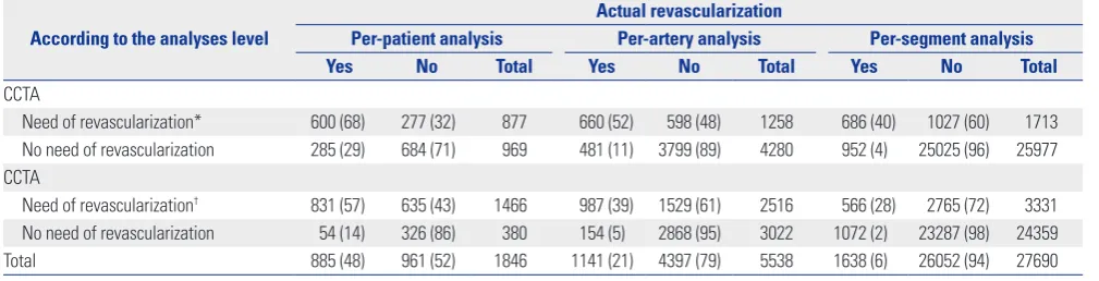
Table 2. The Ability of CCTA to Predict the Therapeutic Decision Making
According to the analyses level
Actual revascularization
Per-patient analysis Per-artery analysis Per-segment analysis
Yes No Total Yes No Total Yes No Total
CCTA
Need of revascularization* 600 (68) 277 (32) 877 660 (52) 598 (48) 1258 686 (40) 1027 (60) 1713 No need of revascularization 285 (29) 684 (71) 969 481 (11) 3799 (89) 4280 952 (4) 25025 (96) 25977 CCTA
Need of revascularization† 831 (57) 635 (43) 1466 987 (39) 1529 (61) 2516 566 (28) 2765 (72) 3331 No need of revascularization 54 (14) 326 (86) 380 154 (5) 2868 (95) 3022 1072 (2) 23287 (98) 24359 Total 885 (48) 961 (52) 1846 1141 (21) 4397 (79) 5538 1638 (6) 26052 (94) 27690 CCTA, coronary computed tomographic angiography.
Data are presented as number (row percentage).
beyond previous investigations because of large number of pa-tients, the multicenter design, and the inclusion of symptomat-ic subjects with stable angina who were referred to the cardiac catheterization laboratory as potential candidates for revascu-larization. This patient population better reflects the sample of
patients seen daily in the clinical setting.
[image:5.595.93.518.151.422.2]In addition to its use as an indicator for appropriate thera-peutic strategies, three previous multicenter studies demon-strated that CCTA has also various range of positive predictive value (64% to 91%) for detection of significant coronary artery
Fig. 2. Representative false positive cases for needing revascularization based on the coronary computed tomographic angiography (CCTA). (A) CCTA falsely identified this patient as a candidate for revascularization, based on the diffuse calcification of left anterior descending artery (LAD). (B) A second false-positive was indicated for revascularization because of a heavily calcified lesion within the LAD. (C) LAD was falsely identified as requiring revascu-larization because of lesion severity overestimation (arrow). (D) This right coronary artery was falsely indicated for revascurevascu-larization because of a motion artifact (arrow). Right images of each panel (A, B, and C) are CCTA and left images are conventional coronary angiography. In panel (D), right and left up-per images are CCTA and left lower image is conventional coronary angiography.
A
C
B
D
Fig. 3. Example of discordance between the coronary computed tomographic angiography (CCTA)-indicated therapy and the actual conducted therapy. Panel (A) and (B) are the CCTA and conventional coronary angiography images, respectively, from the same patient. (A) This patient was originally re-ferred for need of revascularization of the left anterior descending artery based on the CCTA images (white arrow); however, revascularization was not performed (black arrow). Right image is CCTA and left image is conventional coronary angiography. (B) The same patient actually underwent revascular-ization of the left circumflex artery, which was not originally indicated based on CCTA images (white arrow); however, revascularrevascular-ization was performed (black arrow with solid line, before revascularization; black arrow with dotted line, after revascularization). Right image is CCTA, and middle and left imag-es are conventional coronary angiography (before and after percutaneous coronary intervention, rimag-espectively).
Left anterior descending artery-false positive Left circumflex artery-false negative
[image:5.595.94.517.503.661.2]disease.1,2,13 Meta-analysis has also demonstrated a false
posi-tive rate of up to 35% for detection of significant coronary artery disease even in a low- to intermediate-risk population.14 These
limitations of using CCTA as a diagnostic tool, as well as the current finding of it’s being insufficient to predict the therapeu-tic decision, may be attributed to several factors. Previous stud-ies demonstrated that the presence of calcium, vessel tortuosity, or a smaller luminal caliber could affect the diagnostic accuracy, and the main cause of higher false-positive values may depend on the existence of calcium in the stenotic lesions.2,15,16 In the
present study, in symptomatic patients with suspected signifi-cant coronary artery stenosis, only 28% of the 1713 segments determined to require revascularization by CCTA were non-cal-cified plaques, while the remaining 72% of these segments con-tained calcified plaques. In addition to the presence of
calcifi-cation, Kruk, et al.17 found that the calcium length, volume, and
thickness were also associated with the inaccuracy of CCTA. This indicates that specific calcium characteristics may impact the accuracy of CCTA. We also found that the accuracy of CCTA to predict the therapeutic decision varied according to the plaque characteristics; the calcified plaques had a lower positive predic-tive value.
In addition to difficulties posed by the presence of calcifica-tion, the resolution of CCTA might also be insufficient to dis-cern the need for revascularization. The resolution of CCTA (200 μm) is inferior to that of invasive coronary imaging modalities (intravascular ultrasound, 100 μm, and optical coherence tomog-raphy, 10–15 μm).18 Precisely delineating the lumen and vessel
[image:6.595.40.546.282.449.2]borders in cross-sectional analysis poses difficulties when us-ing CCTA. Therefore, inaccurate measurements of the lumen
Table 4. The Accuracy of CCTA to Predict the Therapeutic Decision Making
Sensitivity (%) Specificity (%) PPV (%) NPV (%)
Per-patient* 68 71 68 71
Per-artery* 58 86 52 89
Per-segment* 42 96 40 96
According to the location of segments
Proximal segments 51 95 46 96
Mid segments 42 92 47 91
Side branch and distal segments 31 97 28 98
According to the plaque characteristics
Calcified plaque 34 92 33 92
Mixed plaque 62 77 41 88
Non-calcified plaque 65 77 43 89
Per-patient† 94 34 57 86
Per-artery† 87 65 39 95
Per-segment† 35 89 28 98
CCTA, coronary computed tomographic angiography; PPV, positive predictive value; NPV, negative predictive value.
[image:6.595.42.543.510.703.2]*The segments with more than 70% luminal narrowing of the coronary artery diameter were considered as a significantly stenosis with need of revasculariza-tion, †The segments with more than 50% luminal narrowing were considered as a significantly stenosis with need of revascularization.
Table 3. The Ability of CCTA to Predict the Therapeutic Decision Making
According to the location of segments
Actual revascularization
Proximal segments Mid segments Side branch/distal segments
Yes No Total Yes No Total Yes No Total
CCTA
Need of revascularization* 302 (46) 356 (54) 658 223 (47) 251 (53) 474 161 (28) 420 (72) 581 No need of revascularization 291 (4) 6435 (96) 6726 302 (9) 2916 (91) 3218 359 (2) 15674 (98) 16033 Total 593 (8) 6791 (92) 7384 525 (14) 3167 (86) 3692 520 (3) 16094 (97) 16614
According to the plaque characteristics Calcified plaque Mixed plaque Non-calcified plaque
Yes No Total Yes No Total Yes No Total
CCTA
Need of revascularization* 127 (33) 255 (67) 382 350 (41) 497 (59) 847 209 (43) 275 (57) 484 No need of revascularization 244 (8) 2856 (92) 3100 219 (12) 1664 (88) 1883 111 (11) 923 (89) 1034 Total 371 (11) 3111 (89) 3482 569 (21) 2161 (79) 2730 320 (21) 1198 (79) 1518 CCTA, coronary computed tomographic angiography.
Data are presented as number (row percentage).
and/or plaque dimensions assessed by CCTA were more fre-quent due to the lower resolution. Further, several factors (i.e., motion artifacts, arrhythmia, coronary calcification, inadequate intravascular contrast and reconstruction artifact) affect CCTA images which are digitally reconstructed and, therefore, recon-structed CCTA images do not accurately examine the various conditions of the lesions (i.e., calcification, severe tortuosity, or segment size). Conversely, the images generated from conven-tional coronary angiogram, intravascular ultrasound, or optical coherence tomography in the cardiac catheterization laboratory are real, direct images. Owning to these differences, disagree-ment regarding lesion stenosis severity frequently occurs be-tween two imaging modalities.18 We showed that this
disagree-ment is accentuated when examining small-sized vessels (i.e., side-branch/distal segments of coronary arteries). The present study also showed that CCTA had a lower positive predictive value in the side-branch/distal segments. Furthermore, intra-venous injection, not intracoronary injection, of contrast dye may result in insufficient filling and in difficulties maintaining constant dye concentration within the coronary artery lumen. In accordance with this, previous studies have reported that at-taining greater contrast enhancement of the lumen indepen-dently lowers the risk of false negative diagnosis.16,19 Recent
im-aging study on CCTA and intravascular ultrasound reported significant limitations of CCTA for delineating the lumen and vessel contour of coronary arteries; the minimal lumen area as-sessed by CCTA exhibited very weak correlations with those ob-tained by intravascular ultrasound intravascular ultrasound (r=0.23, 0.24, 0.15, 0.25, and 0.28, respectively).20 In aspects of
clinical benefits, one randomized study reported the use of CCTA to screen for coronary artery disease in high-risk patients with diabetes mellitus did not reduce the composite rate of death, nonfatal myocardial infarction or unstable angina re-quiring hospitalization.21 Recent randomized study with 10003
symptomatic patients also showed that, compared with func-tional testing (n=5007), a strategy of initial CCTA (n=4996) did not improve clinical outcomes over a median follow-up of 2 years.22
This study had several limitations. First, this was a retrospec-tive study. However, patients were enrolled consecuretrospec-tively in or-der to minimize selection bias. Second, there is no physiologic assessment by ischemic measurement such as fractional flow reserve to determine the need of revascularization, which could be important, particularly for intermediate lesions, 50–70% ste-nosis. However, we defined the necessities of revascularization as the more than 70% stenosis in the coronary angiography and CCTA images to avoid an underestimation of CCTA accuracy. In addition, measurement of fractional flow reserve with pres-sure-wire is reasonable to assess angiographic intermediate le-sions (50 to 70% diameter stenosis), not in significant lele-sions (more than 70% diameter stenosis) in current practical guideline for percutaneous coronary intervention.23 More importantly, we
excluded all asymptomatic patients. Thus, all decisions
regard-ing the necessities of revascularizations were made for symp-tomatic patients with objective evidence of positive stress test.
In conclusion, CCTA without conventional coronary angiog-raphy may be insufficient to assess coronary artery stenosis in symptomatic patients with stable angina. Conventional coro-nary angiography is needed to decide the need of revasculariza-tion in this patient popularevasculariza-tion.
ACKNOWLEDGEMENTS
This study was supported by a grant from the Korea Healthcare Technology R&D Project, Ministry for Health, Welfare & Family Affairs, Republic of Korea (Nos. A085136 and A102064) and the Cardiovascular Research Center, Seoul, Korea.
REFERENCES
1. Miller JM, Rochitte CE, Dewey M, Arbab-Zadeh A, Niinuma H, Gottlieb I, et al. Diagnostic performance of coronary angiography by 64-row CT. N Engl J Med 2008;359:2324-36.
2. Meijboom WB, Meijs MF, Schuijf JD, Cramer MJ, Mollet NR, van Mieghem CA, et al. Diagnostic accuracy of 64-slice computed to-mography coronary angiography: a prospective, multicenter, multivendor study. J Am Coll Cardiol 2008;52:2135-44.
3. Schuetz GM, Zacharopoulou NM, Schlattmann P, Dewey M. Me-ta-analysis: noninvasive coronary angiography using computed tomography versus magnetic resonance imaging. Ann Intern Med 2010;152:167-77.
4. Taylor AJ, Cerqueira M, Hodgson JM, Mark D, Min J, O’Gara P, et al. ACCF/SCCT/ACR/AHA/ASE/ASNC/NASCI/SCAI/SCMR 2010 Appropriate Use Criteria for Cardiac Computed Tomogra-phy. A Report of the American College of Cardiology Foundation Appropriate Use Criteria Task Force, the Society of Cardiovascular Computed Tomography, the American College of Radiology, the American Heart Association, the American Society of Echocar-diography, the American Society of Nuclear Cardiology, the North American Society for Cardiovascular Imaging, the Society for Cardiovascular Angiography and Interventions, and the Society for Cardiovascular Magnetic Resonance. Circulation 2010;122: e525-55.
5. Min JK, Berman DS, Dunning A, Achenbach S, Al-Mallah M, Bu-doff MJ, et al. All-cause mortality benefit of coronary revascular-ization vs. medical therapy in patients without known coronary artery disease undergoing coronary computed tomographic an-giography: results from CONFIRM (COronary CT Angiography EvaluatioN For Clinical Outcomes: an InteRnational Multicenter Registry). Eur Heart J 2012;33:3088-97.
6. de Feyter PJ, Nieman K. CCTA to guide revascularization for high-risk CAD: a ‘cliff hanger’. Eur Heart J 2012;33:3011-3.
7. Shaw LJ, Hausleiter J, Achenbach S, Al-Mallah M, Berman DS, Budoff MJ, et al. Coronary computed tomographic angiography as a gatekeeper to invasive diagnostic and surgical procedures: results from the multicenter CONFIRM (Coronary CT Angiogra-phy Evaluation for Clinical Outcomes: an International Multi-center) registry. J Am Coll Cardiol 2012;60:2103-14.
8. Piers LH, Dikkers R, Willems TP, de Smet BJ, Oudkerk M, Zijlstra F, et al. Computed tomographic angiography or conventional coro-nary angiography in therapeutic decision-making. Eur Heart J 2008;29:2902-7.
PL, Meyer M, et al. Coronary CT angiography versus conventional cardiac angiography for therapeutic decision making in patients with high likelihood of coronary artery disease. Radiology 2012; 265:385-92.
10. Austen WG, Edwards JE, Frye RL, Gensini GG, Gott VL, Griffith LS, et al. A reporting system on patients evaluated for coronary artery disease. Report of the Ad Hoc Committee for Grading of Coronary Artery Disease, Council on Cardiovascular Surgery, American Heart Association. Circulation 1975;51(4 Suppl):5-40. 11. Achenbach S, Raggi P. Imaging of coronary atherosclerosis by
computed tomography. Eur Heart J 2010;31:1442-8.
12. Groothuis JG, Beek AM, Meijerink MR, Brinckman SL, Heymans MW, van Kuijk C, et al. Positive predictive value of computed to-mography coronary angiography in clinical practice. Int J Cardiol 2012;156:315-9.
13. Budoff MJ, Dowe D, Jollis JG, Gitter M, Sutherland J, Halamert E, et al. Diagnostic performance of 64-multidetector row coronary computed tomographic angiography for evaluation of coronary artery stenosis in individuals without known coronary artery dis-ease: results from the prospective multicenter ACCURACY (As-sessment by Coronary Computed Tomographic Angiography of Individuals Undergoing Invasive Coronary Angiography) trial. J Am Coll Cardiol 2008;52:1724-32.
14. Mowatt G, Cook JA, Hillis GS, Walker S, Fraser C, Jia X, et al. 64-Slice computed tomography angiography in the diagnosis and assessment of coronary artery disease: systematic review and meta-analysis. Heart 2008;94:1386-93.
15. Arbab-Zadeh A, Miller JM, Rochitte CE, Dewey M, Niinuma H, Gottlieb I, et al. Diagnostic accuracy of computed tomography coronary angiography according to pre-test probability of coro-nary artery disease and severity of corocoro-nary arterial calcification. The CORE-64 (Coronary Artery Evaluation Using 64-Row Multi-detector Computed Tomography Angiography) International Multicenter Study. J Am Coll Cardiol 2012;59:379-87.
16. Yan RT, Miller JM, Rochitte CE, Dewey M, Niinuma H, Clouse ME,
et al. Predictors of inaccurate coronary arterial stenosis assess-ment by CT angiography. JACC Cardiovasc Imaging 2013;6:963-72.
17. Kruk M, Noll D, Achenbach S, Mintz GS, Pre˛gowski J, Kaczmarska E, et al. Impact of coronary artery calcium characteristics on ac-curacy of CT angiography. JACC Cardiovasc Imaging 2014;7:49-58.
18. Arbab-Zadeh A, Hoe J. Quantification of coronary arterial steno-ses by multidetector CT angiography in comparison with conven-tional angiography methods, caveats, and implications. JACC Cardiovasc Imaging 2011;4:191-202.
19. Cademartiri F, Maffei E, Palumbo AA, Malagò R, La Grutta L, Meiijboom WB, et al. Influence of intra-coronary enhancement on diagnostic accuracy with 64-slice CT coronary angiography. Eur Radiol 2008;18:576-83.
20. Kim C, Hong SJ, Shin DH, Kim JS, Kim BK, Ko YG, et al. Limita-tions of coronary computed tomographic angiography for delin-eating the lumen and vessel contours of coronary arteries in pa-tients with stable angina. Eur Heart J Cardiovasc Imaging 2015; 16:1358-65.
21. Muhlestein JB, Lappé DL, Lima JA, Rosen BD, May HT, Knight S, et al. Effect of screening for coronary artery disease using CT an-giography on mortality and cardiac events in high-risk patients with diabetes: the FACTOR-64 randomized clinical trial. JAMA 2014;312:2234-43.
22. Douglas PS, Hoffmann U, Patel MR, Mark DB, Al-Khalidi HR, Ca-vanaugh B, et al. Outcomes of anatomical versus functional test-ing for coronary artery disease. N Engl J Med 2015;372:1291-300. 23. Levine GN, Bates ER, Blankenship JC, Bailey SR, Bittl JA, Cercek B,