INTRODUCTION
Most microorganisms in natural ecosystems exist in the form of biofilms. The biofilms of microorganisms are formed over many stages, the first two of which are adherence to a biotic or abiotic surface and production of a structure to increase ad-herence. In the maturing biofilm, extracellular polymers are
formed by microorganisms in the structure and are known to protect microorganisms from changes in the surrounding en-vironment, to participate in supplying nutrients and discharg-ing metabolic waste, and to gather cells in closer proximity in order to facilitate cell-to-cell interactions. Biofilms can consist of multi-species, including coexisting bacteria and fungi.1-3
Candida albicans (C. albicans) is a resident species of healthy human mucous membranes that is also an opportunistic pa-thogen that induces superficial and systemic infection via the mucous epithelium when a patient suffers from severe disease or when the immune state is deficient. C. albicans has virulence factors that allow it to invade host tissue and to evade the host defense mechanism.4,5 C. albicans grows in three different forms: budding yeast (or blastoconidia), pseudohyphae, and hy-phae. The expression of C. albicans genes differ according to the C. albicans form. Candida hyphae are known to be essen-tial to pathogenicity and disease dissemination,6-8 and several Received: July 11, 2017 Revised: August 21, 2017
Accepted: August 22, 2017
Corresponding author: Dr. Joo Young Park, Department of Microbiology, Yonsei University Wonju College of Medicine, 20 Ilsan-ro, Wonju 26426, Korea. Tel: 82-33-741-0322, Fax: 82-33-742-5034, E-mail: [email protected]
•The authors have no financial conflicts of interest.
© Copyright: Yonsei University College of Medicine 2017
This is an Open Access article distributed under the terms of the Creative Com-mons Attribution Non-Commercial License (http://creativecomCom-mons.org/licenses/ by-nc/4.0) which permits unrestricted non-commercial use, distribution, and repro-duction in any medium, provided the original work is properly cited.
Proteus vulgaris
and
Proteus mirabilis
Decrease
Candida albicans
Biofilm Formation by Suppressing
Morphological Transition to Its Hyphal Form
Kyoung-Ho Lee, Su Jung Park, Sun Ju Choi, and Joo Young Park
Department of Microbiology, Yonsei University Wonju College of Medicine, Wonju, Korea.
Purpose: Candida albicans (C. albicans) and Proteus species are causative agents in a variety of opportunistic nosocomial infec-tions, and their ability to form biofilms is known to be a virulence factor. In this study, the influence of co-cultivation with Proteus vulgaris (P. vulgaris) and Proteus mirabilis (P. mirabilis) on C. albicans biofilm formation and its underlying mechanisms were ex-amined.
Materials and Methods: XTT reduction assays were adopted to measure biofilm formation, and viable colony counts were per-formed to quantify yeast growth. Real-time reverse transcriptase polymerase chain reaction was used to evaluate the expression of yeast-specific genes (rhd1 and rbe1), filament formation inhibiting genes (tup1 and nrg1), and hyphae-related genes (als3, ece1, hwp1, and sap5).
Results: Candida biofilm formation was markedly inhibited by treatment with either living or heat-killed P. vulgaris and P. bilis. Proteus-cultured supernatant also inhibited Candida biofilm formation. Likewise, treatment with live P. vulgaris or P. mira-bilis or with Proteus-cultured supernatant decreased expression of hyphae-related C. albicans genes, while the expression of yeast-specific genes and the filament formation inhibiting genes of C. albicans were increased. Heat-killed P. vulgaris and P. mira-bilis treatment, however, did not affect the expression of C. albicans morphology-related genes.
Conclusion: These results suggest that secretory products from P. vulgaris and P. mirabilis regulate the expression of genes related to morphologic changes in C. albicans such that transition from the yeast form to the hyphal form can be inhibited.
Key Words: Candida albicans, Proteus vulgaris, Proteus mirabilis, biofilm
pISSN: 0513-5796 · eISSN: 1976-2437 Yonsei Med J 2017 Nov;58(6):1135-1143
hyphae-specific genes are known, including hwp1, als3, als8, ece1, and sap4-6.9-11 The yeast-specific genes include rbe1, ywp1, and rhd1, and filament formation inhibiting genes include tup1 and nrg1.12-15
Proteus vulgaris (P. vulgaris) and Proteus mirabilis (P. mirabi-lis) exist in both human and animal small intestines and in the natural environment. P. vulgaris and P. mirabilis also can form biofilms on surfaces of various objects, including insertion ap-paratuses in humans. Proteus infections, especially urinary tract infections, are common in immunosuppressed patients.16-18 In recent years, C. albicans has been highlighted as one of the most common etiologic agents of acquired hospital infections, and Candida biofilms play an important role in initiating infections. Biofilms that exist in the human body are comprised of hun-dreds of different bacterial species: fungi and bacteria can also co-exist in these biofilms.19 Both the microorganism relation-ships with the host and the interactions between various mi-croorganisms are important, the latter of which is not being ac-tively researched.20,21
We previously reported that bacteria had a negative effect on the formation of C. albicans biofilms, and a more distinct de-crease in C. albicans biofilm formation was shown when cul-tivated with P. vulgaris.22 In the present study, the influence of co-culture of C. albicans, a chief agent of hospital-acquired infection, with P. vulgaris and P. mirabilis on the formation of C. albicans biofilm and its underlying mechanisms were examined.
MATERIALS AND METHODS
Organisms
Clinical isolates of C. albicans were obtained: one commensal strain was isolated from the blood of a patient, and P. vulgaris and P. mirabilis were isolated from the urine of another pa-tient. The identity of each microorganism was confirmed with the commercially-available identification systems (BioMeriéux, Marcy I’Etoile, France): API 32C for C. albicans and API 20E for P. vulgaris and P. mirabilis.
Culture conditions and experimental conditions Prior to each experiment, C. albicans isolates were cultured at 30ºC for 48 hours on Sabouraud’s dextrose agar (SDA, DifcoTM, Becton Dickinson, Spark, MD, USA), and one colony of yeast was inoculated into yeast nitrogen base (DifcoTM, Becton Dick-inson) medium supplemented with 50 mM glucose. P. vulgaris and P. mirabilis were first subcultured at 37ºC for 18 hours on tryptic soy agar. One colony each of P. vulgaris and P. mirabilis was then inoculated into tryptic soy broth (DifcoTM, Becton Dickinson) and incubated at 37ºC for 18 hours. The experi-mental conditions were as follows: 1) the microorganism was cultured alone; 2) C. albicans was co-cultured with live P. garis or P. mirabilis; 3) C. albicans was co-cultured with P. vul-garis or P. mirabilis killed at 100ºC for 30 minutes; or 4) C.
al-bicans was treated with bacteria-cultured supernatants of P. vulgaris or P. mirabilis diluted four times, in which the bacte-ria were removed.
XTT reduction assays
Biofilm formation was quantified using the method developed by Ramage, et al.23 Biofilms were formed on commercially available pre-sterilized, polystyrene, flat-bottomed, 96-well microtiter plates (Costar, Cambridge, MA, USA). Microorgan-isms were prepared for each condition and transferred to se-lected wells of a microtiter plate. The plate was incubated for 90 minutes at 37ºC in an orbital shaker at 75 rpm. After the ini-tial adhesion phase, the cell suspensions were aspirated, and each well was washed twice with phosphate-buffered saline (PBS) to remove loose adherent cells. A volume of 200 µL of medium was added to each well, and the plate was then incu-bated for another 72 hours. After biofilm formation, the medi-um was aspirated, and non-adherent cells were removed by thoroughly washing the biofilm three times with PBS. A quan-titative measure of biofilm formation was calculated using the XTT [2,3-bis(2-methyoxy-4-nitro-5-sulfo-phenyl)-2H-tetrazo-lium-5-carboxanilide] reduction assay. A 200-µL aliquot of XTT (1 mg/mL, Sigma, St. Louis, MO, USA) and menadione (0.4 mM, Sigma) solution was added to each well containing the prewashed biofilm and the control well. The plates were then incubated in the dark for up to 3 hours at 37ºC. A colorimetric change resulting from XTT reduction was measured using a microtiter plate reader (EMax, Molecular Devices, Sunnyvale, CA, USA) at 490 nm.
C. albicans cell counts
After biofilm formation, the medium was aspirated, and non-adherent cells were removed by thoroughly washing the bio-film three times with PBS. Then, 1 mL of PBS was transferred to each well, and biomass was meticulously scraped off. The resultant solution containing the detached biofilm cells was gently vortexed for 1 minute to disrupt the aggregates and in-oculated on an SDA plate. The colony forming units (CFUs) of C. albicans were quantified after 48 hours of incubation at 30ºC.
Scanning electron microscopy
Relative quantitation by real-time reverse transcriptase polymerase chain reaction
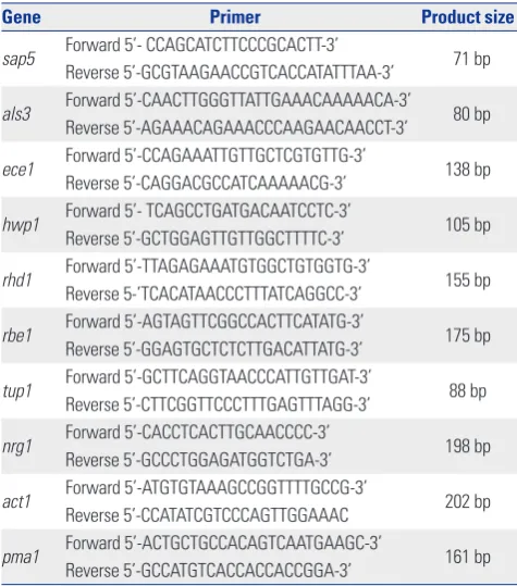
RNA was isolated from C. albicans cells using the MasterPure Yeast RNA Extraction kit (Epicentre Biotechnologies, Madi-son, WI, USA). RNA was treated with amplification grade DN-ase I (Epicentre Biotechnologies) and used for cDNA synthesis with random hexamer primer (Invitrogen Life Technologies, Carlsbad, CA, USA) using Superscript II reverse transcriptase reagents (Invitrogen Life Technologies). Each reaction con-tained 1 µg of total RNA, 1 µL of 50 µM hexamer, and 1 µL of 10 mM dNTP in a final volume of 10 µL. Reactions were incubat-ed at 65ºC for 5 minutes and coolincubat-ed on ice. To each reaction tube, 10 µL of the following mixture was added: 4 µL of 5x First-Strand Buffer, 2 µL of 10 mM MgCl2, 2 µL of 0.1 M DTT, 1.4 µL of RNase inhibitor, and 1 µL of Superscript II. Reactions were incubated at 42ºC for 50 minutes and then at 70ºC for 15 min-utes. Real-time polymerase chain reaction (PCR) contained 10 µL of Power SYBR Green Master Mix (Applied Biosystems, Foster City, CA, USA), as well as forward and reverse primers (1 µL of each) (Table 1)22 and sterile water, at a final volume of 20 µL. The PCR was run on MicroAmp® Optical 384-well reac-tion plates in an ABI 7900 Real-Time PCR system (Applied Bio-systems). Real-time PCR reactions were performed at 95ºC for 5 minutes, followed by 40 cycles of 15 seconds at 95ºC and 1 minute at 60ºC. Dissociation curves were analyzed for all re-actions to verify single peaks/products. Expression levels were analyzed using ABI 7900 System SDS software (Applied Biosys-tems). Real-time PCR data were normalized with the geometric mean of two reference genes. The ACT1 and PMA1 genes were
used for this purpose.
Statistical analysis
All experiments were performed in triplicate on three differ-ent occasions. All data are expressed as mean values with cor-responding standard deviations (SDs). Student’s t-tests and Mann-Whitney U-tests were used to compare the differences between Candida only and Candida co-cultured with P. vul-garis or P. mirabilis. All p-values <0.05 were considered statis-tically significant.
RESULTS
The effect of P. vulgaris or P. mirabilis on C. albicans
biofilm formation
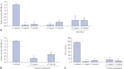
The biofilm value generated when each type of microorgan-ism was incubated separately was 0.07±0.007 for P. vulgaris, 0.120±0.004 for P. mirabilis, and 1.403±0.103 for C. albicans. When culturing C. albicans and P. vulgaris or P. mirabilis togeth-er, biofilm formation was reduced by more than 80%, com-pared to the C. albicans culture alone. To assess the mechanism by which the bacteria impeded Candida biofilm formation, bacteria were initially treated for 30 minutes at 100ºC to elimi-nate biological activity, followed by co-culture with C. albicans. Biofilm formation of C. albicans cultured together with heat-killed P. vulgaris or P. mirabilis elicited a 70% reduction, com-pared to the control group. These data indicate that dead bac-teria interfere with the structural formation of C. albicans biofilms (Fig. 1A).
[image:3.595.56.294.454.724.2]When C. albicans and P. vulgaris or P. mirabilis were co-cul-tured, biofilm formation of C. albicans was significantly re-duced. In order to determine if this reduction was due to the effect of the bacteria or of secretory products when cultured, P. vulgaris and P. mirabilis were cultured for 72 hours, and remain-ing bacteria were removed by filtration. As a result of treatment with P. vulgaris and P. mirabilis culture supernatants, C. albi-cans biofilm formation was reduced by 60−70%, compared to C. albicans cultured alone (Fig. 1B). To determine whether this effect was due to a depletion of nutrients in the medium or to the secretory products of the bacteria, we diluted the cultured supernatants and tested the concentration effect on C. albicans biofilm formation. C. albicans biofilm formation decreased in proportion to the concentration of the bacterial-cultured su-pernatants (data not shown). To determine whether P. vulgar-is and P. mirabilvulgar-is inhibited the growth of C. albicans, C. albi-cans and P. vulgaris or P. mirabilis or bacterial-cultured super-natants were cultured together for 72 hours, and C. albicans CFUs were calculated. When C. albicans was cultured alone, the count was 2.85×10⁸ CFU/mL; when cultured together with P. vulgaris, the count of C. albicans was reduced to 9×10⁶ CFU/ mL; and when C. albicans was cultured together with P. mira-bilis, the count of C. albicans dropped to 2.4×10⁷ CFU/mL (Fig. Table 1. Primers Used for Real-Time RT-PCR
Gene Primer Product size
sap5 Forward 5’- CCAGCATCTTCCCGCACTT-3’ 71 bp Reverse 5’-GCGTAAGAACCGTCACCATATTTAA-3’
als3 Forward 5’-CAACTTGGGTTATTGAAACAAAAACA-3’ 80 bp Reverse 5’-AGAAACAGAAACCCAAGAACAACCT-3’
ece1 Forward 5’-CCAGAAATTGTTGCTCGTGTTG-3’ 138 bp Reverse 5’-CAGGACGCCATCAAAAACG-3’
hwp1 Forward 5’- TCAGCCTGATGACAATCCTC-3’ 105 bp Reverse 5’-GCTGGAGTTGTTGGCTTTTC-3’
rhd1 Forward 5’-TTAGAGAAATGTGGCTGTGGTG-3’ 155 bp Reverse 5-’TCACATAACCCTTTATCAGGCC-3’
rbe1 Forward 5’-AGTAGTTCGGCCACTTCATATG-3’ 175 bp Reverse 5’-GGAGTGCTCTCTTGACATTATG-3’
tup1 Forward 5’-GCTTCAGGTAACCCATTGTTGAT-3’ 88 bp Reverse 5’-CTTCGGTTCCCTTTGAGTTTAGG-3’
nrg1 Forward 5’-CACCTCACTTGCAACCCC-3’ 198 bp Reverse 5’-GCCCTGGAGATGGTCTGA-3’
act1 Forward 5’-ATGTGTAAAGCCGGTTTTGCCG-3’ 202 bp Reverse 5’-CCATATCGTCCCAGTTGGAAAC
pma1 Forward 5’-ACTGCTGCCACAGTCAATGAAGC-3’ 161 bp Reverse 5’-GCCATGTCACCACCACCGGA-3’
1C). It seems that P. vulgaris and P. mirabilis inhibit C. albi-cans biofilm formation and also interfere with its growth. As a result, we investigated the C. albicans CFUs after treating C. al-bicans with bacterial-cultured supernatants and culturing for 72 hours. In the case of culturing C. albicans alone, the count was 2.85×10⁸ CFU/mL. Meanwhile, the count from the treated culture supernatants of P. vulgaris was 3.6×10⁷ CFU/mL, and the count from the treated culture supernatants of P. mirabilis was reduced to 2.0×10⁷ CFU/mL (Fig. 1C).
The effect of P. vulgaris or P. mirabilis on C. albicans
morphology-related gene expression
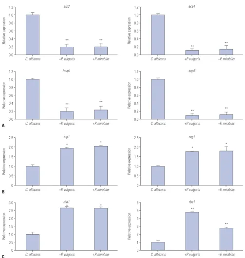
C. albicans morphology changes to the hyphae form from the yeast form as biofilm formation progresses, and the expres-sion pattern of genes related to morphology at these stages was identified. Previous studies have shown that expression pat-terns of Candida species-related genes are significantly in-creased during hyphae-specific gene expression over the du-ration of biofilm formation. On the contrary, the expression of genes suppressing the formation of yeast-specific genes and filament are decreased.22 These results suggest that Candida present in yeast form adhere to a surface to increase the expres-sion of hyphae-related genes and promote the formation of biofilms by decreasing the expression of genes that inhibit the yeast form and filament formation. To clarify the effect of co-cultures on biofilm formation, we analyzed changes in C.
albi-cans gene expression levels in biofilms co-cultured with P. vul-garis or P. mirabilis. In contrast to C. albicans cultured alone, als3 and hwp1 showed a reduction in expression by 80% and ece1 and sap5 by 90% (Fig. 2A). When C. albicans was cultured together with P. vulgaris or P. mirabilis, the expression of tup1 and nrg1, which are genes known to suppress filament forma-tion, increased by about two-fold, compared to when C. albi-cans was cultured alone (Fig. 2B). Regarding the expression of yeast-specific rhd1 and rbe1, the expression levels of rhd1 in-creased by 2.5-fold and rbe1 by about more than 3-fold (Fig. 2C). When C. albicans was co-cultured with heat-killed P. vulgaris and P. mirabilis, biofilm formation was decreased. We con-firmed that these results were due to changes in the expression of biofilm-related genes in C. albicans. First, ece1, hwp1, and sap5 gene levels, which are associated with the formation of hyphae, did not increase. While als3 level was slightly incr-eased, the change was not significant (Fig. 3A). There were also no significant changes in tup1 and nrg1, which are filament for-mation inhibiting genes (Fig. 3B). Yeast-specific genes also sh-owed no difference in expression levels (Fig. 3C). The reduc-tion in C. albicans biofilm formareduc-tion in co-cultures with killed bacteria is considered to be caused not by changes in gene ex-pression, but by the interference of killed bacteria acting as small particles that fit between C. albicans cells and, thus, sup-press the structural formation of biofilm.
1.6 1.4 1.2 1.0 0.8 0.6 0.4 0.2 0.0
Biofilm formation (OD 490 nm)
B
C. albicans
Cultured supernatants +P. vulgaris +P. mirabilis
**
**
350 300 250 200 150 100 50 0
CFU (1×10
6/mL)
C
C. albicans +P. vulgaris P. mirabilis +P. vulgaris P. mirabilis Cultured supernatants
** ** ** **
1.6 1.4 1.2 1.0 0.8 0.6 0.4 0.2 0.0
Biofilm formation (OD 490 nm)
A C. albicans P. vulgaris P. mirabilis +P. vulgaris+P. mirabilis +P. vulgaris Heat-killed
+P. mirabilis
** ** **
**
**
[image:4.595.41.539.409.689.2]**
Fig. 1. The effect of P. vulgaris or P. mirabilis on C. albicans biofilm formation. (A) The co-culturing effect of live or heat-killed P. vulgaris and P. mirabilis
on C. albicans biofilm formation. The effects of P. vulgaris and P. mirabilis cultured supernatant on biofilm formation (B) and growth (C) of C. albicans.
The effect of cultured P. vulgaris or P. mirabilis
supernatants on expression of C. albicans
morphology-related genes
As described above, treatment of P. vulgaris and P. mirabilis cultured supernatants inhibited C. albicans biofilm formation. We examined how this treatment affected the expression of
various genes involved in biofilm formation. The hyphae-spe-cific genes als3, ece1, hwp1, and sap5 all showed a significant reduction in expression in contrast to the cultured supernatants in which C. albicans was cultured alone (Fig. 4A). In contrast, the filament formation inhibiting genes tup1 and nrg1 slightly increased (Fig. 4B). The yeast-specific gene rhd1 increased by
1.2
1.0
0.8
0.6
0.4
0.2
0.0
Relative expression
C. albicans
als3
+P. vulgaris +P. mirabilis
** **
1.2
1.0
0.8
0.6
0.4
0.2
0.0
Relative expression
C. albicans
ece1
+P. vulgaris +P. mirabilis
** **
1.2
1.0
0.8
0.6
0.4
0.2
0.0
Relative expression
A C. albicans
hwp1
+P. vulgaris +P. mirabilis
** **
1.2
1.0
0.8
0.6
0.4
0.2
0.0
Relative expression
C. albicans
sap5
+P. vulgaris +P. mirabilis
** **
2.5
2.0
1.5
1.0
0.5
0
Relative expression
B C. albicans
tup1
+P. vulgaris +P. mirabilis
* *
2.5
2.0
1.5
1.0
0.5
0
Relative expression
C. albicans
nrg1
+P. vulgaris +P. mirabilis
* *
3.0
2.5
2.0
1.5
1.0
0.5
0
6
5
4
3
2
1
0 Relative expression Relative expression
C C. albicans C. albicans
rhd1 rbe1
+P. vulgaris +P. mirabilis +P. vulgaris +P. mirabilis *
** *
[image:5.595.53.557.156.687.2]**
2-fold, and rbe1 slightly increased (Fig. 4C). This suggests that secretory products that are formed and released with growth of P. vulgaris and P. mirabilis inhibit the growth of C. albicans and regulate the expression of biofilm-related genes, thereby
inhibiting biofilm formation in C. albicans.
Scanning electron microscopy of biofilms
It is known that biofilms are not formed with a simple
struc-1.4 1.2 1.0 0.8 0.6 0.4 0.2 0
1.4 1.2 1.0 0.8 0.6 0.4 0.2 0 Relative expression Relative expression
A
C. albicans C. albicans
hwp1 sap5
+P. vulgaris +P. mirabilis +P. vulgaris +P. mirabilis
1.4 1.2 1.0 0.8 0.6 0.4 0.2 0
1.4 1.2 1.0 0.8 0.6 0.4 0.2 0
Relative expression
Relative expression
B
C
C. albicans
C. albicans
tup1
rhd1 +P. vulgaris
+P. vulgaris
+P. mirabilis
+P. mirabilis
1.4 1.2 1.0 0.8 0.6 0.4 0.2 0
1.2
1.0
0.8
0.6
0.4
0.2
0
Relative expression
Relative expression
C. albicans
C. albicans
nrg1
rbe1 +P. vulgaris
+P. vulgaris
+P. mirabilis
[image:6.595.42.546.144.703.2]+P. mirabilis
Fig. 3. The effects of killed P. vulgaris or P. mirabilis cells on expression of C. albicans morphology-related genes. Relative quantitation of expression of hyphae-specific (A), filament formation inhibiting (B), and yeast-specific (C) genes. Expression of genes was determined by quantitative real-time re-verse transcriptase polymerase chain reaction. The data represent the average and standard deviation of three separate experiments.
1.6 1.4 1.2 1.0 0.8 0.6 0.4 0.2 0
Relative expression
C. albicans
als3
+P. vulgaris +P. mirabilis Heat-killed
1.6 1.4 1.2 1.0 0.8 0.6 0.4 0.2 0
Relative expression
C. albicans
ece1
+P. vulgaris +P. mirabilis Heat-killed
Heat-killed
Heat-killed
Heat-killed
Heat-killed
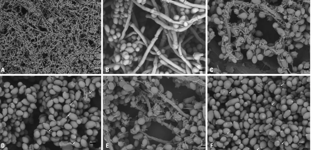
ture and that Candida exist in the yeast form in the basal layer and in the hyphae and pseudohyphae form in the layer above. These layers form a network with a three-dimensional struc-ture. The structural difference between the biofilm when C.
albicans is cultivated separately and when co-cultured with P. vulgaris or P. mirabilis was investigated via scanning electron microscopy (Fig. 5). The biofilm of C. albicans when cultured alone was high in density and was a multi-layer solid (Fig. 5A
1.2
1.0
0.8
0.6
0.4
0.2
0.0
1.2
1.0
0.8
0.6
0.4
0.2
0.0 Relative expression Relative expression
A
C. albicans C. albicans
hwp1 sap5
+P. vulgaris +P. mirabilis +P. vulgaris +P. mirabilis
1.6 1.4 1.2 1.0 0.8 0.6 0.4 0.2 0.0
3.0
2.5
2.0
1.5
1.0
0.5
0.0
Relative expression
Relative expression
B
C
C. albicans
C. albicans
tup1
rhd1
+P. vulgaris
+P. vulgaris
+P. mirabilis
+P. mirabilis
2.0
1.5
1.0
0.5
0.0
1.6 1.4 1.2 1.0 0.8 0.6 0.4 0.2 0.0
Relative expression
Relative expression
C. albicans
C. albicans
nrg1
rbe1 +P. vulgaris
+P. vulgaris
+P. mirabilis
[image:7.595.58.554.141.696.2]+P. mirabilis
Fig. 4. The effects of cultured P. vulgaris and P. mirabilis supernatants on expression of C. albicans morphology-related genes. Relative quantitation of hyphae-specific (A), filament formation inhibiting (B), and yeast-specific (C) gene expression. Expression of genes was determined by quantitative re-al-time reverse transcriptase polymerase chain reaction. The data represent the average and standard deviation of three separate experiments. *p<0.05, **p<0.01 compared with the biofilm formation of C. albicans only.
1.2
1.0
0.8
0.6
0.4
0.2
0.0
Relative expression
C. albicans
als3
+P. vulgaris +P. mirabilis Cultured supernatants
1.2
1.0
0.8
0.6
0.4
0.2
0.0
Relative expression
C. albicans
ece1
+P. vulgaris +P. mirabilis Cultured supernatants
Cultured supernatants
Cultured supernatants
Cultured supernatants
Cultured supernatants
Cultured supernatants Cultured supernatants *
*
** **
** ** ** **
**
** *
and B). The biofilm formed when C. albicans and P. vulgaris or P. mirabilis were cultured together showed that the P. vul-garis or P. mirabilis were attached to the hyphae of C. albicans and inserted between the C. albicans cells. The thickness of the biofilm in these cases also appeared to be thin with a low density (Fig. 5C and E). The structure of biofilms formed after culturing heat-killed bacteria and C. albicans together was thinner and showed lower density than biofilms formed after separate culture of C. albicans (Fig. 5D and F). However, the heat-killed bacteria biofilm was thicker than the biofilm formed after C. albicans had been cultured together with live bacteria, and more of the C. albicans was in the yeast form. The above results agree with the outcome of XTT reduction assays, which quantitatively confirmed biofilm formation (Fig. 1A).
DISCUSSION
Microorganisms that are fixed inside a biofilm show resis-tance to the immune system and have a strong tolerance to antibiotics relative to planktonic microorganisms.24 Many hu-man infections are the result of microorganisms in biofilms. Most studies on biofilm formation and the interrelation be-tween microorganisms in them have focused on bacteria.25 However, biofilms affected by the relationship between bacte-ria and fungi are clinically crucial, because these biofilms in-crease the morbidity and mortality of infections.19,26
The research presented here verified the influence of the coexistence of C. albicans and P. vulgaris and P. mirabilis on C. albicans biofilm formation and whether the correlation
be-tween C. albicans and P. vulgaris and P. mirabilis was compet-itive or symbiotic. The architecture and functioning of com-plex biofilms are very intricate and were not clearly elucidated. In addition, the correlation between microorganisms inside the complex cultivated biofilm was not identified.
When C. albicans was cultivated with P. vulgaris and P. mi-rabilis, both biofilm formation and number of C. albicans cells decreased, compared to when C. albicans was cultured alone (Fig. 1). Even the diluted supernatants of P. vulgaris and P. mi-rabilis cultivation were confirmed to hinder biofilm formation, which implies that it was not the depletion of nutrients due to mixed culture of P. vulgaris and P. mirabilis plus C. albicans that decreased biofilm formation, but that the bacteria direct-ly hindered formation due to secretory products (Fig. 1). The Proteus-specific products inhibiting the growth of C. albicans or biofilm formation are not yet clarified, and further research is needed.
Interestingly, even when C. albicans was cultivated with heat-treated P. vulgaris and P. mirabilis, biofilm formation de-creased (Fig. 1A). It is considered that both the secretory prod-ucts from P. vulgaris and P. mirabilis and the bacterial archi-tecture itself induced structural changes and hindered the ability of C. albicans to form biofilms. Further, we verified bio-film structures of C. albicans alone or when cultivated with P. vulgaris and P. mirabilis via scanning electron microscopy. The biofilm formed normally only when C. albicans was cul-tured alone, showing high density and numerous layers (Fig. 5A). In contrast, biofilms that formed when P. vulgaris and P. mirabilis were cultured together showed low-density, thin bio-films with bacteria among the C. albicans cells, and a
notice-Fig. 5. Scanning electron microscopy of biofilms. C. albicans biofilm (A, ×500; B, ×4000). Biofilm of co-culture with C. albicans and live P. vulgaris (C) or heat-killed P. vulgaris (D); biofilm of co-culture with C. albicans and live P. mirabilis (E) or heat-killed P. mirabilis (F). Arrows point to examples of heat-killed bacteria. Magnifications are ×500 (scale bar; 20 μm) or ×4000 (scale bar; 2 μm).
A
D
B
E
C
[image:8.595.45.540.462.702.2]ably decreased number of mycelia (Fig. 5). It is clear that both living and dead bacteria particles influenced structural matu-ration of the biofilm. Thus, P. vulgaris and P. mirabilis suppress the growth of C. albicans and can function as structural obst-ructive factors to the maturation of Candida biofilms.
The formation of hyphae is essential to C. albicans biofilm formation, so it is also important to understand the genetic ba-sis of the morphological changes in C. albicans.10,11 The mature biofilm enables the Candida yeast to fix the biofilm on to the extracellular surface, and the hyphae form a cross-sectional st-ructure with structural frames.27 Inhibition of the hyphae trans-genes of C. albicans led to biofilm formation with the basal layer only, whereas inhibition of the yeast transgene of C. albi-cans led to biofilm formation with only the outer layer among the existing biofilm structures.11,28 In this study, the expression of hyphae-related genes C. albicans was significantly inhibited in the presence of live Proteus or by Proteus-cultured superna-tant. The expression of both yeast-related and filament forma-tion inhibiting genes in C. albicans was up-regulated by treat-ment with live Proteus or Proteus-cultured supernatant (Figs. 2 and 4); however, the expression of morphology-related genes was not affected by heat-killed P. vulgaris and P. mirabilis (Fig. 3). These results suggest that secretory products of P. vulgaris and P. mirabilis regulate the expression of genes that are related to morphologic changes, which could be the crucial factor in C. albicans biofilm formation, inhibiting hyphal transition from the yeast form to the hyphal form. Due to an increase in only the yeast form and the lack of hyphal form, the C. albicans biofilm would not form a solid 3D structure, but only a thick, basal-layered structure.
REFERENCES
1. Potera C. Forging a link between biofilms and disease. Science 1999;283:1837-9.
2. Donlan RM, Costerton JW. Biofilms: survival mechanisms of clini-cally relevant microorganisms. Clin Microbiol Rev 2002;15:167-93. 3. Fuqua C, Winans SC, Greenberg EP. Census and consensus in
bac-terial ecosystems: the LuxR-LuxI family of quorum-sensing tran-scriptional regulators. Annu Rev Microbiol 1996;50:727-51. 4. Ghannoum MA. Potential role of phospholipases in virulence and
fungal pathogenesis. Clin Microbiol Rev 2000;13:122-43.
5. Sugita T, Kurosaka S, Yajitate M, Sato H, Nishikawa A. Extracellular proteinase and phospholipase activity of three genotypic strains of a human pathogenic yeast, Candida albicans. Microbiol Immu-nol 2002;46:881-3.
6. Hornby JM, Jensen EC, Lisec AD, Tasto JJ, Jahnke B, Shoemaker R, et al. Quorum sensing in the dimorphic fungus Candida albicans is mediated by farnesol. Appl Environ Microbiol 2001;67:2982-92. 7. Douglas LJ. Candida biofilms and their role in infection. Trends
Microbiol 2003;11:30-6.
8. Budtz-Jørgensen E, Mojon P, Rentsch A, Deslauriers N. Effects of an oral health program on the occurrence of oral candidosis in a long-term care facility. Community Dent Oral Epidemiol 2000;28: 141-9.
9. Staab JF, Bradway SD, Fidel PL, Sundstrom P. Adhesive and mam-malian transglutaminase substrate properties of Candida albi-cans Hwp1. Science 1999;283:1535-8.
10. Hoyer LL. The ALS gene family of Candida albicans. Trends Mi-crobiol 2001;9:176-80.
11. Liu H. Transcriptional control of dimorphism in Candida albicans. Curr Opin Microbiol 2001;4:728-35.
12. Granger BL. Insight into the antiadhesive effect of yeast wall pro-tein 1 of Candida albicans. Eukaryot Cell 2012;11:795-805. 13. Sohn K, Urban C, Brunner H, Rupp S. EFG1 is a major regulator
of cell wall dynamics in Candida albicans as revealed by DNA mi-croarrays. Mol Microbiol 2003;47:89-102.
14. Znaidi S, Nesseir A, Chauvel M, Rossignol T, d’Enfert C. A com-prehensive functional portrait of two heat shock factor-type tran-scriptional regulators involved in Candida albicans morphogene-sis and virulence. PLoS Pathog 2013;9:e1003519.
15. Braun BR, Head WS, Wang MX, Johnson AD. Identification and characterization of TUP1-regulated genes in Candida albicans. Genetics 2000;156:31-44.
16. Rózalski A, Sidorczyk Z, Kotełko K. Potential virulence factors of Proteus bacilli. Microbiol Mol Biol Rev 1997;61:65-89.
17. Jacobsen SM, Stickler DJ, Mobley HL, Shirtliff ME. Complicated catheter-associated urinary tract infections due to Escherichia coli and Proteus mirabilis. Clin Microbiol Rev 2008;21:26-59. 18. Jacobsen SM, Shirtliff ME. Proteus mirabilis biofilms and
cathe-ter-associated urinary tract infections. Virulence 2011;2:460-5. 19. Thein ZM, Seneviratne CJ, Samaranayake YH, Samaranayake LP.
Community lifestyle of Candida in mixed biofilms: a mini review. Mycoses 2009;52:467-75.
20. Adam B, Baillie GS, Douglas LJ. Mixed species biofilms of Candida albicans and Staphylococcus epidermidis. J Med Microbiol 2002; 51:344-9.
21. Costerton JW, Geesey GG, Cheng KJ. How bacteria stick. Sci Am 1978;238:86-95.
22. Park SJ, Han KH, Park JY, Choi SJ, Lee KH. Influence of bacterial presence on biofilm formation of Candida albicans. Yonsei Med J 2014;55:449-58.
23. Ramage G, Vandewalle K, Wickes BL, López-Ribot JL. Character-istics of biofilm formation by Candida albicans. Rev Iberoam Micol 2001;18:163-70.
24. Fux CA, Costerton JW, Stewart PS, Stoodley P. Survival strategies of infectious biofilms. Trends Microbiol 2005;13:34-40.
25. Hall-Stoodley L, Costerton JW, Stoodley P. Bacterial biofilms: from the natural environment to infectious diseases. Nat Rev Mi-crobiol 2004;2:95-108.
26. Costerton JW, Lewandowski Z, Caldwell DE, Korber DR, Lappin-Scott HM. Microbial biofilms. Annu Rev Microbiol 1995;49:711-45. 27. Hansen SK, Rainey PB, Haagensen JA, Molin S. Evolution of
spe-cies interactions in a biofilm community. Nature 2007;445:533-6. 28. Nett J, Andes D. Candida albicans biofilm development,