Diffusion-Weighted MR Imaging before and after Contrast
Enhancement with Superparamagnetic Iron Oxide
for Assessment of Hepatic Metastasis
Hana Kim,
1Jeong-Sik Yu,
1Dae Jung Kim,
2Jae-Joon Chung,
1Joo Hee Kim,
1and Ki Whang Kim
3 1Department of Radiology, Gangnam Severance Hospital, Yonsei University College of Medicine, Seoul;2Department of Radiology, CHA Bundang Medical Center, CHA University, Seongnam;
3Department of Radiology, Severance Hospital, Yonsei University College of Medicine, Seoul, Korea.
Received: June 28, 2011 Revised: September 18, 2011 Accepted: October 13, 2011
Corresponding author: Dr. Jeong-Sik Yu, Department of Radiology,
Gangnam Severance Hospital, Yonsei University College of Medicine, 211 Eonju-ro, Gangnam-gu, Seoul 135-720, Korea.
Tel: 82-2-2019-3510, Fax: 82-2-3462-5472 E-mail: [email protected]
∙ The authors have no financial conflicts of interest.
© Copyright:
Yonsei University College of Medicine 2012
This is an Open Access article distributed under the terms of the Creative Commons Attribution Non-Commercial License (http://creativecommons.org/ licenses/by-nc/3.0) which permits unrestricted non-commercial use, distribution, and reproduction in any medium, provided the original work is properly cited.
Purpose: The purpose of our study was to validate diffusion-weighted MRI (DWI) before and after superparamagnetic iron oxide (SPIO) injection for assessment of hepatic metastases. Materials and Methods: Eighty-six hepatic metastases (size range, 0.3-4.7 cm; mean, 1.5 cm) verified pathologically or by follow-up imaging studies in 22 consecutive patients (17 men and 5 women; 44-83 years; mean age, 60 years) during a 13-month period were enrolled. Hepatic MRI, including DWI (b-factors=50, 400, 800 s/mm2) with breath-holding technique of single-shot
spin-echo spin-echo-planar imaging (TR/TE=1000/69 ms, average=2) before and after SPIO administration, were retrospectively reviewed by two independent radiologists with a 5-point scale confidence score for each hepatic lesion on pre-contrast DWI (pre-DWI), SPIO-enhanced DWI (SPIO-(pre-DWI), and SPIO-enhanced T2*-weighted im-aging (SPIO-T2*wI). Results: For all lesions, SPIO-T2*wI showed significantly higher confidence score in the diagnosis of hepatic metastases than pre-contrast or SPIO-DWI regardless of the size of b-factors (p<0.05) with only one exception; us-ing b-factor=50 s/mm2, the score of SPIO-T2*wI was still higher than SPIO-DWI
but there was no statistical significance given by observer 1 (p=0.730). For the sub-centimeter lesions (n=37), SPIO-T2*wI showed the highest score, and using b-fac-tor=50 or 400 s/mm2 SPIO-DWI showed similar confidence scores to SPIO-T2*wI
by both observers (p>0.05). Pre-DWI using b-factor=50 sec/mm2 was also
compa-rable with SPIO-T2*wI by observer 1 (p=0.060). Conclusion: Pre-DWI has a lim-ited value for the assessment of hepatic metastases, however, the repetition of DWI after SPIO injection using small b-factors could complement SPIO-T2*wI, espe-cially for subcentimeter lesions.
Key Words: Liver, metastasis, diffusion magnetic resonance imaging, superpara-magnetic iron oxide (SPIO)
INTRODUCTION
for informed consent from individual patients. During a 13-month period from March 2007 to March 2008, 299 consecutive patients underwent hepatic MRI with DWI, gadolinium-enhanced dynamic imaging, and subsequent SPIO-enhanced MRI for the assessment of focal liver le-sions in our institution. To be enrolled as subjects in this study, patients should have one or more metastatic lesions in the liver from extrahepatic malignancy and the lesions should be verified pathologically or by follow-up imaging studies. Electronically stored clinical records and imaging interpretation notes obtained from all 299 patients were re-viewed by a study coordinator, and a total of 22 patients (17 men and 5 women; 44-83 years; mean age, 60 years) were finally selected. Among 86 lesions found in the subjected patients. 21 lesions were confirmed by pathology from op-eration or biopsy (n=21) or follow-up imaging studies (n=65). At follow-up imaging (range to 5 to 13 months), a lesion was deemed malignant if it showed 20% or greater in size. The primary cancer sites of these patients were as follows: 13 colorectal cancers, 3 gastric cancers, 2 breast cancers, 2 renal cell carcinomas, 1 pancreatic cancer, and 1 nasopha-ryngeal cancer. There was only 1 lesion in nine patients, 2 nodules in three patients, 3 nodules in one patient, 5 nod-ules in three patients, and six patients had 6, 7, 8, 9, 10 or 13 nodules in each patient.
MRI techniques
MRI was performed using a 1.5-T system (Magnetom Avan-to; Siemens, Erlangen, Germany) equipped with high-per-formance gradients (maximum amplitude 45 mT/m) and a six-element phased-array surface coil.
The routine MRI protocol of our institute consisted of spectral fat suppression, T2-weighted, half-Fourier acquisi-tion, single-shot turbo spin-echo images, T1-weighted dou-ble-echo chemical shift gradient-echo (GRE) sequence, and dynamic contrast-enhanced imaging, using a 3D GRE quence in the axial plane consisting of one pre-contrast se-ries, followed by three successive post-contrast series (early arterial, late arterial, and portal phase imaging), and by 5-min delayed phase imaging after administration of a bo-lus injection of gadopentetate dimeglumine (0.1 mmol/kg of Magnevist; Bayer HealthCare, Berlin, Germany). After the completion of dynamic imaging, SPIO (8 µmol iron/kg of Resovist; Bayer HealthCare, Berlin, Germany) was intra-venously injected as the second contrast agent for the SPIO-enhanced T2-weighted imaging and T2*wI. After 10 min-utes, free-breathing navigator-triggered T2wI was obtained cancer, it has been established that complete resection of
these lesions is beneficial to the patient’s prognosis and sur-vival, and imaging assessment of disease extent is crucial for treatment planning.1,2 Among the various imaging
tech-niques using computed tomography or magnetic resonance imaging (MRI), superparamagnetic iron oxide (SPIO)-en-hanced MRI has been regarded as one of the most reliable methods for the detection of hepatic metastases.3 Utilizing
recent advances in MR technology such as parallel imaging with the use of a phased-array multicoil, diffusion-weighted MRI (DWI) has been successfully applied in the assess-ment of focal hepatic lesions.4,5
Recently, some researchers have suggested that hepatic metastases tend to show higher signal intensities on DWI compared with fast spin-echo T2-weighted imaging (T2wI), and DWI is superior to the conventional T2wI in the detec-tion of hepatic metastases.6 Despite a previous investigation
by Nasu, et al.,7 which showed that DWI is comparable to
SPIO-enhanced T2wI in the assessment of hepatic metasta-ses, there are several unsolved problems in the clinical ap-plications of DWI for examining focal liver lesions.8 One
problem is that some metastatic liver tumors are invisible on DWI due to apparent diffusion coefficient (ADC) values similar to that of normal liver parenchyma.9 Another is the
difficulty of standardization of the optimal b-factors in dif-ferent machines made by various venders. Thirdly, it is common to have image degradation due to inherent motion artifacts in the left lobe of the liver.
As for SPIO-enhanced MRI, there is a preliminary study with a limited number of patients that DWI performed after SPIO injection could improve the recognition of malignant focal lesions by increasing the lesion-to-liver contrast which depends on the signal drop of background parenchyma with normal phagocytic function of Kupffer cells after SPIO injection compared with conventional pre-contrast DWI (pre-DWI).10 The purpose of our present study was to
vali-date DWI with the use of various b-factors before and after SPIO injection for MRI assessment of hepatic metastases and compare them with the conventional SPIO-enhanced T2*-weighted imaging (SPIO-T2*wI).
MATERIALS AND METHODS
Patient group
Two observers (10 years and 2 years of experience in ab-dominal imaging, respectively) independently reviewed sev-en differsev-ent imaging sets consisted of SPIO-T2*wI, three pre-DWI with different b-factors (50, 400, 800 s/mm2) and
three SPIO-DWI with different b-factors (50, 400, 800 s/ mm2), and scored the individual arrow-indicated lesions
ac-cording to the 5-point confidence scale (1, no distinguish-able abnormal signals from background parenchyma; 2, poorly defined abnormal signals but not well distinguished from background parenchyma; 3, poorly defined but well distinguished from background parenchyma; 4, well defined with marginal blurring; 5, well defined with sharp margin).
Statistical analyses
According to the longest dimension, each lesion was cate-gorized into one of the two groups (1 cm or greater, less than 1 cm) and separately analyzed. Lesion location was also divided into two categories (right versus left hepatic lobes) and separately analyzed. For convenience of statisti-cal analysis, caudate lobe lesions were included in right lobe lesions in the present study. By using Wilcoxon signed rank test, the confidence scores of each lesion in pre-DWI vs. SPIO-DWI, pre-DWI vs. SPIO-T2* weighted images, and SPIO-DWI vs. SPIO-T2*-weighted images were com-pared to each other. SPSS version 15.0 (SPSS Inc., Chica-go, IL, USA) was used for statistical analysis, and a p value of less than 0.05 was considered statistically significant. In-terobserver variability was assessed for lesion scoring (0.00-0.20, indicated slight agreement; 0.21-0.40, fair agreement; 0.41-0.60, moderate agreement; 0.61-0.80, substantial agree-ment; 0.81-1.00, almost perfect agreement) using Cohen Kappa tests.
RESULTS
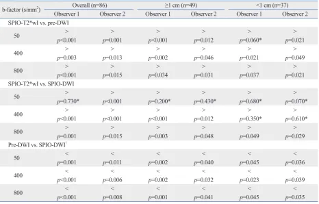
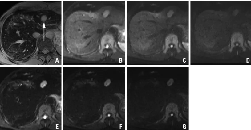
For all 86 lesions (0.3-4.7 cm; mean, 1.5 cm) studied, the mean confidence scores on each of the seven image sequenc-es were calculated and compared with each other (Fig. 1). The mean confidence score of SPIO-T2*wI was always higher than those of pre-contrast or SPIO-DWI, regardless of the b-factors, as rated by the two observers; all mean confi-dence score of SPIO-T2*wI was statistically significant (p<0.05), except SPIO-DWI using a b-factor of 50 s/mm2
scored by observer 1 (p=0.730) (Table 1) (Fig. 2). Compared with pre-DWI, the mean confidence scores of SPIO-DWI were always higher using the same b-factors (p<0.05). using turbo spin-echo sequence (TR 3000-6000 ms, TE 60
ms, flip angle 150°, echo train length 13, bandwidth 260 kHz, matrix 384×193, FOV 273×379 mm, slice thickness 6 mm, interslice gap 1.2 mm, slices 30), followed by a breath-hold 2D GRE sequence (TR 196 ms, TE 10 ms, flip angle 30°, matrix 320×168, slice thickness 6 mm, interslice gap 1.2 mm) for SPIO-T2*wI.
DWI was performed using a single-shot spin-echo echo-planar imaging sequence that combined two diffusion (mo-tion-probing) gradients before and after 180º pulse along all three directions - the section-select, phase-encoding, and fre-quency-encoding directions - and data acquisition with an echo-planar imaging (EPI) readout by applying three differ-ent b-factors of 50, 400, and 800 s/mm2. Other technical
pa-rameters were: TR 1000 ms, TE 69 ms, matrix 128×192, FOV 308×379 mm, slices 27 (9 slices for each b-factor), thickness 6 mm, interslice gap 30%, average 2, bandwidth 1735 Hz/pixel, and acquisition time 22 s. Parallel imaging algorithms (GRAPPA) with an acceleration factor of 2 were added to reduce acquisition time. Spectral fat saturation was employed systematically to suppress chemical-shift artifacts. The sequence was obtained within a single breath-holding period for upper half of the liver and another single breath-holding period for lower half of the liver, and a total of 18 slices were acquired for each b-factor. Two different DWI sets (pre- and post-SPIO) were obtained with the same im-aging parameters. Pre-DWI was performed before the dy-namic imaging, and SPIO-enhanced DWI (SPIO-DWI) was performed just before the acquisition of SPIO-enhanced T2 and T2*wI. All scans were sent to picture archiving and communication system (PACS) for interpretation on PACS workstations.
Image analyses
es except SPIO-DWI using a b-factor of 50 s/mm2 (observer
1, p=0.200; observer 2, p=0.430) (Fig. 3). When the confi-dence scores were compared to each other between the pre-contrast and DWI with corresponding b-factors, SPIO-DWI always showed higher scores regardless of the different For the lesions greater than or equal to 1 cm (n=49), the
mean confidence score of SPIO-T2*wI was always higher than those of pre- or SPIO-DWI irrespective of the different b-factors; all mean confidence scores of SPIO-T2*wI were statistically significantly higher (p<0.05) than other
sequenc-Table 1. Comparison of Mean Confidence Scores according to Lesion Size on Seven Different Sequences
b-factor (s/mm2) Overall (n=86) ≥1 cm (n=49) <1 cm (n=37)
Observer 1 Observer 2 Observer 1 Observer 2 Observer 1 Observer 2
SPIO-T2*wI vs. pre-DWI
50 p<0.001> p=0.001> p<0.001> p=0.012> p=0.060*> p=0.021>
400 p=0.003> p=0.013> p=0.002> p=0.046> p=0.021> p=0.049>
800 p > > > > > >
<0.001 p=0.015 p=0.034 p=0.031 p=0.037 p=0.021
SPIO-T2*wI vs. SPIO-DWI
50 p=0.730*> p<0.001> p=0.200*> p=0.430*> p=0.680*> p=0.070*>
400 p<0.001> p<0.001> p<0.001> p=0.012> p=0.350*> p=0.610*>
800 p > > > > > >
=0.001 p=0.015 p=0.003 p=0.048 p=0.049 p=0.029
Pre-DWI vs. SPIO-DWI†
50 < < < < < <
p=0.001 p=0.011 p=0.002 p=0.040 p=0.045 p=0.036
400 p<0.001< p=0.006< p=0.002< p=0.032< p=0.023< p=0.039<
800 p<0.001< p=0.008< p=0.001< p=0.041< p=0.045< p=0.035<
T2*wI, T2*-weighted imaging; DWI, diffusion-weighted MR imaging; SPIO, superparamagnetic iron oxide.
The direction of inequality signs means the relative superiority between two sequences in comparison for the mean confidence score accompanied with p
values.
*A p value >0.05 was not considered statistically significant.
[image:4.595.83.498.150.301.2]†For comparison of pre-DWI with SPIO-DWI, only the corresponding sequence images using same b-factor were compared each other.
Fig. 1. Mean confidence scores with standard deviations of each MRI sequence for overall lesions. SPIO-T2*wI, SPIO-enhanced T2*wI; Pre-DWI_50, pre-contrast DWI with a b-factor of 50 s/mm2; pre-DWI_400, pre-contrast DWI with a b-factor of 400 s/mm2; pre-DWI_800,
pre-contrast DWI with a b-factor of 800 s/mm2; SPIO-DWI_50, SPIO-enhanced DWI with a b-factor of 50 s/mm2; DWI_400,
SPIO-enhanced DWI with a b-factor of 400 s/mm2; SPIO-DWI_800, SPIO-enhanced DWI with a b-factor of 800 s/mm2. SPIO, superparamagnetic
iron oxide; DWI, diffusion-weighted MRI; MRI, magnetic resonance imaging; T2*wI, T2* weighted imaging.
SPIO-T2*wI pre-DWI_50 pre-DWI_400 pre-DWI_800 SPIO-DWI_50 SPIO-DWI_400 SPIO-DWI_800
M
RI
se
qu
en
ce
s
0 1 2 3 4
Scores
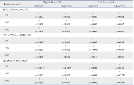
[image:4.595.59.526.381.679.2]1.3±1.0 cm) in the right hepatic lobe including two caudate lobe lesions and 28 lesions (mean size±standard deviation, 1.9±1.3 cm) in the left hepatic lobe. Even in the left hepatic lobe lesions, SPIO-T2*wI had highest confidence score compared with pre-DWI or SPIO-DWI in most cases ex-cept for the comparison with SPIO-DWI by observer 1 who showed higher confidence score of SPIO-DWI with (b-fac-tors of 50 s/mm2, p<0.001) or without statistical
signifi-cance (b-factors of 400 s/mm2, p=1.000) (Table 2). Like the
right lobe lesions, left lobe lesions also showed the superi-ority of SPIO-DWI compared to pre-DWI with (all b-fac-tors by observer 1 and b-factor of 50 s/mm2 by observer 2,
b-factors (p<0.05).
For lesions less than 1 cm (n=37), SPIO-T2*wI showed the highest mean qualitative score and SPIO-DWI (b-fac-tors of 50 and 400 s/mm2) showed confidence scores
simi-lar to SPIO-T2*wI (observer 1, p=0.680 and 0.350, respec-tively; observer 2, p=0.070 and 0.610, respectively). Pre-DWI using a b-factor of 50 s/mm2 was also comparable with
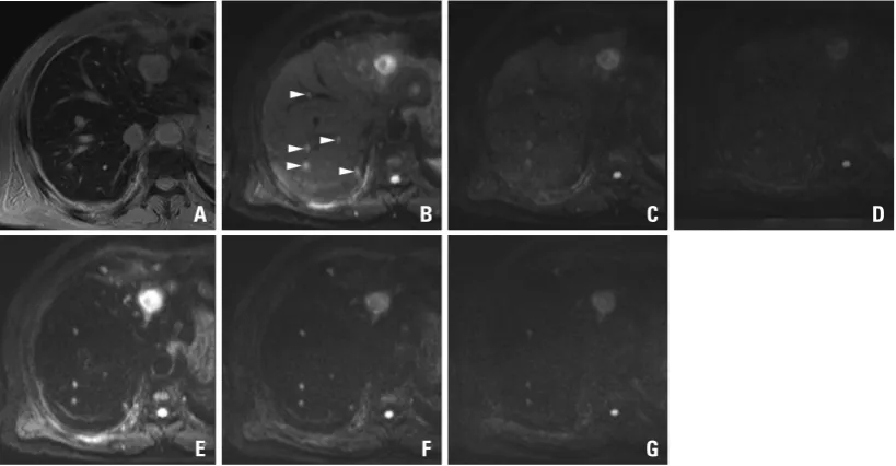
SPIO-T2*wI as rated by observer 1 (p=0.060). Compared with pre-DWI, the mean confidence scores of SPIO-DWI us-ing the correspondus-ing b-factors were always greater, even for the subcentimeter lesions (p<0.05) (Figs. 4, 5 and 6).
[image:5.595.100.508.67.279.2]There were 58 lesions (mean size±standard deviation,
Fig. 3. Mean confidence scores with standard deviations of each MRI sequence for 1 cm or larger lesions. SPIO-T2*wI, SPIO-enhanced T2*wI; Pre-DWI_50, pre-contrast DWI with a b-factor of 50 s/mm2; pre-DWI_400, pre-contrast DWI with a b-factor of 400 s/mm2;
pre-DWI_800, pre-contrast DWI with a b-factor of 800 s/mm2; SPIO-DWI_50, SPIO-enhanced DWI with a b-factor of 50 s/mm2; SPIO-DWI_400,
SPIO-enhanced DWI with a b-factor of 400 s/mm2; SPIO-DWI_800, SPIO-enhanced DWI with a b-factor of 800 s/mm2. SPIO,
[image:5.595.104.513.344.494.2]superpara-magnetic iron oxide; DWI, diffusion-weighted MRI; MRI, superpara-magnetic resonance imaging; T2*wI, T2* weighted imaging.
Fig. 2. A 48-year-old man with sigmoid colon cancer. SPIO-enhanced T2*-weighted image (A) shows a metastatic lesion (arrow) in the lateral segment. On the pre-contrast DWIs using b-factor (s/mm2) of 50 (B), 400 (C) and 800 (D), relative signal intensities of the lesions are
gradually decreased with the increase of b-factors. On the SPIO-enhanced DWIs using b-factor (s/mm2) of 50 (E), 400 (F) and 800 (G), the
lesions are more conspicuously seen, especially on the images using smaller b-factors. With an increase of b-factors, marginal blurring of the lesion looks more severe. SPIO, superparamagnetic iron oxide; DWI, diffusion-weighted MRI.
A B C D
E F G
SPIO-T2*wI pre-DWI_50 pre-DWI_400 pre-DWI_800 SPIO-DWI_50 SPIO-DWI_400 SPIO-DWI_800
M
RI
se
qu
en
ce
s
0 1 2 3 4
Scores
DISCUSSION
When dealing with contrast-enhanced hepatic MRI, SPIO reduces the signal intensity of normal liver parenchyma in a manner dependent on the phagocytic effect of Kupffer cells, resulting in an increase of lesion-to-liver contrast in cases of hepatic malignancy and, therefore, SPIO-enhanced MRI has been thought to be one of the best imaging tools in the as-sessment of hepatic metastases.11-13 DWI creates image
con-p<0.05) or without statistical significance (b-factors of 400 and 800 s/mm2 by observer 2, p>0.3) (Table 2).
The linear-weighted kappa values for interobserver agree-ment in the confidence scoring showed moderate or sub-stantial agreements for all MRI sequences (SPIO-T2*wI, 0.50; SPIO-DWI with a b-factor of 50 s/mm2, 0.47;
SPIO-DWI with a b-factor of 400 s/mm2, 0.51; SPIO-DWI with a
b-factor of 800 s/mm2, 0.52; pre-DWI with a b-factor of 50 s/
mm2, 0.63; pre-DWI with a b-factor of 400 s/mm2, 0.55;
[image:6.595.86.498.65.217.2]pre-DWI using a b-factor of 800 s/mm2, 0.52).
Fig. 4. Mean confidence scores with standard deviations of each MRI sequence for lesions smaller than 1 cm. T2*wI, SPIO-enhanced T2*wI; Pre-DWI_50, pre-contrast DWI with a b-factor of 50 s/mm2; pre-DWI_400, pre-contrast DWI with a b-factor of 400 s/
mm2; pre-DWI_800, pre-contrast DWI with a b-factor of 800 s/mm2; SPIO-DWI_50, SPIO-enhanced DWI with a b-factor of 50 s/mm2;
SPIO-DWI_400, SPIO-enhanced DWI with a b-factor of 400 s/mm2; SPIO-DWI_800, SPIO-enhanced DWI with a b-factor of 800 s/mm2. SPIO,
su-perparamagnetic iron oxide; DWI, diffusion-weighted MRI; MRI, magnetic resonance imaging; T2*wI, T2* weighted imaging.
Fig. 5. A 69-year-old man with colon cancer and numerous metastases throughout the liver including the largest one in the left hemiliver. On the SPIO-enhanced T2*-weighted image (A), metastatic lesions are not well distinguished from the background liver due to small le-sion size and masking effect of the intrahepatic vasculature with high signal intensity. On the pre-contrast DWIs using b-factor (s/mm2) of
50 (B), at least 5 metastases (arrowheads) in a single level of right hemiliver are well delineated from the background liver due to ‘ black-blood’ effect. The confidence is worsened on the images using higher b-factors of 400 (C) or 800 (D) with low signal intensity and margin-al blurring of the lesions. On the SPIO-enhanced DWIs using b-factor (s/mm2) of 50 (E) or 400 (F), all five lesions are conspicuously seen,
compared with poor marginal definition on the image using the b-factor of 800 (G). Compared with pre-contrast DWI, SPIO-enhanced DWI using corresponding b-factors shows consistently higher lesion-to-liver contrasts. SPIO, superparamagnetic iron oxide; DWI, diffu-sion-weighted MRI.
A B C D
E F G
SPIO-T2*wI pre-DWI_50 pre-DWI_400 pre-DWI_800 SPIO-DWI_50 SPIO-DWI_400 SPIO-DWI_800
M
RI
se
qu
en
ce
s
0 1 2 3 4
Scores
[image:6.595.87.497.282.495.2]Table 2. Comparison of Mean Confidence Scores according to Lesion Location on Seven Different Sequences
b-factor (s/mm2) Right lobe (n= 58)† Left lobe (n=28)
Observer 1 Observer 2 Observer 1 Observer 2
SPIO-T2*wI vs. pre-DWI
50 p > > > >
=0.002 p<0.001 p=0.029 p=0.004
400 > > > >
p<0.001 p<0.001 p=0.002 p=0.002
800 p<0.001> p<0.001> p<0.001> p<0.001>
SPIO-T2*wI vs. SPIO-DWI
50 p > > < >
=0.083* p<0.001 p=0.005 p=0.033
400 > > < >
p=0.013 p<0.001 p=1.000* p=0.003
800 p<0.001> p<0.001> p=0.016> p<0.001>
Pre-DWI vs. SPIO-DWI‡
50 p < < < <
=0.053* p=0.445* p<0.001 p=0.049
400 < < < <
p=0.005 p=0.002 p=0.004 p=0.373*
800 < < < <
p=0.003 p=0.005 p=0.006 p=0.308*
T2*wI, T2*-weighted imaging; DWI, diffusion-weighted MR imaging; SPIO, superparamagnetic iron oxide.
The direction of inequality signs means the relative superiority between two sequences in comparison for the mean confidence score accompanied with p
values.
*A p value >0.05 was not considered statistically significant.
†Including two caudate lobe lesions.
[image:7.595.75.541.369.666.2]‡For comparison of pre-DWI with SPIO-DWI, only the corresponding sequence images using same b-factor were compared each other.
Fig. 6. A 69-year-old man with multiple hepatic metastases in the left hemiliver. A small metastatatic lesion (arrowhead) on SPIO-enhanced T2*-weighted image (A) is not well distinguished from the background liver due to small size and the masking effect of the in-trahepatic vasculature. On the pre-contrast DWIs using b-factor (s/mm2) of 50 (B), 400 (C) and 800 (D), there is severe motion artifact and
no focal lesion is defined. On the SPIO-enhanced DWIs using b-factor (s/mm2) of 50 (E), 400 (F) and 800 (G), the lesion (arrowhead) is well
delineated due to the increased lesion-to-liver contrast in spite of the motion artifact. SPIO, superparamagnetic iron oxide; DWI, diffu-sion-weighted MRI.
A B C D
with the SPIO-T2*wI in the present study. In a recent study by Coenegrachts, et al.16 comparing SPIO-enhanced T2wI
with pre-contrast and SPIO-DWI for various benign and ma-lignant lesions, lesion visibility of hepatic metastases from colorectal cancers was comparable between the pre-contrast and SPIO-DWI because of increased lesion-to-liver contrast after injection of SPIO despite degradation of overall image quality due to the size of the b-factors. In the present study, the inferiority of pre-DWI might be related to inhomogeneous group of extrahepatic primary lesion sites, which could be less conspicuous on the precontrast T2wI or DWI using a small b-factor than with the SPIO-enhanced images.
For smaller (<1 cm) lesions, however, pre-DWI with a low b-factor (50 s/mm2) in addition to the SPIO-DWI with
relatively low b-factors (50 and 400 s/mm2) showed quality
scores comparable to SPIO-T2*wI in the present study. Due to the fact that small lesions can be obscured by vascular structures on SPIO-T2*wI, pre-DWI could possess a com-parable ability in the detection of small lesions despite the inherent limitations mentioned above (Fig. 4). For SPIO-DWI in the detection of subcentimeter lesions, Kiryu, et al.17
also concluded that SPIO-DWI is able to recognize subcen-timeter hepatic metastases because of suppression effect of vascular signals, masking the small lesions on SPIO-en-hanced T2wI. Even though the previous study by Kiryu, et al.17 used the PROPELLER T2 fast spin echo sequence
in-stead of the spin-echo EPI used in the present study, our re-sults are similar to theirs. Although not analyzed in this study, the quality of SPIO-DWI can also be limited by increased blooming effect, the susceptibility artifact from iron parti-cles, which is worsened by increasing the b-factors.18
De-spite diminution in the size of lesions by the blooming ef-fect, however, the b-factor of 400 s/mm2 showed comparable
confidence scores with increased lesion-to-liver contrast of T2*wI after SPIO injection.
This study has several limitations. First of all, due to retro-spective nature of this study, pathologic proof was not possi-ble for all lesions. Secondly, this study focused on the com-parative conspicuity of each metastatic lesion from various primary malignancies on the imaging sequences studied, and the sensitivity or the specificity of each sequence was not calculated. Thirdly, there was a high potential of unpre-dictable image degradation for the left lobe lesions related to pulsation artifacts.7 However, even in the left hepatic lobe
lesions, the mean confidence score of SPIO-DWI was sig-nificantly higher than pre-DWI, especially on the images, using small b-factor in the present study. Fourthly, according trast by the difference in the random, thermally-agitated
dif-fusion of water molecules in different tissues and has recently been included in the routine protocol of hepatic MRI with recent technical advances in reducing image distortion and increasing the signal-to-noise ratio.14
According to some recent investigations, DWI provids better sensitivity and comparable ability of lesion character-ization, compared to the conventional breath-hold T2wI, in the assessment of focal hepatic lesions.6 In a comparative
study between SPIO-enhanced imaging and DWI, Nasu, et al.7 showed that the combined interpretation of DWI, fast
T2wI, and dual-echo gradient T1-weighted images yielded better accuracy than SPIO-enhanced T2wI in the evaluation of hepatic metastases, and suggested the potential of DWI to replace SPIO-enhanced T2wI in the preoperative evaluation of hepatic metastases.
Meanwhile, Naganawa, et al.10 applied DWI along with
high b-factors (600 and 1000 s/mm2) before and after the
ad-ministration of SPIO during hepatic MRI for the assessment of focal liver lesions in 5 patients with hepatic metastases, and concluded that SPIO-DWI could enhance the recogni-tion of hepatic metastases. In their study, the number of le-sions detected on SPIO-DWI was larger than other imaging sequences including SPIO-T2, T2* and pre-DWI, however, none of the lesions was verified pathologically or by follow-up studies.10 In the present study, we attempted to explore
whether pre-DWI would be sufficient to replace SPIO-T2*wI and whether SPIO-DWI would be helpful in improving the lesion-to-liver contrast, depending on the lesion size and various b-factors that are available, with the use of a number of verified lesions of hepatic metastases.
In the present study, SPIO-T2*wI showed significantly better confidence scores than pre-DWI, regardless of the le-sion size or the different b-factors, and only SPIO-DWI showed comparable confidence levels when the small b-fac-tor (50 s/mm2) was used. Based on the present results,
pre-DWI would not be sufficient to replace SPIO-T2*wI due to limited spatial resolution and image artifacts in addition to inherently unpredictable lesion-to-liver contrast of various hepatic metastases. With smaller b-factors, diffusion effect is not so distinctively reflected on the images, especially for the solid lesions, and the imaging characteristics of DWI were similar to those of T2wI except for the black blood ef-fect.15 In this situation, injection of SPIO would increase the
S, et al. Hepatic metastases: diffusion-weighted sensitivity-encod-ing versus SPIO-enhanced MR imagsensitivity-encod-ing. Radiology 2006;239: 122-30.
8. Müller MF, Prasad PV, Siewert B, Edelman RR. [The in-vivo dif-fusion measurements of the liver, kidneys, spleen and m. erector with an echo-planar imaging system in normal subjects]. Rofo 1994;161:233-6.
9. Ichikawa T, Haradome H, Hachiya J, Nitatori T, Araki T. Diffu-sion-weighted MR imaging with a single-shot echoplanar se-quence: detection and characterization of focal hepatic lesions. AJR Am J Roentgenol 1998;170:397-402.
10. Naganawa S, Sato C, Nakamura T, Kumada H, Ishigaki T, Miura S, et al. Diffusion-weighted images of the liver: comparison of tu-mor detection before and after contrast enhancement with super-paramagnetic iron oxide. J Magn Reson Imaging 2005;21:836-40. 11. Senéterre E, Taourel P, Bouvier Y, Pradel J, Van Beers B, Daures
JP, et al. Detection of hepatic metastases: ferumoxides-enhanced MR imaging versus unenhanced MR imaging and CT during arte-rial portography. Radiology 1996;200:785-92.
12. Vogl TJ, Schwarz W, Blume S, Pietsch M, Shamsi K, Franz M, et al. Preoperative evaluation of malignant liver tumors: comparison of unenhanced and SPIO (Resovist)-enhanced MR imaging with biphasic CTAP and intraoperative US. Eur Radiol 2003;13:262-72. 13. Strotzer M, Gmeinwieser J, Schmidt J, Fellner C, Seitz J, Albrich
H, et al. Diagnosis of liver metastases from colorectal adenocarci-noma. Comparison of spiral-CTAP combined with intravenous contrast-enhanced spiral-CT and SPIO-enhanced MR combined with plain MR imaging. Acta Radiol 1997;38:986-92.
14. Taouli B, Koh DM. Diffusion-weighted MR imaging of the liver. Radiology 2010;254:47-66.
15. Hussain SM, De Becker J, Hop WC, Dwarkasing S, Wielopolski PA. Can a single-shot black-blood T2-weighted spin-echo echo-planar imaging sequence with sensitivity encoding replace the re-spiratory-triggered turbo spin-echo sequence for the liver? An op-timization and feasibility study. J Magn Reson Imaging 2005;21: 219-29.
16. Coenegrachts K, Matos C, ter Beek L, Metens T, Haspeslagh M, Bipat S, et al. Focal liver lesion detection and characterization: comparison of non-contrast enhanced and SPIO-enhanced diffu-sion-weighted single-shot spin echo echo planar and turbo spin echo T2-weighted imaging. Eur J Radiol 2009;72:432-9. 17. Kiryu S, Watanabe M, Kabasawa H, Akahane M, Aoki S, Ohtomo
K. Evaluation of super paramagnetic iron oxide-enhanced diffu-sion-weighted PROPELLER T2-fast spin echo magnetic reso-nance imaging: preliminary experience. J Comput Assist Tomogr 2006;30:197-200.
18. Liang L, Korogi Y, Sugahara T, Shigematsu Y, Okuda T, Ikushima I, et al. Detection of intracranial hemorrhage with susceptibility-weight-ed MR sequences. AJNR Am J Neuroradiol 1999;20:1527-34.
to inherent limitation of breath-holding technique for DWI, the number of excitation was limited and TR was too short to avoid T1 effect, thus resulting in lowering the signal-to-noise ratio and lesion conspicuity, especially on pre-DWI. Increasing the number of excitation and lengthening of TR would improve the quality of pre-DWI by using free-breath-ing data acquisition techniques which was not performed during our study period. Lastly, due to limited number of pa-tients, separate analysis according to the nature of primary extrahepatic lesions was not possible and ADC value was not measured for each lesion.
In conclusion, compared with SPIO-T2*wI, pre-DWI could not reach a comparable confidence score for the de-tection of hepatic lesions, except subcentimeter lesions, on pre-DWI with a small b-factor (50 s/mm2). Despite the
limi-tations mentioned above, using breath-holding technique, pre-DWI is insufficient to replace SPIO-enhanced imaging for hepatic metastases from various primary origins. De-pending on the increased confidence score, additional DWI after SPIO injection could complement SPIO-T2*wI in the assessment of small hepatic metastases.
REFERENCES
1. Ward J. New MR techniques for the detection of liver metastases. Cancer Imaging 2006;6:33-42.
2. Penna C, Nordlinger B. Colorectal metastasis (liver and lung). Surg Clin North Am 2002;82:1075-90.
3. Robinson PJ. Imaging liver metastases: current limitations and fu-ture prospects. Br J Radiol 2000;73:234-41.
4. Oner AY, Celik H, Oktar SO, Tali T. Single breath-hold diffusion-weighted MRI of the liver with parallel imaging: initial experi-ence. Clin Radiol 2006;61:959-65.
5. Erturk SM, Ichikawa T, Sano K, Motosugi U, Sou H, Araki T. Diffusion-weighted magnetic resonance imaging for characteriza-tion of focal liver masses: impact of parallel imaging (SENSE) and b value. J Comput Assist Tomogr 2008;32:865-71.
6. Parikh T, Drew SJ, Lee VS, Wong S, Hecht EM, Babb JS, et al. Focal liver lesion detection and characterization with diffusion-weighted MR imaging: comparison with standard breath-hold T2-weighted imaging. Radiology 2008;246:812-22.