Citation:
Emiola, A and Andrews, SS and Heller, C and George, J (2016) Crosstalk between the
lipopolysac-charide and phospholipid pathways during outer membrane biogenesis in Escherichia coli.
Proceedings of the National Academy of Sciences of USA, 113 (11).
pp.
3108-3113.
ISSN
1091-6490 DOI: https://doi.org/10.1073/pnas.1521168113
Link to Leeds Beckett Repository record:
http://eprints.leedsbeckett.ac.uk/4109/
Document Version:
Article
Creative Commons: Attribution 4.0
The aim of the Leeds Beckett Repository is to provide open access to our research, as required by
funder policies and permitted by publishers and copyright law.
The Leeds Beckett repository holds a wide range of publications, each of which has been
checked for copyright and the relevant embargo period has been applied by the Research Services
team.
We operate on a standard take-down policy.
If you are the author or publisher of an output
and you would like it removed from the repository, please
contact us
and we will investigate on a
case-by-case basis.
Crosstalk between the lipopolysaccharide and
phospholipid pathways during outer membrane
biogenesis in
Escherichia coli
Akintunde Emiolaa,1,2, Steven S. Andrewsb, Carolin Hellera, and John Georgea
aSchool of Health, Sport and Bioscience, University of East London, Stratford Campus, London E15 4LZ, United Kingdom; andbFred Hutchinson Cancer
Research Center, Seattle, WA 98109-1024
Edited by David W. Russell, University of Texas Southwestern Medical Center, Dallas, TX, and approved January 29, 2016 (received for review October 26, 2015)
The outer membrane of gram-negative bacteria is composed of phospholipids in the inner leaflet and lipopolysaccharides (LPS) in the outer leaflet. LPS is an endotoxin that elicits a strong immune response from humans, and its biosynthesis is in part regulated via degradation of LpxC (EC 3.5.1.108) and WaaA (EC 2.4.99.12/13) enzymes by the protease FtsH (EC 3.4.24.-). Because the synthetic pathways for both molecules are complex, in addition to being pro-duced in strict ratios, we developed a computational model to in-terrogate the regulatory mechanisms involved. Our model findings indicate that the catalytic activity of LpxK (EC 2.7.1.130) appears to be dependent on the concentration of unsaturated fatty acids. This is biologically important because it assists in maintaining LPS/phos-pholipids homeostasis. Further crosstalk between the phospho-lipid and LPS biosynthetic pathways was revealed by experimental observations that LpxC is additionally regulated by an unidentified protease whose activity is independent of lipid A disaccharide con-centration (the feedback source for FtsH-mediated LpxC regulation) but could be induced in vitro by palmitic acid. Further experimental analysis provided evidence on the rationale for WaaA regulation. Overexpression ofwaaAresulted in increased levels of 3-deoxy-D -manno-oct-2-ulosonic acid (Kdo) sugar in membrane extracts, whereas Kdo and heptose levels were not elevated in LPS. This im-plies that uncontrolled production of WaaA does not increase the LPS production rate but rather reglycosylates lipid A precursors. Overall, the findings of this work provide previously unidentified insights into the complex biogenesis of theEscherichia coliouter membrane.
lipopolysaccharide
|
fatty acids|
computational model|
bacterial membrane regulationT
he outer membrane of gram-negative bacteria is decorated with a potent endotoxin (called lipid A), which plays a sig-nificant role in bacterial pathogenicity and immune evasion (1). It also acts as a physical barrier protecting the cell from chemical attack and represents a significant obstacle for the effective delivery of numerous antimicrobial agents (2, 3). The outer membrane is composed of phospholipids in the inner leaflet and lipopolysac-charides (LPS) in the outer leaflet (4). Phospholipids consist of a glycerol molecule, a phosphate group, and two fatty acid moieties (except for cardiolipins) (5) (see reviews (5, 6) andSI Appendixfor the biosynthesis and regulation of phospholipids). LPS, on the other hand, contains three distinct components: lipid A, core oligosac-charides, and O-antigen (7, 8). Lipid A is the sole essential com-ponent of LPS, and its biosynthesis involves nine enzyme-catalyzed reactions (8). The lipid A pathway has been widely investigated, and we recently produced a pathway model that incorporates all of the known regulatory mechanisms (9). Briefly, the first reaction step catalyzed by LpxA is highly unfavorable, which makes the proceeding enzyme, LpxC, the first committed enzyme (10). LpxC is regulated by the protease FtsH (11, 12), and we recently postulated that the negative feedback signal arises from lipid A disaccharide, the substrate for LpxK (9). Furthermore, FtsH regulates WaaA(formerly called KdtA), an enzyme downstream of LpxC (13). The exact rationale for WaaA regulation remains unknown.
A wealth of research exists for either LPS or phospholipids biosynthesis; however, our current understanding on the cross-talk between both pathways is limited at the moment. Because both pathways are synchronized to ensure a proper balance of membrane components (11, 14), studies underpinning the un-derlying mechanisms would appear valuable. There are a number of experimental findings that indicate the existence of strong links between both biosynthetic pathways (11, 15, 16). Thus, in the context of outer membrane biogenesis, the role involving phospholipids cannot be ignored in the study of LPS regulation. Furthermore, during membrane synthesis,∼20 million molecules of fatty acids are synthesized inEscherichia coli(8). Yu et al. (17) reconstituted an in vitro steady-state kinetic system of fatty acid biosynthesis using purified enzymes and observed that the max-imum fatty acid production rate obtainable was 100 μM/min. This production rate falls far below the amount of fatty acids required by a cell in vivo [if one assumes a cell volume of 6.7× 10⁻¹⁶L (18) and a generation time of 30 min (19)]. Therefore, to test the consistency of reported in vitro parameters and in-vestigate the role of the biosynthetic enzymes on fatty acids turnover rate, a“systems”approach is necessary. Similarly, ever since the regulation of WaaA by FtsH was first reported (13), no
Significance
This work examines the relationship between bacterial phos-pholipid biosynthesis and lipopolysaccharides (LPS) regulation. Because LPS is a potent endotoxin in addition to being essential for the survival of gram-negative bacteria, our experimental findings are of importance to the fields of microbiology, immu-nology, and drug design. In addition, the computational aspect of this work represents an in-depth kinetic model comprising 81 chemical reactions; hence, computational and systems biologists would find our work useful. Furthermore, recent interests in the field of biofuel production by bacteria also imply that our study can help elucidate mechanisms for increased saturated or un-saturated fatty acids synthesis. Consequently, our work appeals to a broad range of disciplines.
Author contributions: A.E. and J.G. designed research; A.E., C.H., and J.G. performed research; S.S.A. contributed new reagents/analytic tools; A.E., S.S.A., and J.G. analyzed data; and A.E. and J.G. wrote the paper.
The authors declare no conflict of interest.
This article is a PNAS Direct Submission.
Freely available online through the PNAS open access option.
Data deposition: Our model has been deposited in the BioModels database and assigned the identifier MODEL1601080000.
1Present address: The Jackson Laboratory for Genomic Medicine, Farmington, CT 06032.
2To whom correspondence should be addressed. Email: a.emiola@uel.ac.uk.
study has investigated the underlying regulatory mechanism to date. This would also appear important because under wild-type conditions, WaaA catalyzes a step that is required for the endo-toxic activity of lipid A (20).
In this work, we present a detailed picture of the crosstalk be-tween the LPS and phospholipids biosynthetic machinery. Our work involves a computational kinetic model spanning 81 chemical re-actions and involving 90 chemical species. Additionally, we used a series ofE. colifatty acid biosynthesis mutants to investigate the effect of substrate flux into the saturated and unsaturated fatty acid pathway on LpxC stability. Our complete model agrees qualitatively with published datasets and with our own experiments. Our results imply that the catalytic activation of LpxK is dependent on un-saturated fatty acids. Furthermore, our experimental investigations have implicated a secondary protease involved in LpxC regulation. Finally, we have provided experimental evidence to explain the rationale for WaaA regulation.
Results
LPS/Phospholipids Model Construction.We developed a computa-tional metabolic model that incorporates genetic regulation and several feedback sources (Fig. 1). The required parameters were mostly derived from experimental literature, whereas others were estimated from published data. We modeled the interactions be-tween substrates and enzymes under steady-state conditions using Michaelis–Menten and mass action kinetics. Our initial set of pa-rameters were unable to reproduce experimentally-observed path-way regulatory behavior. This was solely due to a limitation in the kinetic parameters of LpxK, as described below. In addition, we made minor modifications to FabZ, FabB, and FabI parameters for fitting purposes. A full description of the model architecture, model construction, and parameters’estimation is presented inSI Appendix.
Model Findings Indicate the Catalytic Activity of LpxK is Dependent on Unsaturated Fatty Acids.It was previously reported in Ray and Raetz (16) that the catalytic activity of LpxK is dependent on phospholipids. Phospholipids inE. colicontain a combination of saturated and unsaturated fatty acids; however, our model indi-cates that the LpxK catalytic activation is driven solely by un-saturated fatty acids. When we initially assumed in our model that all fatty acids (i.e., both saturated and unsaturated fatty acids)
present in phospholipids could catalytically activate LpxK, the model findings deviated substantially from experimental results. For instance, inhibiting either FabA or FabZ in our model had no effect on LpxC stability, whereas LpxC is rapidly degraded under both conditions in vivo (Fig. 2Aand ref. 15, respectively). From our observations, there were sufficient levels of saturated fatty acids to catalytically activate LpxK, which prevented an accumu-lation of lipid A disaccharide, the feedback source for LpxC degradation. However, when the LpxK activation signal arose solely from unsaturated fatty acids, our model agreed qualita-tively with all our experimental results.
Indeed, it was reported by Ray and Raetz (16) that of all phospholipids species tested, cardiolipins had the most effect at activating LpxK. Although the authors did not provide a rationale for their observation, we suspect their use of bovine heart car-diolipins, which are known to contain at least 94% unsaturated fatty acids of the total fatty acids, were implicated (21). Similarly, cardiolipins of E. coli are characterized with more unsaturated fatty acids relative to other phospholipids moieties (22). In this regard, we propose that the activity of LpxK is dependent on the presence of unsaturated fatty acids. This major model adjustment subsequently provided a framework to interpret the experimental results presented below.
[image:3.585.299.539.48.241.2]Excess Substrate Flux into the Saturated Fatty Acid and LPS Pathway Stimulates LpxC Degradation. We recently reported that lipid A disaccharide is a feedback source for FtsH-mediated LpxC deg-radation (9); however, other evidence suggests that there may be additional FtsH feedback signals. In particular, it was observed in Ogura et al. (11) that LpxC was highly stabilized in a fabI (ts)
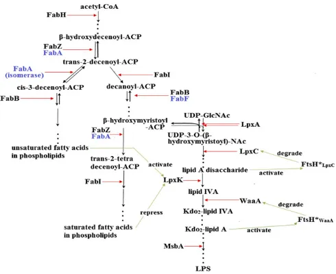
[image:3.585.39.284.487.687.2]Fig. 1. Model of theE. coliLPS and phospholipids biosynthesis pathway. Green arrows represent pathway interactions that were derived from our current and previous work (9). A detailed model schematic is presented inSI Appendix.
Fig. 2. Role of fatty acid biosynthesis on LpxC regulation. Growth condi-tions are described inSI Appendix. (A) Effect of fatty acid biosynthetic en-zyme inhibition on LpxC stability. Blue bars on the right represent our computational simulations under steady-state conditions. Here, model en-zyme concentrations were reduced arbitrarily to mimic the nonpermissive temperature because there was no available information in the literature on residual enzyme activity in these mutants. We reduced the model concen-trations of FabF and FabB by 10- and 20-fold, respectively, to simulate mu-tant strain CY288. FabI and FabA concentrations were reduced by 100- and 500-fold to simulate strains JP1111 and CY53, respectively. (B) Fatty acid composition in strains JP1111, CY288, and CY53. Black and red bars repre-sent growth conditions at 30 °C and 42 °C, respectively. Error bars reprerepre-sent SE of mean. Fatty acids notation are C14, myristic acid; C15, pentadecanoic acid; C16:1, palmitoleic acid; C16, palmitic acid; C18:1,cis-vaccenic acid; and C18, stearic acid. (C) LPS quantification. LPS was quantified by measuring the amount of Kdo in the bacterial membrane. Black bars represent experi-mental results, whereas blue bars are simulation results using model con-ditions as inA. Error bars represent SE of mean.
Emiola et al. PNAS | March 15, 2016 | vol. 113 | no. 11 | 3109
SYSTEMS
BIO
mutant, implying a link between fatty acid and LPS biosynthesis. To understand the role of fatty acids synthesis on LpxC regulation, we analyzed a number of fatty acid biosynthesis mutants.
WhenE. colistrains JP1111 [fabI(ts)] and CY288 [fabF, fabB
(ts)] were grown at the nonpermissive temperature, LpxC was highly stabilized by ∼threefold, whereas LpxC was rapidly de-graded in strain CY58 [fabA(ts)] mutant (Fig. 2A). We next analyzed the saturated and unsaturated fatty acid distribution in these mutants to determine if the regulatory signal arose from lipid A disaccharide or from sources outside of the LPS pathway. In other words, an increment of substrate flux into the saturated fatty acid pathway is an indirect indication of increased flux into the LPS pathway. This is because LPS substrates are derived from the saturated fatty acid pathway arm (Fig. 1).
When strain JP1111 was grown at the nonpermissive tempera-ture, the total proportion of unsaturated fatty acids was sub-stantially increased (Fig. 2B). Clearly, inhibition of FabI, which catalyzes the first committed step in saturated fatty acid synthesis (23), enabled the isomerase activity of FabA to divert substrates toward unsaturated fatty acid synthesis. The observed increment in unsaturated fatty acid is indicative of a sufficient reduction of substrate influx into the LPS pathway. This was further confirmed from LPS quantification assay described below. Therefore, LpxC stability in strain JP1111 is as a result of decreased levels of lipid A disaccharide. It should be noted that C16:1 levels were the same in both wild-type and fabImutant, which suggests that the rate of elongation of C16:1 to C18:1 by FabF is dependent on substrate availability (Fig. 2B).
Similarly, we analyzed fatty acid distribution in strain CY288. This strain had a lesion in thefabFgene in addition to having a temperature-sensitive FabB protein (24). When grown at 30 °C, the fatty acid profile of the cells were characterized with significant levels of medium-chain fatty acids (Fig. 2B). This partial inhibition of fatty acid elongation is a result of the lesion in FabF (24), al-though cells remain viable due to a functional FabB. However, when grown at 42 °C, we observed a further increment in medium-chain fatty acids (especially pentadecanoic acid, an odd-medium-chain fatty acid) and reduction in long-chain fatty acid moieties (Fig. 2B). Although increased levels of saturated fatty acids were generally observed at 42 °C, these were mainly odd-chain fatty acids. Odd-chain fatty acyl-ACPs can be used for phospholipids production (25) but are unsuitable substrates for LPS synthesis due to a strict requirement of LpxA for β-hydroxymyristoyl–ACP (26). There-fore, these metabolites would not be expected to be shunted down the LPS pathway. Consequently, LpxC stability in this strain can
be attributed to low levels of the lipid A disaccharide. Again, this was confirmed by LPS quantification, as described below.
Furthermore, we observed a concomitant decrease in both long-chain saturated and unsaturated fatty acids (stearic andcis- vac-cenic acids) in strain CY53 when grown at the nonpermissive temperature (Fig. 2B). The only reasonable explanation from the pathway schematic in Fig. 1 is that the rate of synthesis oftrans 2-decenoyl–ACP was reduced, given FabA is the major dehydratase involved in this step (23). As a result, limited substrates would be diverted into both saturated and unsaturated arms of the pathway, resulting in reduced palmitoyl-ACP and palmitoleoyl-ACP con-centrations. Increased competition for low levels of palmitoyl-ACP and palmitoleoyl-palmitoyl-ACP by phospholipid acyltransferases (PlsB and PlsC) (27, 28) would further ensure that only a small proportion of these acyl pools are elongated to stearic andcis-vaccenic acids by FabB and FabF, respectively (29). Although substrate flux into the saturated fatty acid pathway was generally reduced, inhibition of FabA resulted in increased LpxC degradation due to an accumu-lation of lipid A disaccharide. Accumuaccumu-lation of lipid A disaccharide would occur under this condition because FabA plays a major role in the dehydration of β-hydroxymyristoyl–ACP due to a higher protein copy number (30) and similar catalytic activities with FabZ (23). In other words, a loss of FabA activity would increase the concentration of β-hydroxymyristoyl–ACP, which is then shunted into the LPS pathway.
As a confirmation that strain CY53 grown at 42 °C was char-acterized with sufficient substrate flux into the LPS pathway, the concentration of LPS was only slightly decreased in comparison with those grown at 30 °C (Fig. 2C). In contrast, we observed a reduction in LPS levels of ∼50% in both strains JP1111 and CY288 when grown at the nonpermissive temperature, which supports the idea that LPS synthesis is impeded as a consequence of low substrate availability.
Together, our findings indicate that the FtsH-mediated LpxC degradation signal arises solely from lipid A disaccharide under fatty acid inhibition conditions.
[image:4.585.43.360.528.727.2]Proteolytic Regulation of LpxC in anftsHKnockout Mutant.Having established from above that excess flux of metabolites into the saturated fatty acid pathway enhances LpxC proteolysis, we ex-amined the likelihood of saturated fatty acids commonly found in membrane phospholipids to directly impact LpxC instability. We observed that in vitro, the addition of 10 and 20 mM of palmitic acid to wild-type (W3110)E. colicell lysates for 10 min resulted in decreased LpxC levels by∼30% and 57%, respectively
Fig. 3. LpxC proteolysis in anftsHknockout mutant. Assay conditions are described inSI Appendix. (A) Effect of fatty acids on LpxC stability in vitro. (B) Effect of palmitic acid on LpxC stability inftsH
(Fig. 3A). This dose-dependent phenomenon was not observed when myristic acid, a shorter-chain fatty acid, was used. Next, we treated cell lysates of anftsHknockout mutant with palmitic acid and observed similar results (Fig. 3B). This therefore indicates that under our in vitro conditions, an unidentified protease con-tributed toward LpxC degradation. Furthermore, proteolysis was inhibited in the presence of EDTA, which indicates the protease belonged to the class of metalloprotease, similar to FtsH (Fig. 3C). However, under normal physiological conditions, LpxC and palmitic acid are localized in different cellular compartments (cytoplasm and bacterial membrane, respectively) and may not be in direct contact with one another, as it was in our in vitro assays. Because palmitic acid exists in the cytoplasm in the forms of palmitoyl-CoA and palmitoyl-ACP (6), we investigated the effect of both forms of palmitic acid on LpxC stability. Palmitic acid is known to be actively transported across the membrane by FadL (31) and immediately converted to palmitoyl-CoA by FadD (32). Interestingly, when we added palmitic acid to the growth medium of wild-typeE. coli(W3110), the levels of LpxC were elevated by 1.7-fold (Fig. 3D). This observation is readily explainable from prior results and our results presented in Fig. 2. Palmitoyl-CoA induces a strong inhibitory effect on FabI with a
Kivalue of about 3μM (33). Due to the inhibition of FabI, LpxC
would be stabilized (Fig. 2A). Thus, elevated cellular palmitoyl-CoA concentrations have an opposite effect on LpxC stability than the free-form of palmitic acid. On the other hand, under in vivo conditions, inhibiting substrate flux into the LPS pathway will increase the cellular concentration of palmitoyl-ACP (34). This is because when LPS substrate influx is inhibited, it leads to elevated levels ofβ-hydroxymyristoyl–ACP, which is subsequently dehydrated by FabZ and shunted toward the production of pal-mitoyl-ACP. We treated cells with sublethal concentrations of an LpxC inhibitor (CHIR-090) to reduce substrate flux into the LPS pathway. Under this condition, we observed LpxC elevation of 7.6-fold in wild-type cells (Fig. 3D). This stability is best explained by reduced level of lipid A disaccharide production. Therefore, in-crement in cellular palmitoyl-ACP concentration has an opposite effect on LpxC stability than the free-form of palmitic acid. However, the presence of palmitic acid or sublethal concentration of CHIR-090 in the culture medium had no effect on LpxC sta-bility in ftsH knockout mutant cells (Fig. 3D). Together, these findings suggest that the direct interaction of LpxC with long-chain free-fatty acids (i.e., fatty acids not bound to ACP or CoA) facil-itated FtsH-independent LpxC proteolysis, as observed in our in vitro assays.
Interestingly, when we monitored the in vivo LpxC degradation rate inftsHknockout cells that were treated with a higher concen-tration of CHIR-090 or other antibiotics that affected the bacterial membrane, we observed an increase in LpxC concentration ranging from 1.3- to 1.9-fold (Fig. 3E). This indicates that in untreated cells, residual LpxC proteolysis occurred, and thus, confirms the presence of an additional regulatory protease. Additionally, these results also indicate that LpxC regulation is crucial to the bacterial cells, which must maintain the desired protein levels irrespective of an accu-mulation of lipid A disaccharide, or presence/absence of a functional FtsH protease.
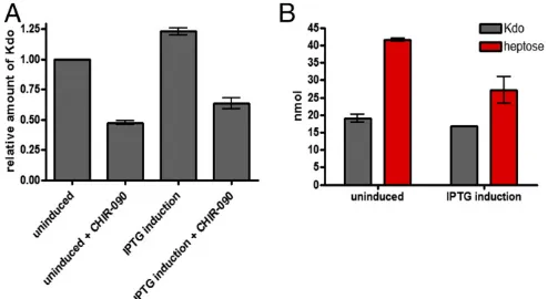
WaaA Regulation.In addition to LpxC, WaaA regulation is also crucial to cellular viability (SI Appendix, Fig. S5). Using compu-tational simulations, we recently postulated that the regulation of WaaA may help prevent reglycosylation of lipid precursors rather than regulating the rate of LPS synthesis (9). In other words, the regulatory role is“qualitative” rather than “quantitative.” To clarify these assumptions, we overexpressedwaaAand quantified LPS using two approaches: first, by quantifying the amount of Kdo in crude membrane extracts, and second, by quantifying the levels of Kdo and heptose from extracted LPS. Under waaA over-expression conditions, the levels of Kdo in crude membrane
extracts were elevated by∼20% (Fig. 4A). However, an increment in Kdo may not indicate increased LPS level under waaA over-expression. This is because the role of WaaA is to add two Kdo residues to its LPS lipid precursor (lipid IVA) (35); therefore, the
possibility still exists that overproduction of WaaA may result in multiple additions of Kdo to lipid IVA(i.e., reglycosylation) or,
alternatively, Kdo may be added to other lipid acceptors (other than lipid IVA) in the membrane (35, 36). This also indicates that
quantification of LPS using the Kdo assay may be inappropriate under waaA overexpression conditions. To further clarify these possibilities, we reduced substrate flux into the LPS pathway using 1/2 MIC of CHIR-090. The rationale for this was that by reducing the influx of substrates, the LPS synthesis rate would be dependent on substrate availability and not WaaA concentration. Similarly, overexpression ofwaaAunder this condition led to a 20% increase in Kdo levels in comparison with cells that were solely treated with CHIR-090 (Fig. 4A). These findings indicate that the rate of gly-cosylation by WaaA is independent of lipid IVAavailability.
Furthermore, it was essential to investigate if the reglycosylated lipid IVAor other alternate lipids (i.e., lipids other than lipid IVA)
that could be glycosylated by WaaA are being consumed for LPS synthesis. Consequently, we quantified the concentration of both Kdo and heptose sugars present in extracted LPS. Heptose sugar is primarily used in the synthesis of core oligosaccharides and is usu-ally present in a 2:1 ratio with Kdo in wild-type cells (20). Under
waaAoverexpression, Kdo and heptose levels from extracted LPS were both decreased by ∼12% and 34%, respectively (Fig. 4B). Together, these results suggest that unregulated WaaA production results in multiple glycosylation of lipid precursors that mostly ac-cumulate within the membrane and are poor substrates for LPS synthesis. However, it does appear that a small proportion of these reglycosylated products are consumed for LPS production, given the observed ratio of Kdo and heptose from extracted LPS (Fig. 4B).
To further elucidate the effect ofwaaAoverexpression on the structural stability of the bacterial membrane, we determined the MICs of different antibiotics for cells that had overexpressed
waaA.Under these conditions, the cells were more susceptible to antimicrobial agents, which implies that the bacterial membrane becomes more permeable under unregulated WaaA conditions (SI Appendix, Table S3).
Discussion
[image:5.585.296.543.52.187.2]In this work, we developed an integrated computational model for the biosynthesis of phospholipids and LPS pathways. One crucial model finding enabled us to pinpoint a key role of un-saturated fatty acids at stimulating LpxK catalysis, whereas on the
Fig. 4. WaaA regulation. Assay conditions are described inSI Appendix, and error bars represent SE of mean. (A) Kdo concentration underwaaA over-expression in membrane extracts. (B) Kdo and heptose quantification from extracted LPS underwaaAoverexpression.
Emiola et al. PNAS | March 15, 2016 | vol. 113 | no. 11 | 3111
SYSTEMS
BIO
other hand, excessive saturated fatty acids represses its catalytic activity. There are a number of published datasets that supports this notion. First, as mentioned previously, the phospholipids used in Ray and Raetz (16) were bovine heart cardiolipins, which are known to consist almost entirely of unsaturated fatty acids (21). Furthermore, Roy and Coleman (37) observed that when LpxD was inhibited, the specific activity of LpxK was decreased, whereas the protein half-lives of LpxK were the same under normal and LpxD inhibition conditions. Further analysis by the authors in-dicated that overexpression of LpxD does not increase the specific activity of LpxK. This means that the decrease in LpxK activity is a secondary effect of the altered LpxD. These observations can readily be interpreted from our model. Because β -hydroxymyr-istoyl–ACP is a substrate used by LpxD, reduced LpxD activity would result in an accumulation of this substrate, which is shunted toward saturated fatty acid production. In return, excess saturated fatty acid would repress the catalytic activation of LpxK.
Because the ratio of unsaturated and saturated fatty acids in-fluences the activation of LpxK, this suggests that LPS synthesis rate is correlated with membrane fluidity. This is due to the fact that unsaturated fatty acids in the membrane increase fluidity, whereas saturated fatty acids decrease fluidity (38). It is probable that under conditions of low membrane fluidity, a reduced LpxK activity may be essential to reduce the amount of LPS produced, which poten-tially would add an extra permeability barrier and subsequently reduce the influx of metabolites from the external environment. The opposite effect seems to occur under conditions of high membrane fluidity. It was reported previously (39) thatE. coligrown in envi-ronments that had increased membrane fluidity through degrada-tion of LPS led to an inducdegrada-tion of PlsB transcripdegrada-tion, which would ultimately enhance the production of phospholipids.
There has been some uncertainty as to the role of acyl-ACPs in the regulation of LpxC. In the first scenario, due to the fact that LPS and phospholipids biosynthetic pathways both derive their precursor molecules fromβ-hydroxymyristoyl–ACP, it was accepted that competition for this common substrate influences the regula-tion of LpxC (11). In other words, LpxC degradaregula-tion would help conserve substrates for phospholipids synthesis. This initially seemed reasonable and is supported by findings that FabZ in-hibition enhances LpxC degradation (15), and FabZ overexpression results in LpxC stability (11). However, our model and experimental results disagree with this“competition model”regardingβ -hydroxy-myristoyl–ACP availability. As an example, LpxC would have been expected to be stabilized under conditions of FabA overexpression, which invariably increases substrate flux into the saturated fatty acid pathway (40), and subsequently elevate the pool of β -hydroxymyr-istoyl–ACP. This should lead to a reduced competition between LpxC and FabZ for substrates. On the contrary, we observed an increased rate of LpxC degradation under this condition (SI Ap-pendix, Fig. S4A). In support of our model’s disagreement with the
“competition model,” Ogura et al. (11) also observed that LpxC levels were elevated infabI(ts) mutants. Although the authors had initially suggested that increased concentration of trans 2-tetradece-noyl–ACP may enhance LpxC stability due to the observation that LpxC levels are also being elevated when FabZ is overexpressed, Ogura et al. also admitted the unlikelihood oftrans2-tetradecenoyl– ACP accumulation underfabI(ts) conditions (41). Thus, they were unable to explain the rationale for LpxC stability under FabI in-hibition conditions. Our results presented in Fig. 2 indicate that the increased levels of LpxC infabI(ts) mutant occurred due to decreased flux of substrates into the saturated fatty acids and LPS pathways, which ultimately depreciated the lipid A disaccharide concentration. The regulation of LpxC becomes more complicated by our observations that an unidentified protease degrades LpxC. Al-though our in vitro results provided evidence that palmitic acid enhances LpxC proteolysis via another metalloprotease, this mechanism appears not to reflect the normal in vivo physiolog-ical setting (Fig. 3). In fact, an increase in the isoforms of palmitic
acid usually found in the cytoplasm (i.e., palmitoyl-ACP and pal-mitoyl-CoA) stabilized LpxC levels (Fig. 3D). Therefore, the ob-served in vitro results were probably due to a direct interaction of the free-form of palmitic acid with LpxC. A possible implication is that during the synthesis of fatty acids, dissociation of the palmitic acid prosthetic group from its carrier protein (perhaps through the action of thioesterases) triggers LpxC proteolysis. This mechanism may be linked to cellular toxicity, as it is well documented that long-chain free-fatty acids (i.e., fatty acids not bound to ACP or CoA) such as palmitic acid are toxic to cells (42). An alternative explanation to the in vitro results could be due to palmitic acid binding to LpxC. Palmitic acid has also been reported to bind directly to the active site of LpxC in vitro (43). Therefore, the actual LpxC substrate and excess palmitic acid possibly competes for the enzyme active site, in which case, palmitic acid may act as an inhibitor. Proteolysis under this condition could be directed at aberrant LpxC proteins. In support of this, Fuhrer et al. (12) had initially suggested a possible involvement of other classes of pro-teases in the degradation of nonfunctional LpxC. Irrespective of the in vitro results, residual degradation of LpxC still occurs under FtsH inactivation conditions in vivo. Interestingly, treatment of bacterial cultures with compounds that are expected to reduce the levels of lipid A disaccharide and stabilize LpxC in wild-type cells also led to LpxC stability inftsHknockout mutants, although much higher concentrations of those compounds were required (Fig. 3E). However, this does not indicate that lipid A disaccharide is the feedback source under those FtsH inactivation conditions because other antibiotics that directly targeted the membrane structure also resulted in LpxC stability (Fig. 3E). Therefore, the regulatory signal for FtsH-independent LpxC proteolysis arises from an unidentified metabolite or pathway. Due to LpxC being localized in the cytoplasm, this unidentified protease would most likely be localized in the cytoplasm or inner membrane. Thus, under condi-tions of direct LPS attack from the environment (i.e., outer mem-brane), it is highly plausible that this protease functions in parallel with a separate adapter protein that senses the vulnerability of the outer membrane and translates such information to the protease. Indeed, in a similar pattern, FtsH-mediated LpxC degradation has been reported to be dependent on an adapter protein YciM (44). As a result of LpxC being the first committed step in LPS synthesis, it is intuitively reasonable that under conditions that directly alter the membrane structure, cells must attain the desired level of LPS through LpxC regulation irrespective of the concentration of lipid A disaccharide or presence of an active FtsH protein.
Furthermore, our experimental findings imply that an un-controlled production of WaaA does not increase LPS level but rather reglycosylates lipid IVA. In support of our claim, a
pre-vious in vitro study (36) observed that under excess concentra-tion of WaaA, there were several unidentified products obtained from lipid IVAthat were higher in molecular weight than Kdo2- lipid
IVA. Because wild-typeE. coliusually possesses two Kdo residues,
this indicates the glycosylation pattern of lipid IVAis crucial and
Methods
Computational Model Construction and Simulation.We simulated our LPS and
phospholipids synthesis model using deterministic methods. Our primary tool was the COPASI software (46), and we assumed a 6.7×10⁻¹⁶liter cell volume
(18). Our simulations represented anE. colicell generation under optimal growth conditions, which is 1800 s (19). We have identified previously (9) that using stochastic simulations and accounting for stochasticity would have a negligible effect on the results due to the high copy numbers of all model components, which justified the use of deterministic simulations. A detailed
description of our parameters’estimation and model construction is presented inSI Appendix.
Experimental Procedures.The procedures used in the preparation of cell extracts, Western blotting, fatty acids, and LPS analyses are described inSI Appendix.
ACKNOWLEDGMENTS.We thank Prof. Franz Narberhaus for sharing theftsH
knockout strain, plasmid pBO110, and LpxC antiserum. We are also grateful to Julia Freeman for technical assistance with the Gas Chromatography-Mass Spectrometry instrument.
1. Walker SL, Redman JA, Elimelech M (2004) Role of Cell Surface Lipopolysaccharides in Escherichia coli K12 adhesion and transport.Langmuir20(18):7736–7746. 2. Nikaido H (1996) Multidrug efflux pumps of gram-negative bacteria.J Bacteriol
178(20):5853–5859.
3. Delcour AH (2009) Outer membrane permeability and antibiotic resistance.Biochim Biophys Acta1794(5):808–816.
4. Nikaido H (2003) Molecular basis of bacterial outer membrane permeability revisited.
Microbiol Mol Biol Rev67(4):593–656.
5. Heath RJ, Jackowski S, Rock CO (2002) Fatty acid and phospholipid metabolism in prokaryotes.Biochemistry of Lipids, Lipoproteins, and Membranes, eds Vance DE, Vance JE (Elsevier Sci., New York), pp 55–92.
6. Janßen HJ, Steinbüchel A (2014) Fatty acid synthesis in Escherichia coli and its applications towards the production of fatty acid based biofuels.Biotechnol Biofuels7(1):7.
7. Raetz CR, Whitfield C (2002) Lipopolysaccharide endotoxins.Annu Rev Biochem71: 635–700.
8. Raetz CR, et al. (2009) Discovery of new biosynthetic pathways: The lipid A story.
J Lipid Res50(Suppl):S103–S108.
9. Emiola A, George J, Andrews SS (2015) A complete pathway model for lipid A bio-synthesis in Escherichia coli.PLoS One10(4):e0121216.
10. Anderson MS, et al. (1993) UDP-N-acetylglucosamine acyltransferase of Escherichia coli. The first step of endotoxin biosynthesis is thermodynamically unfavorable.J Biol Chem268(26):19858–19865.
11. Ogura T, et al. (1999) Balanced biosynthesis of major membrane components through regulated degradation of the committed enzyme of lipid A biosynthesis by the AAA protease FtsH (HflB) in Escherichia coli.Mol Microbiol31(3):833–844.
12. Führer F, Langklotz S, Narberhaus F (2006) The C-terminal end of LpxC is required for degradation by the FtsH protease.Mol Microbiol59(3):1025–1036.
13. Katz C, Ron EZ (2008) Dual role of FtsH in regulating lipopolysaccharide biosynthesis in Escherichia coli.J Bacteriol190(21):7117–7122.
14. Galloway SM, Raetz CR (1990) A mutant of Escherichia coli defective in the first step of endotoxin biosynthesis.J Biol Chem265(11):6394–6402.
15. Zeng D, et al. (2013) Mutants resistant to LpxC inhibitors by rebalancing cellular homeostasis.J Biol Chem288(8):5475–5486.
16. Ray BL, Raetz CR (1987) The biosynthesis of gram-negative endotoxin. A novel kinase in Escherichia coli membranes that incorporates the 4′-phosphate of lipid A.J Biol Chem262(3):1122–1128.
17. Yu X, Liu T, Zhu F, Khosla C (2011) In vitro reconstitution and steady-state analysis of the fatty acid synthase from Escherichia coli. Proc Natl Acad Sci USA108(46): 18643–18648.
18. Cayley S, Lewis BA, Guttman HJ, Record MT, Jr (1991) Characterization of the cyto-plasm of Escherichia coli K-12 as a function of external osmolarity. Implications for protein-DNA interactions in vivo.J Mol Biol222(2):281–300.
19. Mackie GA (2013) RNase E: at the interface of bacterial RNA processing and decay.
Nat Rev Microbiol11(1):45–57.
20. Meredith TC, Aggarwal P, Mamat U, Lindner B, Woodard RW (2006) Redefining the requisite lipopolysaccharide structure in Escherichia coli.ACS Chem Biol1(1):33–42. 21. Schlame M, Brody S, Hostetler KY (1993) Mitochondrial cardiolipin in diverse eukaryotes.
Comparison of biosynthetic reactions and molecular acyl species.Eur J Biochem212(3): 727–735.
22. Yokota K, Kanamoto R, Kito M (1980) Composition of cardiolipin molecular species in Escherichia coli.J Bacteriol141(3):1047–1051.
23. Heath RJ, Rock CO (1996) Roles of the FabA and FabZ beta-hydroxyacyl-acyl carrier protein dehydratases in Escherichia coli fatty acid biosynthesis.J Biol Chem271(44): 27795–27801.
24. Garwin JL, Klages AL, Cronan JE, Jr (1980) Structural, enzymatic, and genetic studies of beta-ketoacyl-acyl carrier protein synthases I and II of Escherichia coli.J Biol Chem
255(24):11949–11956.
25. Heath RJ, Rock CO (1996) Inhibition of beta-ketoacyl-acyl carrier protein synthase III (FabH) by acyl-acyl carrier protein in Escherichia coli.J Biol Chem271(18):10996–11000.
26. Anderson MS, Raetz CR (1987) Biosynthesis of lipid A precursors in Escherichia coli. A cytoplasmic acyltransferase that converts UDP-N-acetylglucosamine to UDP-3-O-(R-3-hydroxymyristoyl)-N-acetylglucosamine.J Biol Chem262(11):5159–5169.
27. Goelz SE, Cronan JE, Jr (1980) The positional distribution of fatty acids in Escherichia coli phospholipids is not regulated by sn-glycerol 3-phosphate levels.J Bacteriol
144(1):462–464.
28. Rock CO, Goelz SE, Cronan JE, Jr (1981) Phospholipid synthesis in Escherichia coli. Characteristics of fatty acid transfer from acyl-acyl carrier protein to sn-glycerol 3-phosphate.J Biol Chem256(2):736–742.
29. Edwards P, Nelsen JS, Metz JG, Dehesh K (1997) Cloning of the fabF gene in an ex-pression vector and in vitro characterization of recombinant fabF and fabB encoded enzymes from Escherichia coli.FEBS Lett402(1):62–66.
30. Ishihama Y, et al. (2008) Protein abundance profiling of the Escherichia coli cytosol.
BMC Genomics9:102.
31. Kumar GB, Black PN (1991) Linker mutagenesis of a bacterial fatty acid transport protein. Identification of domains with functional importance.J Biol Chem266(2): 1348–1353.
32. Weimar JD, DiRusso CC, Delio R, Black PN (2002) Functional role of fatty acyl-coenzyme A synthetase in the transmembrane movement and activation of exogenous long-chain fatty acids. Amino acid residues within the ATP/AMP signature motif of Escherichia coli FadD are required for enzyme activity and fatty acid transport.J Biol Chem277(33): 29369–29376.
33. Bergler H, Fuchsbichler S, Högenauer G, Turnowsky F (1996) The enoyl-[acyl-carrier-protein] reductase (FabI) of Escherichia coli, which catalyzes a key regulatory step in fatty acid biosynthesis, accepts NADH and NADPH as cofactors and is inhibited by palmitoyl-CoA.Eur J Biochem242(3):689–694.
34. Zhu K, Zhang YM, Rock CO (2009) Transcriptional regulation of membrane lipid ho-meostasis in Escherichia coli.J Biol Chem284(50):34880–34888.
35. Belunis CJ, Raetz CR (1992) Biosynthesis of endotoxins. Purification and catalytic properties of 3-deoxy-D-manno-octulosonic acid transferase from Escherichia coli.
J Biol Chem267(14):9988–9997.
36. Brozek KA, Hosaka K, Robertson AD, Raetz CR (1989) Biosynthesis of lipopolysac-charide in Escherichia coli. Cytoplasmic enzymes that attach 3-deoxy-D-manno-octu-losonic acid to lipid A.J Biol Chem264(12):6956–6966.
37. Roy AM, Coleman J (1994) Mutations in firA, encoding the second acyltransferase in lipopolysaccharide biosynthesis, affect multiple steps in lipopolysaccharide bio-synthesis.J Bacteriol176(6):1639–1646.
38. Mansilla MC, Cybulski LE, Albanesi D, de Mendoza D (2004) Control of membrane lipid fluidity by molecular thermosensors.J Bacteriol186(20):6681–6688. 39. Wahl A, My L, Dumoulin R, Sturgis JN, Bouveret E (2011) Antagonistic regulation of
dgkA and plsB genes of phospholipid synthesis by multiple stress responses in Es-cherichia coli.Mol Microbiol80(5):1260–1275.
40. Clark DP, DeMendoza D, Polacco ML, Cronan JE, Jr (1983) Beta-hydroxydecanoyl thio ester dehydrase does not catalyze a rate-limiting step in Escherichia coli unsaturated fatty acid synthesis.Biochemistry22(25):5897–5902.
41. Heath RJ, Rock CO (1995) Enoyl-acyl carrier protein reductase (fabI) plays a de-terminant role in completing cycles of fatty acid elongation in Escherichia coli.J Biol Chem270(44):26538–26542.
42. Lennen RM, et al. (2011) Membrane stresses induced by overproduction of free fatty acids in Escherichia coli.Appl Environ Microbiol77(22):8114–8128.
43. Barb AW, Zhou P (2008) Mechanism and inhibition of LpxC: An essential zinc-dependent deacetylase of bacterial lipid A synthesis.Curr Pharm Biotechnol9(1):9–15. 44. Mahalakshmi S, Sunayana MR, SaiSree L, Reddy M (2014) yciM is an essential gene
required for regulation of lipopolysaccharide synthesis in Escherichia coli.Mol Microbiol
91(1):145–157.
45. Reynolds CM, Raetz CR (2009) Replacement of lipopolysaccharide with free lipid A molecules in Escherichia coli mutants lacking all core sugars.Biochemistry48(40): 9627–9640.
46. Mendes P, et al. (2009) Computational modeling of biochemical networks using COPASI.
Methods Mol Biol500:17–59.
Emiola et al. PNAS | March 15, 2016 | vol. 113 | no. 11 | 3113
SYSTEMS
BIO