INTRODUCTION
Children with cerebral palsy (CP) are at risk for respiratory dysfunction due to various causes, such as pneumonia, atel-ectasis, bronchiatel-ectasis, sleep apnea, and chronic obstructive lung disease. Restrictive lung disease is also common in pa-tients with CP.1,2 In addition, poor nutritional status, drooling, aspiration, gastroesophageal reflux, impairment of airway clearance due to muscular weakness or incoordination, and poor pulmonary reserve increase the risk of morbidity and
mortality as a result of respiratory infection.3-8 According to a recently reported study, children with spastic CP have lower pulmonary function than normal, healthy children.9 Poor chest mobility, trunk extensibility, and weak respiratory muscle st-rength are related to poor respiratory function in children with CP.3,10,11
Incentive spirometer exercise (ISE) is widely used in chest physiotherapy, and it encourages the patient to perform slow and deep inspiration through visual feedback, allowing for the stretching and opening of collapsed airways. ISE is useful as it is inexpensive and simple to use with no known side effects, and also it does not require supervision once the child is tr-ained in its use. Furthermore, achievement of the visual target encourages the children to try their best and thus promotes pa-tient compliance.12 Previous reports demonstrated some ben-eficial effects of ISE on pulmonary function and arterial blood gases in patients with chronic obstructive pulmonary disease,13-15 as well as in patients with ankylosing spondylitis.12
According to a recently published study, feedback respira-tory training (FRT) in children with CP leads to improvement in pulmonary function.16 However, there is still a paucity of data
Change in Pulmonary Function after Incentive
Spirometer Exercise in Children with Spastic
Cerebral Palsy: A Randomized Controlled Study
Ja Young Choi, Dong-wook Rha, and Eun Sook Park
Department of Rehabilitation Medicine, Severance Hospital, Research Institute of Rehabilitation Medicine, Yonsei University College of Medicine, Seoul, Korea.
Purpose: The aim of this study was to investigate the effect of incentive spirometer exercise (ISE) on pulmonary function and maxi-mal phonation time (MPT) in children with spastic cerebral palsy (CP).
Materials and Methods: Fifty children with CP were randomly assigned to two groups: the experimental group and the control
group. Both groups underwent comprehensive rehabilitation therapy. The experimental group underwent additional ISE. The forced vital capacity (FVC), forced expiratory volume at one second (FEV1), FEV1/FVC ratio, peak expiratory flow (PEF), and MPT were assessed as outcome measures before and after 4 weeks of training.
Results: There were significant improvements in FVC, FEV1, PEF, and MPT in the experimental group, but not in the control group. In addition, the improvements in FVC, FEV1, and MPT were significantly greater in the experimental group than in the control group.
Conclusion: The results of this randomized controlled study support the use of ISE for enhancing pulmonary function and breath control for speech production in children with CP.
Key Words: Pulmonary function, incentive spirometer, cerebral palsy
pISSN: 0513-5796 · eISSN: 1976-2437
Received: May 26, 2015 Revised: July 23, 2015 Accepted: September 3, 2015
Corresponding author: Dr. Eun Sook Park, Department of Rehabilitation Medi-cine, Severance Hospital, Research Institute of Rehabilitation MediMedi-cine, Yonsei University College of Medicine, 50-1 Yonsei-ro, Seodaemun-gu, Seoul 03722, Korea. Tel: 82-2-2228-3712, Fax: 82-2-363-2795, E-mail: [email protected]
•The authors have no financial conflicts of interest.
© Copyright: Yonsei University College of Medicine 2016
This is an Open Access article distributed under the terms of the Creative Com-mons Attribution Non-Commercial License (http://creativecomCom-mons.org/licenses/ by-nc/3.0) which permits unrestricted non-commercial use, distribution, and repro-duction in any medium, provided the original work is properly cited.
regarding the effectiveness of respiratory training on pulmo-nary function in children with CP.
Maximum phonation time (MPT) represents the ability to maximally sustain a vowel sound after having taken a maximal inspiration. It is widely used to evaluate the efficiency of respi-ratory mechanisms during phonation, because it is a quick, non-invasive, and inexpensive method. According to a previous report, MPT has been used to objectively assess the degree of severity of dysphonia and to determine the effects of voice th-erapy.17 Additionally, it has proven to be a highly reliable mea-sure in voice assessment.18 To the best of our knowledge, the effects of respiratory training on MPT have not yet been inves-tigated in children with CP.
Therefore, the aims of this study were to evaluate the effect of ISE on pulmonary function tests (PFT) and MPT in children with spastic CP.
MATERIALS AND METHODS
This was a prospective, randomized, case-control study (reg-istration number NCT02406404). Ethical approval was grant-ed by the Institutional Review Board and ethics committee of Severance Hospital. Since all of the children in this study were younger than 18 years, informed consent was obtained from the parents of the children who agreed to participate in this study.
Participants
This study was conducted in a rehabilitation hospital affiliated with a university between May 2013 and April 2015. Among the children who were admitted to our hospital for intensive ther-apy, those who met the following inclusion and exclusion crite-ria were selected for this study: the inclusion critecrite-ria included 1) children with spastic CP between the ages of 8 and 15 years, 2) level I to IV on the Gross Motor Function Classification Sys-tem (GMFCS), who have the ability to maintain antigravity head and trunk postures, 3) cognitive and cooperative function al-lowing for pulmonary function measurements, and 4) no
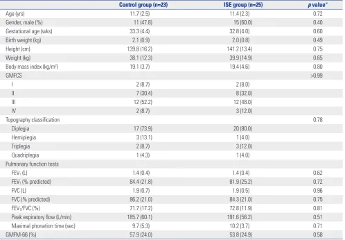
his-tory of psychiatric or neurological disorders other than CP. The exclusion criteria included 1) any uncontrolled, clinically sig-nificant medical condition, such as coexistent cardiac disease or respiratory disease, 2) children with cognitive impairment who are unable to comply with the protocol-required proce-dure, 3) children with the presence or history of tracheostomy, 4) children who are taking medications that can affect respira-tory function, and 5) children with bone deformities of the spine, such as kyphosis or scoliosis. As a result, 60 children met the criteria, and 10 eligible participants declined to partici-pate in the study. Thus, 50 children participartici-pated in this study. A computerized random generator was used to assign each par-ticipant to the experimental group (n=25) or the control group (n=25) (Fig. 1).
Study design
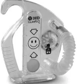
Participants in the experimental group were treated using a flow-oriented incentive spirometer (DHD CliniFLO®, Smiths
[image:2.595.345.501.360.530.2]medical ASD, Inc., Rockland, MA, USA) (Fig. 2), which has a ch-amber-containing ball. The ball is raised in its chamber upon generation of different inspiratory flow rates (six flow settings from 100 mL/sec to 600 mL/sec) by participants breathing
Fig. 1. Flow chart of participant enrollment.
Screened (n=60)
Randomized (n=50)
Conventional exercise+
incentive spirometer exercise completers (n=25) Conventional exercise+
incentive spirometer exercise (n=25)
Conventional exercise completers (n=23) Conventional exercise (n=25)
[image:2.595.74.510.560.721.2]Two were excluded due to follow-up loss Consent withdrawn (n=10)
through a mouthpiece. The children in the ISE group were inst-ructed on how to use the incentive spirometer. After a quiet ex-piration, they were encouraged to close their lips tightly around the mouthpiece and then inhale slowly and deeply until the ball in the spirometer lifted. They were then instructed to hold their breath for as long as possible, or for at least 5 seconds, and then to breathe out slowly. Corresponding to the inspira-tory flow, the balls lifted and remained suspended by the sus-tained inspiratory flow, which served as visible feedback. The flow rate progressively increased at 100 mL/sec intervals from 100 mL/sec to 600 mL/sec, if the participants could maintain the balls lifted at least 5 seconds.
The participants were encouraged to use the device for 10– 15 breaths per session. Ten training sessions were performed daily for 4 weeks. They trained themselves with checklist in a hospital room with regular supervision twice a week. Children in both groups received conventional physical therapy and oc-cupational therapy that focused on gross and fine motor tasks five times per week for a period of four weeks.
Outcome measures
The Gross Motor Function Measure (GMFM)-66 was used to assess improvement in gross motor function. GMFM-66 has been shown to be a reliable and valid measure of gross motor function and useful for measuring the effect of intervention programs.19
The PFT was done in the resting period at least 1 hour after physical and occupational therapy and ISE. The PFT was per-formed before training and at the end of the 4-week training period by the same investigator. PFT was performed using a portable spirometer (Micro Spirometer MS01; Micro Medical Ltd., Kent, UK) assessing forced expiratory volume at one sec-ond (FEV1) and forced vital capacity (FVC). A peak flow meter (ASSESS® Respironics International Inc., Murrysville, PA, USA)
[image:3.595.57.556.349.695.2]was also used to measure peak expiratory flow (PEF). The chil-dren were seated on a chair with the head and trunk straight and the hip and knee joints flexed to 90° with use of external supporting pad. For the children at GMFCS level IV, the test was performed in a chair sitting with external supporting pad and also the shoulder supported by their parent’s hands. We used
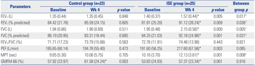
Table 1. Demographic and Baseline Characteristics of the Control Group and the Incentive Spirometer Treatment Group
Control group (n=23) ISE group (n=25) p value*
Age (yrs) 11.7 (2.5) 11.4 (2.3) 0.72
Gender, male (%) 11 (47.8) 15 (60.0) 0.40
Gestational age (wks) 33.3 (4.4) 32.8 (4.0) 0.60
Birth weight (kg) 2.1 (0.9) 2.0 (0.8) 0.49
Height (cm) 139.8 (16.2) 141.2 (13.4) 0.75
Weight (kg) 38.1 (12.3) 39.9 (14.9) 0.65
Body mass index (kg/m2) 19.1 (3.7) 19.4 (4.6) 0.80
GMFCS >0.99
I 2 (8.7) 2 (8.0)
II 7 (30.4) 8 (32.0)
III 12 (52.2) 12 (48.0)
IV 2 (8.7) 3 (12.0)
Topography classification 0.78
Diplegia 17 (73.9) 20 (80.0)
Hemiplegia 3 (13.1) 1 (4.0)
Triplegia 2 (8.7) 3 (12.0)
Quadriplegia 1 (4.3) 1 (4.0)
Pulmonary function tests
FEV1 (L) 1.4 (0.4) 1.4 (0.4) 0.62
FEV1 (% predicted) 84.4 (21.8) 81.9 (25.2) 0.72
FVC (L) 1.9 (0.7) 1.9 (0.5) 0.96
FVC (% predicted) 86.2 (21.0) 84.3 (21.0) 0.75
FEV1/FVC (%) 71.7 (17.2) 72.8 (11.9) 0.81
Peak expiratory flow (L/min) 185.7 (60.1) 191.6 (56.2) 0.51
Maximal phonation time (sec) 9.7 (5.3) 10.2 (3.7) 0.71
GMFM-66 (%) 57.9 (24.0) 53.8 (24.9) 0.58
ISE, incentive spirometry exercise; GMFCS, Gross Motor Function Classification System; FVC, forced vital capacity; FEV1, forced expiratory volume in 1 sec; GMFM,
Gross Motor Function Measure.
Values are expressed as mean (SD) or number of participants (percentage).
various chairs in consideration of each child’s size. The chil-dren were told to inhale as deeply as possible and to blow their entire lung volume through the spirometer. This process was repeated at least three times, and the highest value was select-ed. PFT data were normalized for age, gender, and height, and the predicted FEV1 and FVC values (%) were calculated using the equations provided by Yoon, et al.20 based on values acqu-ired from healthy Korean children. These calculations were used to obtain percent predicted values for FVC (FVC, %) and FEV1 (FEV1, %).
Maximal phonation time (MPT) testing was conducted in a quiet room with children seated on a chair in the same body position with measurement of PFT. The children were asked to inhale deeply and then produce a sustained phonation of the vowel /a:/ at a comfortable pitch and loudness in the seated position for as long as possible. The children were allowed three trials in a row with a 30-second break in between. Each pho-nation was video recorded (Handycam DCR-SR67; Sony Co., Tokyo, Japan), and the longest value was used for analysis.
Statistical analysis
The baseline characteristics of the two groups were compared using the Student’s t-test or Mann-Whitney test for continuous variables and the chi-square test for categorical variables. Com-parisons of pulmonary function before and after training within each group were made using the paired samples t-test or Wil-coxon signed rank test. Differences between the experimental group and the control group were assessed using an indepen-dent samples t test or Mann-Whitney test. Statistical software SPSS 20.0 (SPSS Inc., Chicago, IL, USA) was used for statistical analysis. The level of significance was chosen to be 0.05.
RESULTS
Although 50 children with CP were recruited for the study, two children in the control group did not complete the study
pro-tocol due to early discharge. As a result, 25 children in experi-mental group and 23 children in control group were included in this study. Their mean age was 11.6 years old (±2.3 years). The demographic and baseline characteristics of the study population are shown in Table 1. The two groups were not sig-nificantly different in gestational age, birth weight, height, or body weight. The PFT parameters and GMFM scores at base-line were not significantly different between the two groups.
FEV1, FVC, PEF, and MPT increased significantly after ISE in the experimental group, compared with baseline data. The im-provements in FEV1, FVC, and MPT were significantly higher in the experimental group than in the control group (Table 2).
Compared to baseline GMFM-66 scores, GMFM scores sig-nificantly increased after 4 weeks of training in both groups, al-though improvements in the GMFM scores were not signifi-cantly different between the two groups.
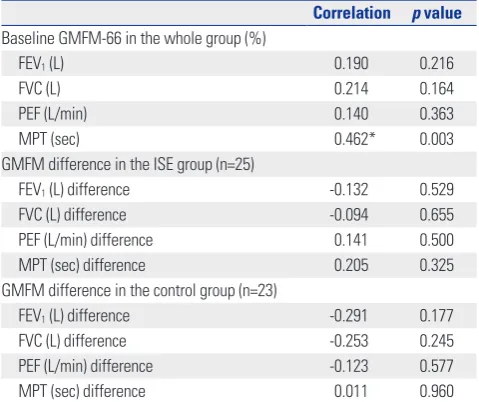
There were no significant relationships between GMFM-66 scores and PFT parameters, such as FEV1, FVC and PEF, but significant correlation with MPT (Table 3); and also the im-provements in GMFM-66 scores did not significantly relate to changes in pulmonary function or MPT.
DISCUSSION
The FEV1, FVC, and FEV1/FVC ratio are useful parameters for assessing restrictive lung disease. On the other hand, PEF is a measure of the maximal or peak flow produced during exha-lation with maximal effort, and it assesses maximal expiratory effort as a surrogate measure for expiratory muscle strength and is useful for assessing obstructive lung disease, such as asthma. These parameters are considered to be potentially very useful in integrated measures of respiratory function in Duchenne muscular dystrophy patients.21 In addition, previ-ous studies have shown that pulmonary function in the chil-dren with CP had characteristics of both obstructive and re-strictive lung disease,2,22-25 and thus, FEV
[image:4.595.41.540.557.684.2]1, FVC, and PEF were Table 2. Comparison of Pre- and Post-Treatment Measurements in the Control Group and the Incentive Spirometer Treatment Group
Parameters Control group (n=23) ISE group (n=25) Between
group p
Baseline Wk 4 p value Baseline Wk 4 p value
FEV1 (L) 1.35 (0.44) 1.35 (0.45) 0.848 1.40 (0.37) 1.57 (0.44)* 0.005 0.017†
FEV1 (% predicted) 84.42 (21.78) 85.59 (24.15) 0.605 81.91 (25.20) 91.12 (28.24)* 0.009 0.036†
FVC (L) 1.94 (0.66) 1.90 (0.69) 0.511 1.95 (0.48) 2.15 (0.56)* 0.000 0.005†
FVC (% predicted) 86.19 (20.95) 83.31 (18.44) 0.685 84.25 (21.03) 92.18 (24.98)* 0.001 0.031†
FEV1/FVC (%) 71.71 (17.23) 73.79 (15.68) 0.563 72.76 (11.91) 74.48 (13.98) 0.443 0.821
PEF (L/min) 185.65 (60.14) 194.78 (55.40) 0.473 191.60 (56.25) 217.60 (67.58)* 0.003 0.085 MPT (sec) 9.65 (5.30) 10.06 (5.75) 0.705 10.16 (3.70) 12.13 (3.61)* 0.001 0.008†
GMFM-66 (%) 57.92 (23.97) 61.58 (24.24)* 0.003 53.83 (24.93) 57.37 (23.34)* 0.001 0.916 ISE, incentive spirometry exercise; FVC, forced vital capacity; FEV1, forced expiratory volume in 1 sec; PEF, peak expiratory flow; MPT, maximal phonation time;
GMFM, Gross Motor Function Measure. Values are expressed as means (SD).
*p<0.05 by paired samples t-test or Wilcoxon signed rank test, baseline versus after four weeks within each group, †p<0.05 by independent samples t-test or
assessed as outcome measures in this study.
Shallow breathing, paradoxical breathing, and low breath-ing volume are commonly noted in children with CP, particu-larly low functioning CP, and these findings may lead to the development of widespread microatelectasis and a decrease in lung distensibility.26,27 Morbidity and mortality associated with CP is mainly related to respiratory compromise, especially in low functioning CP. Therefore, interventions for enhancing respiratory function should be included as part of the compre-hensive management for children with CP.
Some previous studies, report positive effects of various ex-ercise programs on vital capacity (VC).11,24,27 However, the ef-fect of respiratory training in children with CP has rarely been reported. In a previous randomized controlled study by Lee, et al.,16 FRT led to significant gains in FEV
1 and FVC, but not in PEF. In that study, the FRT group consisted of only nine chil-dren, and thus, the improvement in PEF after FRT did not reach a statistically significant level. On the other hand, our random-ized controlled study revealed significant improvements in FEV1, FVC, and PEF after ISE training. FRT consists of repetitive, continuous performance of both maximal inspiration and ex-piration training, while ISE is designed to achieve and sustain maximal inspiration for a prolonged period using slow inspi-ration and deep breaths.16,28,29 Although there are some differ-ences between FRT and ISE, both ISE and FRT are effective in enhancing pulmonary function. The ISE, which increases transpulmonary pressure, inspiratory volumes, and inspiratory muscle performance, by encouraging the patient to take long, slow, deep breaths,30 led to improvement in these parameters. The results of our study suggest that ISE is useful for enhanc-ing both inspiratory and expiratory muscle strengthenenhanc-ing.
According to another previous study by Kwon and Lee,9 the children at GMFCS level III had a higher gain in FVC after FRT than the children at GMFCS level I or II.31 They suggested that children at GMFCS level I or II may have already reached the upper level of their respiratory function capacity, and thus, their improvement is not significant after FRT. The normal val-ues of FVC and FEV1 can vary depending on various factors such as height, weight, and sex, and thus, predicted values (%) for FVC or FEV1 are preferred to compare the results across studies. In Kwon and Lee9 and Lee, et al.’s16 previous studies, predicted values for FVC and FEV1 were not assessed. In addi-tion, the responses of respiratory training on pulmonary func-tion in children with CP may differ between unilateral CP and bilateral CP. Further studies are needed to determine the best candidates for respirator training.
Respiration is an essential physiologic component for main-taining vital function and for performing physical activity in daily life.32 Respiration is also one of the key elements of phys-ical fitness, along with muscular function and cardiovascular function.33 A recent study pointed out that respiratory muscle strength in children with CP is positively correlated with the activities of daily living, self-care, and social function.10 From this perspective, it is interesting to note whether improvement in respiratory function can lead to better exercise endurance, perception of dyspnea, and quality of life. Further studies to address this issue may be helpful for understanding the role of respiratory exercise in children with CP.
According to a previous study, MPT increases in normal chil-dren as they grew older, and MPT of normal chilchil-dren in the United States at the age of 8 to 15 years is between 17.1 and 20.7 sec.34 On the other hand, MPT of normal Korean children aged from 8 to15 years reportedly range from 12.6 to 15.8 sec.35 Compared to those values, the MPTs of our children are short-er. In a previous study, children with spastic CP had a shorter MPT than the normal control group.36 Also, a significant posi-tive correlation between MPT and peak VO2 and also between MPT and the severity of chronic heart failure (CHF) were re-ported in patients with CHF.37,38 On the other hand, there was no significant relationship between MPT and VC in adults with normal speech and voice.39 The proportion of VC used by individual speakers for the MPT task can vary, leading to no significant relationship between VC and MPT.40-42 Also, in our study, there were no significant relationships between MPT and the parameters of PFT. ISE is a breathing technique in which deep breathing exercises are performed through a de-vice that offers visual feedback in response to inspired flow and volume. These exercises allow patients to maximally inflate their lungs and to sustain that inflation. Thus, through this ex-ercise, improvement in breath control appears to result in the observed increase in MPT. Breathy speech, weak breath con-trol, and short utterances are major speech and voice concerns in children with CP.43 The results of our study suggest that ISE may be helpful for enhancing breath control for speech pro-Table 3. Pearson Correlation Coefficients between Gross Motor
Func-tion Measure (GMFM) and Pulmonary FuncFunc-tion Tests
Correlation p value
Baseline GMFM-66 in the whole group (%)
FEV1 (L) 0.190 0.216
FVC (L) 0.214 0.164
PEF (L/min) 0.140 0.363
MPT (sec) 0.462* 0.003
GMFM difference in the ISE group (n=25)
FEV1 (L) difference -0.132 0.529
FVC (L) difference -0.094 0.655
PEF (L/min) difference 0.141 0.500
MPT (sec) difference 0.205 0.325
GMFM difference in the control group (n=23)
FEV1 (L) difference -0.291 0.177
FVC (L) difference -0.253 0.245
PEF (L/min) difference -0.123 0.577
MPT (sec) difference 0.011 0.960
ISE, incentive spirometry exercise; FVC, forced vital capacity; FEV1, forced
ex-piratory volume in 1 sec; PEF, peak exex-piratory flow; MPT, maximal phonation time.
[image:5.595.56.296.96.297.2]duction in children with CP. Further studies are needed to de-lineate the clinical relevance of the improvement in MPT in terms of speech and voice quality in children with CP.
There are some limitations of our study. Most of children of our study were at level II or III, and thus, the effects of ISE ac-cording to GMFCS level were not investigated in our study. Fur-ther studies are needed to outline optimal candidates for ISE in terms of GMFCS level. Another limitation comes from the duration of respiratory training. According to a previous re-port,16 significant improvements in pulmonary function were obtained with 4 weeks of respiratory training in children with CP. Four weeks is the maximal duration of stay in our hospital in general, and thus, 4 weeks of respiratory training was se-lected for our study. However, the optimal duration of training in order to maximize pulmonary function needs to be clari-fied in children with CP through further studies.
In conclusion, this randomized controlled study demon-strated significant benefits of ISE in terms of pulmonary func-tion and MPT in children with spastic CP. ISE is a simple and inexpensive tool for children to use, and therefore, wider use of ISE may be helpful for children at risk of pulmonary dysfunc-tion and poor breath control in speech producdysfunc-tion.
ACKNOWLEDGEMENTS
This work was supported by the Research Institute of Rehabil-itation Medicine at Yonsei University College of Medicine.
REFERENCES
1. Strauss D, Cable W, Shavelle R. Causes of excess mortality in cere-bral palsy. Dev Med Child Neurol 1999;41:580-5.
2. Seddon PC, Khan Y. Respiratory problems in children with neu-rological impairment. Arch Dis Child 2003;88:75-8.
3. Ersöz M, Selçuk B, Gündüz R, Kurtaran A, Akyüz M. Decreased chest mobility in children with spastic cerebral palsy. Turk J Pediatr 2006;48:344-50.
4. Toder DS. Respiratory problems in the adolescent with develop-mental delay. Adolesc Med 2000;11:617-31.
5. O’Donnell DM. Pulmonary complications in neuromuscular dis-ease. Adolesc Med 2000;11:633-45.
6. Sullivan PB, Lambert B, Rose M, Ford-Adams M, Johnson A, Griffiths P. Prevalence and severity of feeding and nutritional prob-lems in children with neurological impairment: Oxford Feeding Study. Dev Med Child Neurol 2000;42:674-80.
7. Reyes AL, Cash AJ, Green SH, Booth IW. Gastrooesophageal re-flux in children with cerebral palsy. Child Care Health Dev 1993;19: 109-18.
8. Saito N, Ebara S, Ohotsuka K, Kumeta H, Takaoka K. Natural histo-ry of scoliosis in spastic cerebral palsy. Lancet 1998;351:1687-92. 9. Kwon YH, Lee HY. Differences of respiratory function according to
level of the gross motor function classification system in children with cerebral palsy. J Phys Ther Sci 2014;26:389-91.
10. Wang HY, Chen CC, Hsiao SF. Relationships between respiratory muscle strength and daily living function in children with cerebral palsy. Res Dev Disabil 2012;33:1176-82.
11. Hutzler Y, Chacham A, Bergman U, Szeinberg A. Effects of a
move-ment and swimming program on vital capacity and water orien-tation skills of children with cerebral palsy. Dev Med Child Neurol 1998;40:176-81.
12. So MW, Heo HM, Koo BS, Kim YG, Lee CK, Yoo B. Efficacy of in-centive spirometer exercise on pulmonary functions of patients with ankylosing spondylitis stabilized by tumor necrosis factor in-hibitor therapy. J Rheumatol 2012;39:1854-8.
13. Scherer TA, Spengler CM, Owassapian D, Imhof E, Boutellier U. Respiratory muscle endurance training in chronic obstructive pul-monary disease: impact on exercise capacity, dyspnea, and quality of life. Am J Respir Crit Care Med 2000;162:1709-14.
14. Basoglu OK, Atasever A, Bacakoglu F. The efficacy of incentive spi-rometry in patients with COPD. Respirology 2005;10:349-53. 15. Igarashi T, Konishi A, Suwa K. [The effects of incentive spirometry
on pulmonary functions]. Masui 1994;43:770-3.
16. Lee HY, Cha YJ, Kim K. The effect of feedback respiratory training on pulmonary function of children with cerebral palsy: a random-ized controlled preliminary report. Clin Rehabil 2014;28:965-71. 17. Speyer R. Effects of voice therapy: a systematic review. J Voice 2008;
22:565-80.
18. Speyer R, Bogaardt HC, Passos VL, Roodenburg NP, Zumach A, Heijnen MA, et al. Maximum phonation time: variability and reli-ability. J Voice 2010;24:281-4.
19. Wang HY, Yang YH. Evaluating the responsiveness of 2 versions of the gross motor function measure for children with cerebral palsy. Arch Phys Med Rehabil 2006;87:51-6.
20. Yoon KA, Lim HS, Koh YY, Kim H. Normal predicted values of pul-monary function test in Korean school-aged children. Korean J Pe-diatr 1993;36:25-37.
21. Mayer OH, Finkel RS, Rummey C, Benton MJ, Glanzman AM, Flickinger J, et al. Characterization of pulmonary function in Duch-enne muscular dystrophy. Pediatr Pulmonol 2015;50:487-94. 22. Blumberg ML. Speech and respiratory impairments and related
therapies in cerebral palsy. Br J Phys Med 1955;18:215-9. 23. Hardy JC. Lung function of athetoid and spastic qusdriplegic
chil-dren. Dev Med Child Neurol 1964;6:378-88.
24. Bjure J, Berg K. Dynamic and static lung volumes of school chil-dren with cerebral palsy. Acta Paediatr Scand Suppl 1970;204:Suppl 204:35+.
25. Kotagal S, Gibbons VP, Stith JA. Sleep abnormalities in patients with severe cerebral palsy. Dev Med Child Neurol 1994;36:304-11. 26. Leopando MT, Moussavi Z, Holbrow J, Chernick V, Pasterkamp H,
Rempel G. Effect of a soft Boston orthosis on pulmonary mechan-ics in severe cerebral palsy. Pediatr Pulmonol 1999;28:53-8. 27. Rothman JG. Effects of respiratory exercises on the vital capacity
and forced expiratory volume in children with cerebral palsy. Phys Ther 1978;58:421-5.
28. Bartlett D Jr. Origin and regulation of spontaneous deep breaths. Respir Physiol 1971;12:230-8.
29. Petz TJ. Physiologic effects of IPPB, blow bottles and incentive spi-rometry. Curr Rev Respir Ther 1979;1:107-11.
30. AARC (American Association for Respiratory Care) clinical practice guideline. Incentive spirometry. Respir Care 1991;36:1402-5. 31. Lee HY, Kim K. Can walking ability enhance the effectiveness of
breathing exercise in children with spastic cerebral palsy? J Phys Ther Sci 2014;26:539-42.
32. Buchanan GF. Timing, sleep, and respiration in health and disease. Prog Mol Biol Transl Sci 2013;119:191-219.
34. Finnegan DE. Maximum phonation time for children with normal voices. J Commun Disord 1984;17:309-17.
35. Her CM. The change of maximum phonation time, articulation di-adochokinetic rate and speech rate of standardized passage: from 8 to 21 years old [Unpublished master’s thesis]. Seoul: Yonsei Uni-versity; 2009.
36. Park ES, Rha DW, Chung HI, Park JE, Nam HS, Kim MJ. Acoustic analysis of vowel sound in children with spastic cerebral palsy. J Korean Acad Rehabil Med 2007;31:103-8.
37. Izawa KP, Watanabe S, Yokoyama H, Hiraki K, Morio Y, Oka K, et al. Muscle strength in relation to disease severity in patients with congestive heart failure. Am J Phys Med Rehabil 2007;86:893-900. 38. Izawa KP, Watanabe S, Tochimoto S, Oka K, Otobe Y, Nemoto S, et
al. Maximum phonation time is related to disease severity in male chronic heart failure patients. Int J Cardiol 2014;174:727-8.
39. Solomon NP, Garlitz SJ, Milbrath RL. Respiratory and laryngeal contributions to maximum phonation duration. J Voice 2000;14: 331-40.
40. Yanagihara N, Koike Y. The regulation of sustained phonation. Folia Phoniatr (Basel) 1967;19:1-18.
41. Yanagihara N, Koike Y, Von Leden H. Phonation and respiration. Function study in normal subjects. Folia Phoniatr (Basel) 1966;18: 323-40.
42. Isshiki N, Okamura H, Morimoto M. Maximum phonation time and air flow rate during phonation: simple clinical tests for vocal function. Ann Otol Rhinol Laryngol 1967;76:998-1007.