0095-1137/08/$08.00⫹0 doi:10.1128/JCM.01548-08
Copyright © 2008, American Society for Microbiology. All Rights Reserved.
Multilocus Sequence Typing Analysis of
Clostridium perfringens
Isolates
from Necrotic Enteritis Outbreaks in Broiler Chicken Populations
䌤
G. Chalmers,
1H. L. Bruce,
2† D. B. Hunter,
1V. R. Parreira,
1R. R. Kulkarni,
1Y.-F. Jiang,
1J. F. Prescott,
1and P. Boerlin
1,3*
Department of Pathobiology, Ontario Veterinary College, University of Guelph, Guelph, Ontario N1G 2W1, Canada1;
Nutreco Canada Agresearch, 473-Sixth Concession Road, R.R. #3, Burford, Ontario N0E 1A0, Canada2; and
Laboratory for Foodborne Zoonoses, Public Health Agency of Canada, Guelph, Ontario N1G 3W4, Canada3
Received 11 August 2008/Returned for modification 13 September 2008/Accepted 10 October 2008
Clostridium perfringens is an important pathogen of animals and humans and is the causative agent of necrotic enteritis (NE) in poultry. This study focuses on the typing of intestinalC. perfringensisolates (nⴝ61) from outbreaks of NE collected from several areas of Southern Ontario, using a recently developed multilocus sequence typing (MLST) technique. For comparison, C. perfringens isolates from healthy birds were also obtained and typed. An additional locus, thepfoSlocus, was included in our analysis, in an attempt to increase the discriminatory ability of the method previously published. Birds were collected from two major poultry processors in Canada, and isolates from processor 2 formed a distinct MLST cluster. Isolates from healthy birds also collected from the outbreak flocks clustered together with isolates from the birds with NE. Although isolates from eight outbreaks clustered together, MLST types were also occasionally different between out-breaks. Strong linkage disequilibrium was observed between loci, suggesting a clonalC. perfringenspopulation structure. Detection assays for toxin genescpb2(beta-2 toxin),tpeL, and the newly describednetB(NetB toxin) were also performed.netBwas almost always found in outbreak isolates, whereascpb2was found exclusively in healthy bird isolates. The toxin genetpeL, which has not been previously identified inC. perfringenstype A strains, was also found, but only in the presence ofnetB. Resistance to bacitracin was found in 34% of isolates from antimicrobial agent-free birds and in 100% of isolates from conventionally raised birds.
Clostridium perfringens is an important pathogen of many animals and can cause a disease known as necrotic enteritis (NE) in avian species. The disease results from extracellular toxin production by the bacterium in the intestinal tract and leads to tissue necrosis and malabsorption and is often fatal (29). Although the disease is generally controlled through the use of antimicrobial agents, it is reemerging in countries that are now banning the use of antimicrobial agents as growth promoters in poultry production (33). The disease generally occurs in outbreaks in which a small percentage of a flock can be affected but can lead to significant losses within a farm.
There are various molecular typing techniques used for as-sessing bacterial population diversity and for outbreak inves-tigation ofC. perfringens. These methods include, among oth-ers, pulsed-field gel electrophoresis (4, 7, 23), multilocus variable number of tandem repeats analysis (5, 25), and mul-tilocus sequence typing (MLST) (16). MLST is a procedure for characterizing bacteria by using the sequences of several “house-keeping” genes (20). Generally, 400 to 500 base pairs of each gene are amplified by PCR and sequenced, and differ-ences in the DNA sequdiffer-ences define the allelic profile for each isolate, known as the sequence type (ST). Genetic events such as point mutations or recombinations result in different alleles
and subsequently different allele combinations (8, 32). Most bacterial species have sufficient variation within house-keeping genes to provide many alleles per locus, allowing multiple STs to be identified using a limited number of loci. An MLST protocol has been developed for C. perfringens by Jost and collaborators (16); however, it is not currently available as a standard protocol on MLST.net (http://www.mlst.net) or PubMLST (http://pubmlst.org). Although MLST is generally expensive, it can be set up as a high-throughput technique which generates data that can be shared between laboratories and al-lows for the creation of a global database of sequences. This global approach would be of great value for the study of NE epidemiology and of other diseases caused byC. perfringens.
In this study, our aim was to characterize the genetic diver-sity ofC. perfringensisolates from outbreaks of NE in broiler chicken populations by using the MLST protocol described by Jost and collaborators (16). Our focus was on investigating the level of clustering of MLST types in NE outbreaks to assess the diversity of isolates within and between outbreaks. Others have shown that only a few pulsed-field gel electrophoresis types of
C. perfringenscan be isolated from field cases of NE, whereas a higher diversity can be found in the intestinal flora of healthy birds (7, 12, 23). With its population genetics implications, MLST data will help provide a clearer picture of the epidemi-ology ofC. perfringensand of NE.
MATERIALS AND METHODS
Collection ofClostridium perfringensisolates.Chickens from farms associated with two major poultry processors were collected in Southern Ontario between 2005 and 2007. Chickens which were sick or had died of NE were collected from processor 1, and whenever possible, healthy birds were collected at the same time
* Corresponding author. Mailing address: Department of Pathobi-ology, Ontario Veterinary College, University of Guelph, Guelph, On-tario N1G 2W1, Canada. Phone: (519) 824-4120, ext. 54647. Fax: (519) 824-5930. E-mail: [email protected].
† Present address: Department of Agriculture, Food and Nutritional Science, University of Alberta, Edmonton, Alberta T6G 2P5, Canada.
䌤Published ahead of print on 22 October 2008.
3957
on May 16, 2020 by guest
http://jcm.asm.org/
from the same farm as the outbreak flock. These birds were raised without antimicrobial agents; the farms had employed antimicrobial agents in feed 2 weeks to 4 months prior to the sampling dates but did not use them thereafter. Birds from processor 2 were raised following conventional methods (non-anti-biotic-free and nonorganic); from the information available, most of the flocks received coccidiostats (salinomycin, narasin, and nicarbazin) in feed, and at least some were given virginiamycin. Live animals were euthanized by following stan-dard protocols approved by the Animal Care Committee of the University of Guelph, and necropsies were conducted on all birds by the Animal Health Laboratory of the University of Guelph to confirm the diagnosis of NE. Non-outbreak, healthy birds were also included from processor 2. A total of 11 different outbreak flocks and seven different normal flocks were sampled.
From each bird, cecal, mid-small-intestinal, and duodenal samples were re-moved aseptically, and the contents were suspended in 5 ml Difco brain heart infusion base (Becton Dickinson Microbiology Systems, Sparks, MD) plus 20%
glycerol (Fisher Scientific, Fair Lawn, NJ) and frozen at⫺70°C. Duodenal
samples were used in NE cases, and mid-small-intestinal and cecal samples were
used only whenC. perfringensisolates could not be recovered from the
duode-num. A 100-l aliquot of each sample was plated onto Shahidi-Ferguson
per-fringens selective medium plates (Becton Dickinson Microbiology Systems). Plates were incubated anaerobically for 24 h at 37°C. Two single colonies with dark centers were subcultured on Shahidi-Ferguson perfringens plates and sub-cultured again for purity. At least three isolates collected from sick or dead birds from each outbreak were used for MLST analysis. Whenever possible, isolates from the matched healthy birds were obtained from the duodenum, but cecal isolates were used if no duodenal isolates could be successfully cultured.
A total of 61C. perfringensisolates were cultured from 45 broiler chickens, and
these isolates were used for MLST analysis. These included 41 isolates from birds with NE, 9 isolates from matched healthy birds, and 11 isolates from
nonout-break birds (Table 1). An additionalC. perfringensisolate, CP4, which is a known
virulent strain isolated from an NE case in Ontario (30), was also included for MLST analysis and toxin typing.
Multilocus sequence typing and analysis. The eight loci described in the MLST protocol by Jost and collaborators (16) were used in this study, with an
additional regulatory protein gene (pfoS) added in an attempt to increase the
discriminatory ability of the method (Table 2). This gene is described in a
publication by Rooney and collaborators (24). InitialpfoSsequencing results
showed allelic diversity in some isolates, and it was therefore included in the method. Lysates for each isolate were prepared using an InstaGene matrix (Bio-Rad Laboratories, Hercules, CA) according to the manufacturer’s
instruc-tions. Each 100-l PCR mixture contained 1⫻PCR buffer, 200M
deoxynucleo-side triphosphates, 1.5 mM MgCl2, 250 nM each primer, 5 UTaqpolymerase
(Promega, Madison, WI), and 2l of template DNA. The PCR conditions were
the same as those used in the publication by Jost and collaborators (16), with only the final extension time modified to 7 min. A 96-well plate format was adopted for all manipulations to facilitate robot-handling of reagents using a Biomek NX automation workstation (Beckman Coulter, Fullerton, CA). PCR product cleanup was performed using a MinElute 96 UF PCR purification kit (Qiagen Inc., Valencia, CA) by following the manufacturer’s instructions. Sequencing was performed using an ABI Prism 3730 sequencer (Applied Biosystems, Foster City, CA), and sequences were analyzed using Sequencher version 4.5 software (Gene Codes Corporation, Ann Arbor, MI).
Concatemers of all nine sequences were analyzed using BioNumerics software version 5.1 (Applied Maths, Austin, TX), and a dendrogram was created using cluster analysis with pairwise similarities and the unweighted-pair group method using arithmetic mean (UPGMA) clustering; neighbor-joining cluster analysis was also performed for comparison. Concatemers of the nine genes from three
sequencedC. perfringensgenomes, strain 13, ATCC 13124, and SM101 (22, 27),
were generated in silico and were included as well. Additionally, sequences
obtained from eight avianC. perfringensisolates previously analyzed by Jost and
collaborators were also included (16). These included seven STs, 6, 7, 26, 50, 61, 68, and 80, which are listed in their publication (B. H. Jost, personal communi-cation).
eBURST analysis of isolates was performed, using eBURSTv3, with STs as-signed based on unique allele profiles (http://eburst.mlst.net) (10). Groups were defined by isolates with at least seven of the nine alleles being identical. All ST numbers were based on this analysis (data not shown).
Presence of toxin genes.Detection of various toxin genes was carried out by
PCR amplification of eachC. perfringensisolate (n⫽61). This included toxin
typing (alpha, beta, epsilon, and iota toxin genes), which was performed as a multiplex reaction according to Yoo and collaborators (35). The beta-2 toxin
gene (cpb2) and the enterotoxin gene (cpe) were detected using protocols
de-scribed by Meer and Songer (21) and Herholz and collaborators (14),
respec-tively. Additionally, a recently described NE beta-like toxin, NetB (netB) (17),
was detected by the primers AKP78 and AKP79 described in the publication.
The presence of thetpeL(toxin C.perfringens large cytotoxin) gene was
detected by PCR amplification of an internal fragment, using primers designed
from theC. perfringenstoxin type C sequence oftpeL(AB262081) supplied by
Amimoto and collaborators (2). Amplifications were performed in a 25-l total
volume containing the following: 5l of template DNA, 1⫻PCR buffer with
Mg2⫹(New England BioLabs, Ipswich, MA), 0.2 mM deoxynucleoside
triphos-phate mixture, 2.5 units ofTaqDNA polymerase (New England BioLabs), and
800 nM of each primer (T2875-F [ATAAGTGCCAATCAATATGA] and T3348-R [TTCTCCTGAATTAATATTAA]). After an initial denaturing step of 3 min at 94°C, the samples were subjected to 30 cycles of denaturation at 94°C for 1 min, annealing at 55°C for 1 min, extension at 72°C for 1 min, and final extension at 72°C for 10 min. All PCR products were visualized by horizontal agarose gel electrophoresis.
OnenetBand threetpeLPCR products were sequenced to confirm the
spec-ificity of the PCR. The entiretpeLgene fromC. perfringensstrain CP4 was
amplified using primers specific to sequences 298 bp upstream of the first
po-tentialtpeLstart site to 370 bp downstream of the stop codon fortpeLinC.
perfringenstype C (AB262081). Both strands of this entire PCR product were sequenced by primer walking, and the DNA sequence was deposited in the
GenBank database (EU848493). A Southern blot to determine iftpeLis
plasmid-borne was performed on plasmid preparations (plasmid midi kit; Qiagen Inc.)
from four isolates (threetpeLpositive and one negative), using a PCR
digoxi-genin-labeled probe (Roche Diagnostics, Mannheim, Germany) synthesized
us-ing the primers and PCR conditions described fortpeLdetection by Amimoto
and collaborators (2).
Susceptibility testing.Susceptibility to bacitracin was determined for each isolate by using Etest strips (AB Biodisk, Solna, Sweden) following the manu-facturer’s instructions. Isolates were grown overnight on blood agar plates, and a 1.0 McFarland standard was suspended in Mueller-Hinton broth (Sensititre,
Westlake, OH).Brucellaagar (150 mm) with vitamin K and Hemin agar plates
(Medox Diagnostics, Ottawa, Ontario, Canada) were swabbed with the suspen-sion, and single strips were applied to the surface. Readings were taken after 24 h of incubation under anaerobic conditions, and end points were determined using
Etest guidelines for anaerobes.C. perfringensstrain ATCC 13124 was used as a
control. Isolates with MICs of⬎16g/ml were considered resistant, according to
Johansson and collaborators (15).
Statistical analysis. Linkage disequilibrium analysis was performed using
Arlequin version 3.11 (9) population genetics software. Polymorphic sites (n⫽
116) from each ST were analyzed (only one isolate per ST was used for this analysis), and linkage disequilibrium testing was estimated for all pairs of
poly-morphic sites. Significance was set at aPvalue of 0.05, with 10,000 steps in the
Markov chain and 1,000 dememorization steps, resulting in a matrix ofPvalues.
A second measure of linkage disequilibrium was performed using the tool pro-vided by MLST.net (http://linux.mlst.net/link_dis/index.htm). This method
cal-culates an index of association (IA), which measures the degree of association
between loci (28).
Nucleotide sequence accession numbers.Each unique sequence for each locus
found in this study (n⫽53) has been uploaded to the GenBank database (http:
//www.ncbi.nlm.nih.gov) under the following accession numbers:
EU605734-EU605739 forplc, the alpha toxin gene; EU605740-EU605748 forddlA, theD
-alanine-D-alanine ligase gene; EU605749-EU605753 for dut, the deoxyuridine
triphosphatase gene; EU605754-EU605759 forglpK, the glycerol kinase gene;
EU605760-EU605764 for gmk, the deoxyguanylate kinase gene;
EU605765-EU605768 forrecA, the recombinase gene; EU605769-EU605773 forsod, the
su-peroxide dismutase gene; EU605774-EU605782 for tpiA, the triose phosphate
isomerase gene; and EU605783-EU605786 forpfoS, the putative membrane protein
gene.
RESULTS
C. perfringensisolates were cultured from birds from all NE outbreaks but were more difficult to recover from healthy, nonoutbreak birds. Duodenal samples were generally sufficient for culture; however, mid-small-intestinal and cecal samples from healthy birds in which concentrations of C. perfringens
were found to be lower were also used (Table 1). Sixty-one isolates were selected for MLST analysis from 45 birds, which included 11 nonoutbreak birds. In a few instances (Table 1),
3958 CHALMERS ET AL. J. CLIN. MICROBIOL.
on May 16, 2020 by guest
http://jcm.asm.org/
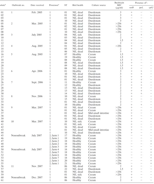
TABLE 1. Complete list ofClostridium perfringensisolates typed by MLST, including outbreak attributes, ST, bacitracin MIC determined by Etest, and presence or absence ofnetB, tpeL, andcpb2genes detected by PCR
Isolateb
Outbreak no. Date received Processora
ST Bird health Culture source
Bacitracin MIC
(g/ml)
Presence ofc:
netB tpeL cpb2
01 1 Feb. 2005 1 01 NE, dead Duodenum 3 ⫹ ⫺ ⫺
02 1 01 NE, dead Duodenum 3 ⫹ ⫺ ⫺
03 1 01 NE, dead Duodenum 3 ⫺ ⫺ ⫺
04 1 10 NE, dead Duodenum 3 ⫹ ⫺ ⫺
05 2 Mar. 2005 1 01 NE, dead Duodenum ⬎256 ⫹ ⫺ ⫺
06 1 02 NE, dead Duodenum ⬎256 ⫹ ⫺ ⫺
07 1 01 NE, dead Duodenum ⬎256 ⫹ ⫺ ⫺
08 1 10 NE, dead Duodenum ⬎256 ⫹ ⫺ ⫺
09 3 July 2005 1 04 NE, sick Duodenum 2 ⫹ ⫹ ⫺
10 1 03 NE, dead Duodenum 2 ⫹ ⫺ ⫺
11 1 04 NE, dead Duodenum 1.5 ⫹ ⫺ ⫺
12 1 04 NE, dead Duodenum 2 ⫹ ⫺ ⫺
13 4 Aug. 2005 1 01 NE, dead Duodenum ⬎256 ⫹ ⫺ ⫺
14 1 05 NE, dead Duodenum 2 ⫹ ⫺ ⫺
15 1 06 NE, dead Duodenum 3 ⫹ ⫺ ⫺
16 5 Aug. 2005 1 07 Healthy Cecum 1.5 ⫺ ⫺ ⫺
17 1 19 Healthy Cecum 1.5 ⫺ ⫺ ⫺
18 1 08 Healthy Cecum 1.5 ⫹ ⫺ ⫺
19 1 08 NE, dead Duodenum 1.5 ⫹ ⫺ ⫺
20 1 09 NE, dead Duodenum 1.5 ⫹ ⫺ ⫺
21 1 08 NE, dead Duodenum 1.5 ⫹ ⫺ ⫺
22 6 Apr. 2006 1 01 Healthy Cecum 3 ⫹ ⫹ ⫺
23 1 10 NE, dead Duodenum 3 ⫹ ⫹ ⫺
24 1 01 NE, dead Duodenum 3 ⫹ ⫹ ⫺
25 1 01 NE, dead Duodenum 2 ⫹ ⫹ ⫺
26 7 Sept. 2006 1 11 Healthy Cecum 2 ⫹ ⫹ ⫺
27 1 12 Healthy Duodenum 2 ⫹ ⫹ ⫺
28 1 13 NE, dead Duodenum 2 ⫹ ⫹ ⫺
29 1 13 NE, dead Duodenum 2 ⫹ ⫹ ⫺
30 1 14 NE, dead Duodenum 2 ⫹ ⫹ ⫺
31 8 Nov. 2006 1 10 NE, dead Cecum 2 ⫹ ⫺ ⫺
32 1 15 NE, dead Duodenum 3 ⫹ ⫺ ⫺
33 1 01 NE, dead Duodenum 2 ⫺ ⫺ ⫺
34 1 10 Healthy Duodenum 2 ⫹ ⫹ ⫺
35 9 Mar. 2007 1 01 NE, dead Cecum ⬎256 ⫹ ⫺ ⫺
36 1 01 NE, dead Cecum ⬎256 ⫹ ⫺ ⫺
37 1 01 NE, dead Mid-small intestine ⬎256 ⫹ ⫺ ⫺
38 1 01 NE, dead Duodenum ⬎256 ⫹ ⫺ ⫺
39 1 01 NE, dead Duodenum ⬎256 ⫹ ⫺ ⫺
40 10 Mar. 2007 1 10 NE, sick Cecum ⬎256 ⫹ ⫺ ⫺
41 1 01 NE, sick Cecum ⬎256 ⫹ ⫺ ⫺
42 1 16 NE, dead Cecum ⬎256 ⫹ ⫺ ⫺
43 1 01 NE, dead Mid-small intestine ⬎256 ⫹ ⫺ ⫺
44 1 10 NE, dead Duodenum ⬎256 ⫹ ⫺ ⫺
45 Nonoutbreak July 2007 2, farm 1 17 Healthy Cecum ⬎256 ⫺ ⫺ ⫺
46 2, farm 1 19 Healthy Cecum ⬎256 ⫺ ⫺ ⫺
47 2, farm 2 18 Healthy Cecum ⬎256 ⫺ ⫺ ⫹
48 2, farm 2 18 Healthy Cecum ⬎256 ⫺ ⫺ ⫹
49 2, farm 3 19 Healthy Cecum ⬎256 ⫺ ⫺ ⫺
50 Nonoutbreak July 2007 2, farm 4 18 Healthy Cecum ⬎256 ⫺ ⫺ ⫹
51 2, farm 5 18 Healthy Cecum ⬎256 ⫺ ⫺ ⫹
52 2, farm 6 18 Healthy Cecum ⬎256 ⫺ ⫺ ⫹
53 2, farm 7 18 Healthy Cecum ⬎256 ⫺ ⫺ ⫹
54 2, farm 1 20 Healthy Cecum ⬎256 ⫺ ⫺ ⫹
55 2, farm 1 21 Healthy Cecum ⬎256 ⫺ ⫺ ⫹
56 11 Nov. 2007 1 06 NE, dead Cecum 2 ⫹ ⫺ ⫺
57 1 22 NE, dead Duodenum 1.5 ⫹ ⫺ ⫺
58 1 01 NE, dead Duodenum ⬎256 ⫹ ⫺ ⫺
59 1 01 NE, sick Cecum ⬎256 ⫹ ⫺ ⫺
60 Nonoutbreak Dec. 2007 1 06 Healthy Cecum 2 ⫹ ⫺ ⫺
61 1 06 Healthy Cecum 2 ⫹ ⫺ ⫺
aProcessor 1, no antimicrobial agent use; processor 2, conventional antimicrobial agent use.
bEach pair of isolates 16 and 17, 23 and 24, 35 and 36, 40 and 41, and 60 and 61 was recovered from the same intestinal sample of a single bird within a given outbreak.
c⫹, present;⫺, absent.
on May 16, 2020 by guest
http://jcm.asm.org/
two isolates were taken from a single sample to assess the genetic diversity between isolates within a single bird.
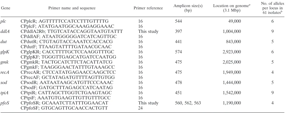
Amplification and sequence analysis of the 61 isolates were successful using the protocol outlined by Jost and collaborators (16). The addition of thepfoSlocus did not create more STs and did not increase the discrimination of the method even though four different alleles were found (Table 2). ThepfoS
amplicon also showed some sequence length polymorphism of up to three base pairs, which was not observed in the other loci (Table 2). One pfoS allele (EU605786) showed a deletion within a long poly(A) sequence, introducing an internal stop codon, and resulted in a truncated PfoS protein.
The average number of alleles per locus was 5.9, with a minimum of 4 and a maximum of 9. Twenty-two STs were identified. With a few exceptions (outbreaks 4, 5, and 11), clustering of isolates within each outbreak was observed (Fig. 1). Isolates obtained from healthy birds in the same outbreak flock (outbreaks 5, 6, 7, and 8) also clustered with isolates from dead birds affected by NE. Multiple isolates obtained from a single bird were either identical (n⫽8) or very similar (n⫽2) (Fig. 1). The isolates from healthy birds originating from the seven farms of processor 2 all clustered tightly together. Al-though a major cluster contained isolates from eight different outbreaks from processor 1 (outbreaks 1, 2, 4, 6, 8, 9, 10, and 11), the isolates from the three remaining outbreaks were part of unrelated clusters. Neighbor-joining cluster analysis pro-duced identical major clusters of isolates compared to the UPGMA clustering.
The eBURST analysis produced 22 STs and was in general agreement with the other clustering analysis. Six complexes (linked by single allelic differences) and two singletons were generated, with ST01 and ST18 considered founders (10) of the two major complexes (data not shown). ST01 included 19 isolates from the eight outbreaks mentioned above, all from processor 1. ST18 contained six isolates, all from processor 2, and included five nonoutbreak farms (Fig. 1).
The isolate JGS4135 from Jost and collaborators (16), which was the only one studied by these authors that clustered tightly with our own isolates, was traced back to a poultry sample from Ontario originally collected in Guelph (Fig. 1). Strain CP4, which was isolated about 10 years ago from a chicken with NE in Ontario, is also part of the major ST01 group (Fig. 1).
Results from the Arlequin linkage disequilibrium analysis (Fig. 2) showed a random distribution of significant values. The extent of significant disequilibrium (P ⱕ0.05) between poly-morphic sites ranged between 7.2% (tpiAandddlA) and 43.9% (glpK and ddlA), with an overall average of 23.6%. Linkage disequilibrium within each locus was higher (38.0% of poly-morphic sites), as was expected. TheIAvalue calculated using
the MLST.net tool was 3.140, which implies significant linkage disequilibrium as well (28).
All isolates from our birds were found to be toxin type A and negative for the enterotoxin gene. ThenetBgene was found only in isolates from processor 1 (92.0%). Conversely, thecpb2
toxin gene was found only in isolates from processor 2 (72.7%). DNA sequencing of the gene amplified from CP4 showed that the tpeL gene consisted of 4,955 bp (EU848493). The translated amino acid sequence of TpeL in strain CP4 has 97% identity with the sequence of TpeL from aC. perfringenstoxin type B strain (NZ_ABDV01000024), and both are 128 amino acids longer than that in the toxin type C strain MC18 (AB262081). The completetpeLgenes from two NE isolates, 23 and 28, were sequenced and found to have 100% identity withtpeLfrom strain CP4. Among the 61 isolates investigated here,tpeLwas detected in 11 outbreak-associated isolates (Ta-ble 1) which included eight STs and four out of eleven out-breaks. All isolates from processor 2 were negative fortpeL(n⫽
13). Southern blot results showed that the tpeL gene was present on a large plasmid of identical size (⬃100 kb) in all threetpeL-positive isolates (13, 30, and CP4) examined (data not shown).
[image:4.585.42.544.82.281.2]Etest values for susceptibility to bacitracin ranged between
TABLE 2. MLST locus properties
Gene Primer name and sequence Primer reference Amplicon size(s)
(bp)
Location on genomea
(3.1 Mbp)
No. of alleles per locus in
61 isolatesb
plc CPplcR; AGTTTTTCCATCCTTTGTTTTG 16 544 49,000 6
CPplcF; ATATGAATGGCAAAGAGGAAAC 16
ddlA CPddlA2Rb; TTGTCATACCAGGTAATGTATTT This study 397 1,004,000 9
CPddlAF; ATAATGGGGGATCATCAGTTGC 16
dut CPdutR; CTGTAGTACCAAATCCACCACG 16 441 843,000 5
CPdutF; TTAAGTATTTTGATAACGCAAC 16
glpK CPglpKR; CACCTTTTGCTCCAAGGTTTGC 16 574 2,923,000 6
CPglpKF; TGGGTTGAGCATGATCCAATGG 16
gmk CPgmkR; TACTGCATCTTCTACATTATCG 16 475 2,025,000 5
CPgmkF; TAAGGGAACTATTTGTAAAGCC 16
recA CPrecAR; CTCCATATGAGAACCAAGCTCC 16 475 1,949,000 4
CPrecAF; GCTATAGATGTTTTAGTTGTGG 16
sod CPsodR; AATAATAAGCATGTTCCCAAAC 16 478 1,444,000 5
CPsodF; GATGCTTTAGAGCCATCAATAG 16
tpiA CPtpiR; CATTAGCTTGGTCTGAAGTAGC 16 451 1,542,000 9
CPtpiF; AAATGTGAAGTTGTTGTTTGCC 16
pfoS CPpfoSR; GCAAATCTTATTTGGAACAT This study 560, 562, 563 1,190,000 4
CPpfoSF; GTGCAGTTGCAACCACTGTT 24
aGene locations were based on the published genome sequence ofC. perfringensstrain 13 (27).
bThe average number of alleles per locus in 61 isolates was 5.9.
3960 CHALMERS ET AL. J. CLIN. MICROBIOL.
on May 16, 2020 by guest
http://jcm.asm.org/
FIG. 1. Dendrogram of concatenated MLST sequences of 61 avian isolates from Southern Ontario generated with pairwise similarities and UPGMA clustering. It includes eight previously published avian C. perfringensMLST sequences (16). Isolates from the study by Jost and collaborators are labeled JGS and shown in gray. CP4 is a virulent NE-associated isolate from another previous study in Ontario (30). The three
C. perfringensstrains with published genome sequences (strains 13, ATCC 13124, and SM101) were also included, and their MLST types were generated in silico (22, 27). Selected bootstrap values are indicated at nodes. Susceptibility to bacitracin was determined using a 16-g/ml breakpoint. STs were determined through eBURSTv3 analysis (10), and ST01 and 18 are labeled in boldface type (founders).
3961
on May 16, 2020 by guest
1.5 and 256g/ml (Table 1). A very distinct bimodal distribu-tion was present with MICs being either between 1.5 and 3
g/ml (susceptible) or⬎256g/ml (resistant). Seventeen re-sistant and 33 susceptible isolates were obtained from proces-sor 1, whereas only bacitracin-resistant isolates (n⫽11) were detected in birds from processor 2. All the bacitracin-resistant isolates were part of the two major clusters containing ST01 and ST18, but not all the isolates in these two clusters were resistant (Fig. 1).
DISCUSSION
In this study, C. perfringenswas typically isolated from the duodenal samples of NE-affected birds, whereas isolates from healthy birds could be recovered frequently from cecal samples only. This has been previously observed in other studies, in which concentrations ofC. perfringensare higher in the
intes-tinal tract of NE-affected birds, where the bacteria have pro-liferated upward into the duodenum and are often associated with coccidial coinfection (1, 3, 26).
The MLST method developed by Jost and collaborators was originally developed for a broad array ofC. perfringensisolates of very diverse origins (16) and may not have been sufficient for this study, where only chicken isolates were used. Therefore, we initially attempted to increase the discriminatory power of this typing scheme by investigating potential additional loci. The loci forgyrAandrplL(24) andrpoD(primers derived from the published C. perfringens SM101 sequence [22]) showed little or no polymorphism at all in a subset of our isolates (data not shown). However, thepfoSlocus did show some diversity and was added to the established protocol. Ultimately, this locus did not contribute any additional discrimination despite the presence of four different alleles at this locus in our iso-lates. It may also not be a suitable MLST locus because of its
FIG. 2. Linkage disequilibrium matrix of polymorphic sites in 22 STs, generated by Arlequin version 3.11 (9). Darker cells refer to more-significantPvalues (black cells represent values ofⱕ0.05 and the lightest-gray cells up to 0.20). Locus names are shown on each axis, in sequential order on the chromosome.
3962 CHALMERS ET AL. J. CLIN. MICROBIOL.
on May 16, 2020 by guest
http://jcm.asm.org/
sequence length variability; one allele of the pfoS locus ap-peared to have resulted from the deletion of an “ATT” tandem repeat in the gene. AnotherpfoS allele encodes a truncated and probably nonfunctional protein. Therefore, it appears that the protocol described by Jost and collaborators (16) already provides a good estimate ofC. perfringensdiversity and was not improved with the addition ofpfoS. When isolates from the three sequenced genomes ofC. perfringenswere included (Fig. 1), as well as the eight avian isolates from the Jost et al. study (16), these isolates generally belonged to different sequence types than to those from this study. Interestingly though, the JGS4135 isolate typed by Jost and collaborators was identical to isolate 10 in our study, and it was later revealed that the former isolate had originated from the University of Guelph about 10 years earlier. This is a convincing indication that this MLST system is epidemiologically viable and also showed that the same C. perfringensclone may have persisted for many years in Southern Ontario.
In previous studies (7, 12, 23), researchers have proposed that there are distinct clones withinC. perfringenspopulations which cause NE outbreaks and that birds within an outbreak are usually affected by only one of these clones. Our results, which showed strong genetic clustering in isolates from NE outbreaks, both within a single bird and between birds of the same outbreak, are in agreement with this hypothesis. How-ever, we did not observe any major differences between isolates from healthy birds and those from NE cases within any out-break. This stresses once more the fact that NE is a multifac-torial disease and that the mere presence of a specific C. perfringens strain in a bird is not a sufficient cause for the development of the disease.
The very strong clustering of isolates originating from birds of seven different farms from geographically separate areas affiliated with processor 2 is striking and was unexpected. Sim-ilarly, despite the higher diversity observed among isolates from farms with NE outbreaks and those associated with pro-cessor 1, the majority of these isolates belonged to a single major cluster (cluster containing ST01, -02, -10, -15, and -16) (Fig. 1). In the case of this processor, the clustering may be related to a higher propensity of a specific clone for causing NE. These clusters also suggest a limited degree of genetic diversity ofC. perfringensthroughout each processor’s poultry operations. This could be the result of the dissemination of clones throughout integrated producer networks, starting at hatcheries and chick distribution systems. Selection of a lim-ited number of resistant clones through antimicrobial agent use, either for prevention (cluster containing ST18) or therapy (cluster that contains ST01 and is associated with NE out-breaks), may also be part of the reason for the observed clus-tering. This latter hypothesis is supported by the fact that bacitracin resistance was clearly associated with these two ma-jor clusters and lineages.
Locus linkage was found to be high in the isolates charac-terized in this study, with significant linkage disequilibrium found between all loci (IA ⫽ 3.140); panmictic populations
have an IAvalue approaching zero. This also shows that
re-combination rates for the core genome ofC. perfringensare low or possibly absent (28). Additionally, only 22 STs were found in the sample population. When considering the number of pos-sible allele combinations (over 5 million), the 22 STs found in
61 isolates may be considered low. These data suggest that the
C. perfringens population under investigation had a highly clonal structure (28).
Since MLST examines core genes encoding house-keeping functions not usually involved in pathogenesis, there is not necessarily a correlation between virulence potential and the overall genomic relationships evidenced by MLST (20, 31). It is therefore possible that more virulent clones do exist within these outbreaks, which carry accessory genes and virulence factors not detected using MLST.
The complete absence ofcpb2in the NE outbreak isolates was surprising, since this toxin had originally been associated with virulence (11, 13, 34). However, there is a growing body of evidence that suggests that the beta-2 toxin is not a critical factor ofC. perfringensinfection in this particular case of poul-try (4, 6, 30). The recently discoverednetBtoxin gene, how-ever, was found exclusively in isolates which were all associated with NE outbreaks and was never detected in healthy control samples from processor 2. AlthoughnetBwas detected in every outbreak, it was still found in healthy matched birds from outbreak flocks as well. Additionally,netBwas not always de-tected in isolates from birds clearly affected with NE (Table 1, isolates 03 and 33). Overall, this study provided a good oppor-tunity to observe the prevalence of the netBgene in a field situation where NE- and non-NE-associated isolates were used. The data suggest that this toxin is an important factor in
C. perfringens infection, facilitating the development of NE. However, our results also show that it is neither a necessary nor a sufficient cause for the disease to occur. As the authors of the netBdiscovery stated (17), there may be many other unidentified factors which are also important for virulence.
TheC. perfringenstoxin TpeL (2) was identified in the cul-ture supernatant of C. perfringens strain CP4, when the se-creted proteins of CP4 were electrophoretically separated and reacted with the serum from broiler chickens immune to NE (18, 19), and was further identified by mass spectrometry (un-published data). This is the first report describing the presence of tpeL genes in C. perfringens type A strains and the first report of the presence of this gene in isolates from chickens with NE. ThetpeLgene identified in these toxin type A isolates is very similar to the tpeL gene in the toxin type B strain (NZ_ABDV01000024). The toxin type C strain (2), however, has a cytosine insert at position 4941, which creates a prema-ture stop codon and thus truncates the protein. The variable presence of this toxin gene within clones and even within clonally related isolates from a single farm suggested that it is located on a mobile genetic element. Our Southern blot results confirmed that it is indeed located on large plasmids. The identification of TpeL among the secreted proteins of CP4 may be related to the greater virulence of this strain in an experi-mental model of NE in comparison to some other strains in which NE originated (30). SincetpeLwas detected in isolates and STs associated with NE outbreaks only, it is reasonable to speculate that it contributes to the pathogenesis of NE, but further work is required to assess its contribution to the viru-lence of NE isolates.
In conclusion, the MLST protocol described by Jost and collaborators (16) was successfully applied in this study for typing avianC. perfringensisolates. The resulting data suggest that clustering of MLST types occurs in association with the
on May 16, 2020 by guest
http://jcm.asm.org/
source of the isolates and with bacitracin resistance. A major clonal lineage containing isolates from eight different out-breaks was identified, which may be either more prone to cause outbreaks than others or selected by the use of antimicrobial agents. Toxin typing suggests that NetB is a far more significant toxin than the beta-2 toxin, although there may be many more unexplored factors that are important for NE development, such as TpeL, which was identified here for the first time in NE isolates.
ACKNOWLEDGMENTS
This work was funded by the Canadian Poultry Research Council and the Poultry Industry Council.
We thank the Animal Health Laboratory at the University of Guelph, the poultry processors, and the Canadian Food Inspection Agency for their assistance in this project. We also thank Teresa Crease for her help with the linkage disequilibrium analysis.
REFERENCES
1.Al-Sheikhly, F., and R. B. Truscott.1977. The interaction ofClostridium perfringensand its toxins in the production of necrotic enteritis of chickens.
Avian Dis.21:256–263.
2.Amimoto, K., T. Noro, E. Oishi, and M. Shimizu.2007. A novel toxin homologous to large clostridial cytotoxins found in culture supernatant of
Clostridium perfringenstype C. Microbiology153:1198–1206.
3.Barnes, E. M., G. C. Mead, D. A. Barnum, and E. G. Harry.1972. The intestinal flora of the chicken in the period 2 to 6 weeks of age, with
particular reference to the anaerobic bacteria. Br. Poult. Sci.13:311–326.
4.Chalmers, G., S. W. Martin, D. B. Hunter, J. F. Prescott, L. J. Weber, and P. Boerlin.2008. Genetic diversity ofClostridium perfringens isolated from
healthy broiler chickens at a commercial farm. Vet. Microbiol.127:116–127.
5.Chalmers, G., S. W. Martin, J. F. Prescott, and P. Boerlin.2008. Typing of
Clostridium perfringensby multiple-locus variable number of tandem repeats
analysis. Vet. Microbiol.128:126–135.
6.Crespo, R., D. J. Fisher, H. L. Shivaprasad, M. E. Fernandez-Miyakawa, and F. A. Uzal.2007. Toxinotypes ofClostridium perfringensisolated from sick
and healthy avian species. J. Vet. Diagn. Investig.19:329–333.
7.Engstro¨m, B. E., C. Fermer, A. Lindberg, E. Saarinen, V. Baverud, and A. Gunnarsson.2003. Molecular typing of isolates ofClostridium perfringens
from healthy and diseased poultry. Vet. Microbiol.94:225–235.
8.Enright, M. C., and B. G. Spratt.1999. Multilocus sequence typing. Trends
Microbiol.7:482–487.
9.Excoffier, L., G. Laval, and S. Schneider.2005. Arlequin ver. 3.0: an inte-grated software package for population genetics data analysis. Evol.
Bioin-form. Online1:47–50.
10.Feil, E. J., B. C. Li, D. M. Aanensen, W. P. Hanage, and B. G. Spratt.2004. eBURST: inferring patterns of evolutionary descent among clusters of re-lated bacterial genotypes from multilocus sequence typing data. J. Bacteriol.
186:1518–1530.
11.Fisher, D. J., K. Miyamoto, B. Harrison, S. Akimoto, M. R. Sarker, and B. A. McClane.2005. Association of beta2 toxin production withClostridium per-fringenstype A human gastrointestinal disease isolates carrying a plasmid
enterotoxin gene. Mol. Microbiol.56:747–762.
12.Gholamiandekhordi, A. R., R. Ducatelle, M. Heyndrickx, F. Haesebrouck, and F. Van Immerseel.2006. Molecular and phenotypical characterization of
Clostridium perfringens isolates from poultry flocks with different disease
status. Vet. Microbiol.113:143–152.
13.Gibert, M., C. Jolivet-Reynaud, and M. R. Popoff.1997. Beta2 toxin, a novel
toxin produced byClostridium perfringens. Gene203:65–73.
14.Herholz, C., R. Miserez, J. Nicolet, J. Frey, M. Popoff, M. Gibert, H. Gerber, and R. Straub.1999. Prevalence of beta2-toxigenicClostridium perfringensin
horses with intestinal disorders. J. Clin. Microbiol.37:358–361.
15.Johansson, A., C. Greko, B. E. Engstro¨m, and M. Karlsson.2004.
Antimi-crobial susceptibility of Swedish, Norwegian and Danish isolates of
Clostrid-ium perfringens from poultry, and distribution of tetracycline resistance
genes. Vet. Microbiol.99:251–257.
16.Jost, B. H., H. T. Trinh, and J. G. Songer.2006. Clonal relationships among
Clostridium perfringensof porcine origin as determined by multilocus
se-quence typing. Vet. Microbiol.116:158–165.
17.Keyburn, A. L., J. D. Boyce, P. Vaz, T. L. Bannam, M. E. Ford, D. Parker, A. Di Rubbo, J. I. Rood, and R. J. Moore.2008. NetB, a new toxin that is
associated with avian necrotic enteritis caused byClostridium perfringens.
PLoS Pathog.4:e26.
18.Kulkarni, R. R., V. R. Parreira, S. Sharif, and J. F. Prescott.2006. Clostrid-ium perfringensantigens recognized by broiler chickens immune to necrotic
enteritis. Clin. Vaccine Immunol.13:1358–1362.
19.Kulkarni, R. R., V. R. Parreira, S. Sharif, and J. F. Prescott.2007.
Immu-nization of broiler chickens againstClostridium perfringens-induced necrotic
enteritis. Clin. Vaccine Immunol.14:1070–1077.
20.Maiden, M. C.2006. Multilocus sequence typing of bacteria. Annu. Rev.
Microbiol.60:561–588.
21.Meer, R. R., and J. G. Songer.1997. Multiplex polymerase chain reaction
assay for genotypingClostridium perfringens. Am. J. Vet. Res.58:702–705.
22.Myers, G. S., D. A. Rasko, J. K. Cheung, J. Ravel, R. Seshadri, R. T. DeBoy, Q. Ren, J. Varga, M. M. Awad, L. M. Brinkac, S. C. Daugherty, D. H. Haft, R. J. Dodson, R. Madupu, W. C. Nelson, M. J. Rosovitz, S. A. Sullivan, H. Khouri, G. I. Dimitrov, K. L. Watkins, S. Mulligan, J. Benton, D. Radune, D. J. Fisher, H. S. Atkins, T. Hiscox, B. H. Jost, S. J. Billington, J. G. Songer, B. A. McClane, R. W. Titball, J. I. Rood, S. B. Melville, and I. T. Paulsen.
2006. Skewed genomic variability in strains of the toxigenic bacterial
patho-gen, Clostridium perfringens. Genome Res.16:1031–1040.
23.Nauerby, B., K. Pedersen, and M. Madsen.2003. Analysis by pulsed-field gel
electrophoresis of the genetic diversity amongClostridium perfringens
iso-lates from chickens. Vet. Microbiol.94:257–266.
24.Rooney, A. P., J. L. Swezey, R. Friedman, D. W. Hecht, and C. W. Maddox.
2006. Analysis of core housekeeping and virulence genes reveals cryptic
lineages ofClostridium perfringensthat are associated with distinct disease
presentations. Genetics172:2081–2092.
25.Sawires, Y. S., and J. G. Songer.2005. Multiple-locus variable-number
tan-dem repeat analysis for strain typing ofClostridium perfringens. Anaerobe
11:262–272.
26.Shane, S. M., J. E. Gyimah, K. S. Harrington, and T. G. Snider III.1985.
Etiology and pathogenesis of necrotic enteritis. Vet. Res. Commun.9:269–
287.
27.Shimizu, T., K. Ohtani, H. Hirakawa, K. Ohshima, A. Yamashita, T. Shiba, N. Ogasawara, M. Hattori, S. Kuhara, and H. Hayashi.2002. Complete
genome sequence ofClostridium perfringens, an anaerobic flesh-eater. Proc.
Natl. Acad. Sci. USA99:996–1001.
28.Smith, J. M., N. H. Smith, M. O’Rourke, and B. G. Spratt.1993. How clonal
are bacteria? Proc. Natl. Acad. Sci. USA90:4384–4388.
29.Songer, J. G.1996. Clostridial enteric diseases of domestic animals. Clin.
Microbiol. Rev.9:216–234.
30.Thompson, D. R., V. R. Parreira, R. R. Kulkarni, and J. F. Prescott.2006. Live attenuated vaccine-based control of necrotic enteritis of broiler
chick-ens. Vet. Microbiol.113:25–34.
31.Turner, K. M., and E. J. Feil.2007. The secret life of the multilocus sequence
type. Int. J. Antimicrob. Agents29:129–135.
32.Urwin, R., and M. C. Maiden.2003. Multi-locus sequence typing: a tool for
global epidemiology. Trends Microbiol.11:479–487.
33.Van Immerseel, F., J. De Buck, F. Pasmans, G. Huyghebaert, F. Haese-brouck, and R. Ducatelle.2004.Clostridium perfringensin poultry: an
emerg-ing threat for animal and public health. Avian Pathol.33:537–549.
34.Waters, M., A. Savoie, H. S. Garmory, D. Bueschel, M. R. Popoff, J. G. Songer, R. W. Titball, B. A. McClane, and M. R. Sarker.2003. Genotyping
and phenotyping of beta2-toxigenicClostridium perfringensfecal isolates
as-sociated with gastrointestinal diseases in piglets. J. Clin. Microbiol.41:3584–
3591.
35.Yoo, H. S., S. U. Lee, K. Y. Park, and Y. H. Park.1997. Molecular typing and
epidemiological survey of prevalence of Clostridium perfringenstypes by
multiplex PCR. J. Clin. Microbiol.35:228–232.
3964 CHALMERS ET AL. J. CLIN. MICROBIOL.