Miguel A. Martínez,aMaría de los Dolores Soto-del Río,a*Rosa María Gutiérrez,bCharles Y. Chiu,c,dAlexander L. Greninger,e Juan Francisco Contreras,fSusana López,aCarlos F. Arias,aPavel Isaa
Departamento de Genética del Desarrollo y Fisiología Molecular, Instituto de Biotecnología UNAM, Cuernavaca, Morelos, Mexicoa; Departamento de Microbiología
Molecular, Instituto de Biotecnología UNAM, Cuernavaca, Morelos, Mexicob; Department of Laboratory Medicine, University of California—San Francisco, San Francisco,
California, USAc; UCSF-Abbott Viral Diagnostics and Discovery Center, University of California—San Francisco, San Francisco, California, USAd; Department of Biochemistry
and Biophysics, University of California—San Francisco, San Francisco, California, USAe; Departamento de Microbiología e Inmunología, Universidad Autónoma de Nuevo
León, Monterrey, Nuevo León, Mexicof
Gastroenteritis is a clinical illness of humans and other animals that is characterized by vomiting and diarrhea and caused by a
variety of pathogens, including viruses. An increasing number of viral species have been associated with gastroenteritis or have
been found in stool samples as new molecular tools have been developed. In this work, a DNA microarray capable in theory of
parallel detection of more than 100 viral species was developed and tested. Initial validation was done with 10 different virus
spe-cies, and an additional 5 species were validated using clinical samples. Detection limits of 1
ⴛ
10
3virus particles of
Human
ade-novirus C
(HAdV),
Human astrovirus
(HAstV), and group A
Rotavirus
(RV-A) were established. Furthermore, when exogenous
RNA was added, the limit for RV-A detection decreased by one log. In a small group of clinical samples from children with
gas-troenteritis (
n
ⴝ
76), the microarray detected at least one viral species in 92% of the samples. Single infection was identified in 63
samples (83%), and coinfection with more than one virus was identified in 7 samples (9%). The most abundant virus species
were RV-A (58%), followed by
Anellovirus
(15.8%), HAstV (6.6%), HAdV (5.3%),
Norwalk virus
(6.6%),
Human enterovirus
(HEV) (9.2%),
Human parechovirus
(1.3%),
Sapporo virus
(1.3%), and
Human bocavirus
(1.3%). To further test the specificity
and sensitivity of the microarray, the results were verified by reverse transcription-PCR (RT-PCR) detection of 5 gastrointestinal
viruses. The RT-PCR assay detected a virus in 59 samples (78%). The microarray showed good performance for detection of
RV-A, HAstV, and calicivirus, while the sensitivity for HAdV and HEV was low. Furthermore, some discrepancies in detection of
mixed infections were observed and were addressed by reverse transcription-quantitative PCR (RT-qPCR) of the viruses
in-volved. It was observed that differences in the amount of genetic material favored the detection of the most abundant virus. The
microarray described in this work should help in understanding the etiology of gastroenteritis in humans and animals.
G
astroenteritis stands among the five principal causes of
mor-tality by disease and morbidity at all ages worldwide. The
most affected population is children under 5 years of age, where it
accounts for the second cause of postneonatal death, with
approx-imately 2.6 million deceased per year (
1
). Although the majority of
deaths occur in developing countries, diarrheal disease is among
the most common causes of illness worldwide, with
approxi-mately 4,620 million episodes annually (
1
). Besides humans, all
vertebrate species suffer from enteric diseases. Infections in farm
animals can lead to large economic losses, while household pets,
such as dogs and cats, are also affected. On the other hand, wild
animals, such as deer, monkeys, bats, foxes, wolves, and boars,
among others, can act as potential reservoirs for pathogens (
2
).
Gastrointestinal (GI) infections are caused by a variety of
patho-gens, including parasites, bacteria, and viruses. The
characteriza-tion of pathogens causing GI infeccharacteriza-tions of viral etiology has led to
the establishment of a main group of pathogens (Rotavirus A
[RV-A],
Norwalk virus
[NV],
Human astrovirus
[HAstV], and
Hu-man adenovirus F
[HAdV-F]) (
3
) for which specific diagnostic
tests were developed (
4
). Tests for secondary or rare viruses are
available but are usually restricted to experimental use. Routine
diagnostic methods for viral gastroenteritis are nowadays based
on the detection of virus components by immunoassays or by
molecular methods (
5
,
6
,
7
,
8
), with the majority of these tests
designed to evaluate only a single pathogen at a time.
The use of two or more specific primer sets (multiplexing) in
PCR allows the amplification of several targets in one test.
Al-though multiplex tests are available for diverse viruses (
9
,
10
,
11
,
12
,
13
), facilitating rapid and sensitive detection of the main GI
disease agents, these assays are still limited in the number of
vi-ruses detected, and the results can be affected by mutations at
primer binding sites. On the other hand, DNA microarrays
repre-sent an alternative to detect hundreds to thousands of potential
pathogens in a single assay. Microarray detection is based on
solid-phase hybridization, in which specific probes are deposited on a
surface and react with a mixture of labeled nucleic acids. So far,
different microarrays have been developed to detect causative
in-fectious agents associated with a number of diseases: respiratory
(
14
,
15
,
16
), hemorrhagic (
17
), blood borne (
18
,
19
), and central
Received8 May 2014Returned for modification9 June 2014
Accepted23 October 2014
Accepted manuscript posted online29 October 2014
CitationMartínez MA, Soto-del Río MDLD, Gutiérrez RM, Chiu CY, Greninger AL,
Contreras JF, López S, Arias CF, Isa P. 2015. DNA microarray for detection of gastrointestinal viruses. J Clin Microbiol 53:136 –145.
doi:10.1128/JCM.01317-14.
Editor:B. A. Forbes
Address correspondence to Pavel Isa, pavel@ibt.unam.mx.
* Present address: María de los Dolores Soto-del Río, Department of Animal Pathology, University of Turin, Grugliasco, Turin, Italy.
Supplemental material for this article may be found athttp://dx.doi.org/10.1128 /JCM.01317-14.
Copyright © 2015, American Society for Microbiology. All Rights Reserved.
doi:10.1128/JCM.01317-14
on May 16, 2020 by guest
http://jcm.asm.org/
nervous system (
20
) syndromes. Other broad microarrays have
been developed for virus discovery (
21
); however, a diagnostic
microarray specific for viruses found in the GI tract is lacking.
Given the recent rise in the number of new viral species (
22
,
23
,
24
,
25
,
26
), diagnostic DNA microarrays represent a possibility for
testing their clinical importance and impact on human and
ani-mal health.
In this work, the development and validation of a DNA
mi-croarray designed to detect in principle more than 100 viral
spe-cies associated with the GI tract in vertebrates is presented. This
microarray was successfully used to identify viruses in a small set
of gastroenteritis clinical samples.
MATERIALS AND METHODS
Cells, viruses, and clinical samples.Viruses were either present in our laboratory or kindly provided by different partner laboratories (Table 1). Clinical samples from children presenting gastroenteritis during the win-ter season from 2004 to 2005 were obtained in Monwin-terrey, Mexico, with the written consent of a parent or guardian. Analysis of human clinical samples was approved by the Bioethics Committee of the Instituto de Biotecnologia. The initial screening of samples for RV-A was performed in Monterrey by silver staining of RV-A segmented double-stranded RNAs separated by SDS-PAGE. No previous screening for bacterial or parasitic agents was performed on the group of samples. Triple-layered particles of RV-A strain RRV were purified with a cesium chloride density gradient as described previously (27).
Microarray probe design.All virus species that have been either asso-ciated with gastroenteritis or found in the gastrointestinal tract were iden-tified by an extensive review of published literature and selected to be included in microarray. All available full-length genomes or complete gene sequences of the selected virus species were obtained from GenBank (up to February 2009), and the proper databases were created. For each virus species, sequence redundancy was eliminated according to sequence
similarity with cutoff values of 95 to 99% using CD-HIT software (28). One sequence for each species was selected as a source for probe produc-tion and was processed as described previously (29). Specifically, se-quences were consecutively split into 70-mers with a shifting window of 3 nucleotides, with each 70-mer corresponding to a potential probe. The 70-mer-length probes have sufficient size to allow for stringent hybridiza-tion condihybridiza-tions while allowing for a certain degree of mismatches, but they are small enough to maintain species specificity (30,31,32). Target probes were selected to be included in the microarray by analysis of BLAST results and calculation of hybridization thermodynamics (⌬G) calculated by the nearest-neighbor method (33). For the probe to be con-sidered a good candidate for the microarray, the⌬Gwas required to be at least⫺70 kcal/mol for homologous sequences and higher than⫺40 kcal/ mol for heterologous sequences. A minimum of 6 nonoverlapping probes from conserved regions in virus genomes were selected for each virus, and each available genome sequence in the target database for given species was recognized by at least two probes. When necessary, due to variability within a species, two or more source sequences were chosen and each single sequence was processed as described above.
Microarray probein silico analysis.The hybridization thermody-namics of RV-A selected probes were evaluatedin silicowith VP1, VP2, and NSP5 segments of RV-A strains representing all full-genome G and P genotypes available. The hybridization⌬G(kcal/mol) between probe and target was calculated by the nearest-neighbor method. The best probe-target⌬Gwas plotted in a heat map using R. Detection of a target is when the⌬Gis⬍⫺50 kcal/mol.
Microarray production.Selected 70-mer probes were synthesized by Illumina Oligator (Illumina Inc., CA, USA). Oligonucleotides were resus-pended to 400 pmol in 3⫻SSC buffer (0.45 M NaCl, 45 mM sodium citrate, pH 7.0) and spotted onto epoxide-coated glass slides in the Mi-croarray Facility of the Prostate Centre at Vancouver General Hospital, Vancouver, British Columbia, Canada. Each spot contained one specific probe to detect one virus species and 4 pmol of spike70 (a 70-mer without a known biological complementary sequence) (34), used to precisely identify probe spot locations on the microarray. Slides were maintained in a humidity-free chamber until their use.
Nucleic acid extraction, amplification, and labeling.Genetic mate-rial from virus lysates (cell culture supernatants from reference strains) was extracted with the PureLink viral RNA/DNA kit according to the manufacturer’s instructions (Invitrogen, USA). For clinical samples and Norwalk and Sapporo virus positive controls, 100g of stool was added to conical screw-cap tubes containing 100 mg of 150- to 212-m glass beads (Sigma, USA), chloroform (100l), and phosphate-buffered saline (PBS) up to 1 ml. Samples were homogenized in a bead beater (Biospec Prod-ucts, USA). After 10 min of centrifugation at 2,000⫻g, supernatants were recovered and filtered in Spin-X 22-m-pore filters (Costar, NY) at 5,000⫻gfor 10 to 20 min. Filtered samples were treated with Turbo DNase (Ambion, USA) and RNase (Sigma, USA) for 30 min at 37°C and immediately chilled on ice. Nucleic acids were then extracted from 200l using the PureLink viral RNA/DNA kit according to the manufacturer’s instructions (Invitrogen, USA). Nucleic acids from virus lysates or clinical samples were eluted in nuclease-free water, aliquoted, quantified in Nano-Drop ND-1000 (NanoNano-Drop Technologies, DE), and stored at⫺70°C until further use.
[image:2.585.39.286.78.271.2]Sample processing and random amplification of nucleic acids were performed essentially as described previously (21,35,36). Briefly, reverse transcription was done using SuperScript III reverse transcriptase (Invit-rogen, USA) and primer A (5=-GTTTCCCAGTAGGTCTCN9-3=). The cDNA strand was generated by two rounds of synthesis with Sequenase 2.0 (USB, USA). The obtained cDNA was then amplified with KlenTaq poly-merase (Sigma, USA) orTaqpolymerase (New England BioLabs, USA) using primer B (5=-GTTTCCCAGTAGGTCTC-3=) by 30 cycles of the following program: 30 s at 94°C, 1 min at 50°C, and 1 min at 72°C. As a last step, the nucleotide analogue aminoallyl-dUTP (TriLink, USA) in a 7:3 ratio with dTTP was incorporated during an additional 20 cycles of PCR
TABLE 1Reference virus species used in microarray validation
Family Genus Species Straina
No. of positive probes/ totalb
Astroviridae Mammastrovirus Human astrovirus Yuc8 4/4 Adenoviridae Mastadenovirus Human adenovirus C Adv5 10/13 Caliciviridae Vesivirus Feline calicivirus F9 14/22
Norovirus Norwalk virusc
8/12 Sapovirus Sapporo virusc 5/14
Flaviviridae Pestivirus Bovine viral diarrhea virus 1
NADL 6/6
Flavivirus Dengue virus 4 9/9
Paramyxoviridae Respirovirus Bovine parainfluenza virus 3
SF-4 9/9
Reoviridae Rotavirus Rotavirus A RRV 22/42 TFR-41 14/42
UK 19/42
Wa 21/42
Orthoreovirus Mammalian orthoreovirus
T1L 11/25
T3D 19/25
aReference strains were provided by Ramon Gonzalez, FC-UAEM (human adenovirus
C), Lorena Gutierrez, CINVESTAV-IPN (feline calicivirus, Norwalk virus, and Sapporo virus), Rosa E. Sarmiento, FMVZ-UNAM (bovine viral diarrhea virus 1 and bovine parainfluenza virus 3), Rosa María Del Angel, CINVESTAV-IPN (dengue virus 4), and Terrence S. Dermody, Vanderbilt University School of Medicine (mammalian orthoreovirus).
bNumber of oligonucleotide probes which recognized virus/total number of
oligonucleotide probes designed to bind viral species.
cClinical reference samples.
on May 16, 2020 by guest
http://jcm.asm.org/
using the same conditions described above and 5l of product from the previous PCR as starting material. The amplified products were purified with the DNA Clean & Concentrator-5 kit (Zymo Research, USA). Cou-pling reactions of sample DNA with Cy3 and probe 70 (70-mer comple-mentary to spike 70) with Cy5 dyes (GE HealthCare, USA) were done as described elsewhere (31). Fluorophore-labeled DNA was purified with the Zymo DNA Clean & Concentrator-5 kit, and label incorporation was quantified with NanoDrop.
Slide preparation, hybridization and scanning.Microarray slides were treated just before their use with an ethanolamine wash solution (50 mM ethanolamine, 0.1% SDS, 0.1 M Tris, pH 9) for 15 min at 50°C, followed by two washes in distilled water, and they were then dried by centrifugation for 5 min at 500 rpm. Processed slides were loaded with 30
l of a combination of Cy3- and Cy5-labeled DNA in 3⫻SSC buffer, and the hybridization was left to proceed in a sealed chamber submerged in a water bath at 65°C for 8 to 12 h. After incubation the slides were washed consecutively in 2⫻SSC (65°C), 2⫻SSC, 1⫻SSC, and 0.2⫻SSC and dried for 5 min at 500 rpm. Hybridization images were acquired with an Axon GenePix 4000B scanner (Molecular Devices, USA) synchronized with GenePix Pro 6.0 software to detect and measure spot intensities.
Data analysis.Hybridization spot intensities were first filtered by the following spot quality control parameters: spot size and shape (denoted as good/bad/absent), channel 532 foreground (F532) signal saturation (% F532 saturated,⬍5), and F532 signal proportion over channel 532 back-ground (B532) signal [(%⬎B532⫹2 standard deviations)⬎50%]. Spots showing good quality were used to generate microarray level back-ground values. Normalization of intensity values was done with the for-mula (F532i/F532m)⫺(B532i-B5532m), where F532i and B532i are the foreground and background signals of spot “i,” respectively, and F532m and B532m are the sums of all foreground or background spots, respec-tively.
The statistical significance of probe intensities in the reference samples was obtained by the rank products algorithm (37) using a minimum of three technical replicates. Rank values from negative-control samples were recorded and used to generate a “spot rank value” included in sub-sequent spot quality analysis. For clinical samples, z-score transformed intensities and theirPvalues were analyzed with the fdr tool package (38) in R (39). Positive virus species were defined as having at least two probes withPvalues of⬍0.05 and false-discovery rates (FDRs) of⬍0.01.
Limit-of-detection assays.In order to determine the amount of virus particles detectable by the microarray, three reference viruses with differ-ent genome types were assayed: RV-A double-stranded RNA (dsRNA), HAstV positive single-stranded RNA (ssRNA⫹), and HadV-C double-stranded DNA (dsDNA). RNA was extracted from purified RV-A strain RRV and MA104 cells. The RV-A genome molecular mass was calculated according to the following formula: (genome length [bp]⫻325)/6.022⫻ 1023(40). Decreasing dilutions of RV-A RNA corresponding to 1⫻108to 10 particles were analyzed alone or mixed with an excess of MA104 cells RNA (50 ng). Similarly, decreasing dilutions of focus-forming unit-titrated cell lysates of HAstV or HAdV-C, corresponding to 1⫻107to 100 virus particles, were extracted, amplified, labeled, and processed using the full microarray protocol as described above.
Conventional diagnostic or confirmatory RT-PCR.Nucleic acids ex-tracted from clinical samples were used to perform diagnostic reverse transcription-PCR (RT-PCR) using Qiagen’s one-step RT-PCR kit (Qiagen, USA) or SuperScript III one-step RT-PCR with PlatinumTaq
polymerase (Invitrogen, USA). For confirmatory RT-PCR, cDNA was generated with SuperScript III reverse transcriptase (Invitrogen, USA), andTaqpolymerase (New England BioLabs) was used for PCRs following the manufacturer’s instructions. Oligonucleotide primers used in diag-nostic or confirmatory RT-PCR are listed in Table S1 in the supplemental material. PCRs for RV-A detection included a 5-min boiling step followed by immediate ice-chilling step just before RT-PCR. Amplification condi-tions for RV-A, HAstV, and calicivirus (CV) were as follows: 30 min at 50°C; 15 min at 95°C; 40 cycles of 30 s at 95°C, 30 s at 50°C, and 1 min at
72°C; and a final extension of 5 min at 72°C. RT-PCR conditions for human adenovirus (HAdV) were as follows: 30 min at 50°C; 15 min at 95°C; 40 cycles of 30 s at 95°C, 30 s at 55°C, and 1 min at 72°C; and a final extension for 5 min at 72°C. The human enterovirus (HEV) amplification program was as follows: 30 min at 50°C; 15 min at 95°C; 40 cycles of 30 s at 95°C, 30 s at 50°C, and 30 s at 72°C; and final extension for 5 min at 72°C.Human parechovirus(HPeV) amplification was as follows: 30 min at 50°C; 15 min at 95°C; 35 cycles of 1 min at 95°C, 1 min at 48°C, and 1 min at 72°C; and a final extension for 5 min at 72°C.Anellovirus(TTV) con-firmation was performed as a seminested PCR. Conditions for the first round were as follows: 2 min at 94°C; 35 cycles of 30 s at 94°C, 30 s at 55°C, and 30 s at 72°C; and a final extension for 5 min at 72°C. The second round used the same program but with only 30 cycles.Human bocavirus(HBoV) was detected by Seeplex RV15 OneStep ACE detection (Seegene, USA). PCR products were visualized in 2.0% agarose gels, except for HEV, which required 3.5% gels due to a small amplicon size.
Semiquantitative RT-PCR and PCR detection of viruses.One-step real-time RT-PCR and real-time PCR were performed using primers tar-geting conserved genomic regions (see Table S1 in the supplemental ma-terial). RV-A detection required previous sample boiling for 5 min and immediate ice chilling. For the RNA viruses (RV-A, HAstV, NV, and HEV), detection was performed as a two-step process. First, 3l of RNA (5 ng) was reverse transcribed with 0.125l (50 U/l) SuperScript III reverse transcriptase (Invitrogen, USA), 0.25l of RNase inhibitor (20 U/l), 12.5l of 2⫻SYBR green master mix (Applied Biosystems, USA), 1l of the primer, and diethyl pyrocarbonate (DEPC)-treated water in a 24-l final volume. Samples were incubated for 30 min at 48°C, followed by enzyme inactivation for 10 min at 90°C. In the second step 1l of second primer was added, and PCR was carried out as follows. The HAstV and RV-A amplification program consisted of 10 min at 95°C and 40 cycles of 15 s at 95°C and 1 min at 60°C. The NV amplification program was 5 min at 95°C and 45 cycles of 10 s at 95°C, 20 s at 48°C, and 45 s at 60°C. The HEV program was 10 min at 95°C, 45 cycles of 20 s at 95°C, 20 s at 55°C, and 1 min at 72°C, and final extension of 5 min at 72°C. In the case of HEV, both specific primers were added before PCR, since the RT step was performed using random hexamers. HAdV amplification reac-tion mixtures consisted of 3l (5 ng) of DNA, 12.5l of 2⫻SYBR green master mix, and 1l of each corresponding primer in a 25-l volume. Conditions were 95°C for 8 min, 45 cycles of 30 s at 95°C, 20 s at 55°C, and 20 s at 72°C, and a final extension of 5 min at 72°C. Amplifications were carried out in an ABI Prism 7500 sequence detector system (Applied Bio-systems). Dissociation curves were evaluated for nonspecific products. Threshold cycle (CT) values corresponding to detection of specific virus sequences were obtained from triplicates of selected samples presenting coinfections and compared for the viruses detected. PCR primer sets for detection of CV, HAdV, and HEV were designed to recognize the target at the genus level (5,6,41).
RESULTS
Selection of viruses related to gastrointestinal infections.
An
ad-vantage of the microarray technology is the capacity to test
hun-dreds and even thousands of targets in a single assay. The main
goal of this study was to develop an assay for detection of all
vi-ruses that have been found in stool samples from vertebrates,
as-sociated or not with gastroenteritis, which should facilitate clinical
and epidemiological studies in humans and animals. A deep
search of the scientific literature available in public databases
re-sulted in a list of 128 species of viruses reported to be present in the
gastrointestinal tract, representing 55 genera that belonged to 17
viral families (see Table S2 in the supplemental material). The list
of virus species includes the well-known human gastroenteritis
viruses (calicivirus group, rotaviruses, human astroviruses, and
enteric adenoviruses), together with some recently described
hu-man viruses (Huhu-man adenovirus G
[
23
],
Human bocavirus
[
42
],
on May 16, 2020 by guest
http://jcm.asm.org/
Cosavirus
[
24
],
Saffold virus
[
43
], and
Salivirus A
[
25
,
44
]).
Clas-sical, nonhuman gastrointestinal viruses (coronavirus, circovirus,
and pestiviruses) and other new discovered viral agents (at the
time of the microarray design) from different animal species, such
as animal anelloviruses (
45
,
46
), bat astroviruses (
47
), and bovine
kobuviruses (
48
), whose participation as pathogens is not well
understood, are also included in the microarray. Thus, the virus
species of interest encompassed a variety of viruses with different
characteristics, such as RNA and DNA genomes,
enveloped/non-enveloped virions, segmented or nonsegmented genomes, and
single- or double-stranded genomes. All available complete gene
or genome sequences were retrieved from a public database
(GenBank) and were organized in a taxonomic hierarchical
data-base following the ICTV classification at the date the microarray
probes were designed (ICTV, 2009) or, for novel species, as
sug-gested by the discoverer.
Probe selection and microarray validation.
A set of 1,256
70-mer microarray probes were selected from conserved regions and
designed to identify 128 viral species associated with the GI tract,
with at least 6 probes designed for each viral species and at least 2
probes corresponding to each sequenced viral genome. To
main-tain stringent experimental conditions (hybridization at 65°C)
while allowing a certain amount of sequence variability, the
probes were designed as 70-mers. The highest number of probes
covered RV-A (42 probes),
Alphacoronavirus
(28 probes), and
mammalian
Orthoreovirus
(25 probes) (see Tables S2 and S3 in the
supplemental material). For some viruses, the design of a
com-plete set of 6 oligonucleotides was not possible due to the lack of
enough complete sequences; nevertheless, available probes were
included for each viral species.
Reference strains for 10 viral species were available for probe
validation. These species represent 6 viral families and include 4
main human pathogens (HAstV, NV, SV, and RV-A), other
hu-man viruses (mammalian
Orthoreovirus, HAdV-C, and
Dengue
virus 4), and three nonhuman viruses (Feline calicivirus,
Bovine
viral diarrhea virus 1, and
Bovine parainfluenza virus 3) (
Table 1
).
All reference strains tested were detected as expected, including
four different RV-A strains (human strain Wa, simian strain RRV,
porcine virus TFR-41, and bovine strain UK) and two different
mammalian
Orthoreovirus
strains (T1L and T3D) (
Table 1
). To
test the
in silico
capacity of probes to recognize different and
vari-able strains, 42 probes specific for rotavirus were analyzed with a
panel of all available G and P genotypes (see Fig. S1 in the
supple-mental material). The only genotype that the microarray probably
would not detect was G22P[35], belonging to a turkey rotavirus
strain.
Sensitivity and specificity of the assay.
To determine the
sen-sitivity limits of the DNA microarray, the virus genetic material
was extracted from lysates of HAstV- or HAdV-C-infected cells or
from CsCl-purified simian strain RRV particles. In a series of cell
lysate dilutions (corresponding from 10
2to 10
7viral particles), the
microarray was able to detect as few as 10
3HAdV-C or HAstV
virus particles. Similarly, RV-A RNA (corresponding to 10 to 10
8viral particles) was amplified before or after addition of a constant
amount of cellular RNA (50 ng). In the absence of cellular RNA,
the detection limit for viral RNA was 10
3genome copies; however,
when the complexity of the sample was augmented by adding
cellular RNA, the detection limit was one logarithm lower,
detect-ing 10
4genome copies.
To evaluate the probe specificity, a rank products algorithm
(
37
) was applied to the results obtained from technical replicates
of reference viruses and mock-infected cell controls (MA104 cells,
A549 cells, and C6/36 cells). Based on the false-discovery rate
(FDR) test included in the software, 16 probes were identified as
presenting nonspecific behavior (marked with asterisks in Table
S3 in the supplemental material). When analyzed, these
nonspe-cific probes did not show any common feature, although some
presented a high GC content (
⬎
70%). In the following
experi-ments, the results obtained with these probes were excluded from
analysis.
Analysis of clinical samples.
To further test the capacity of the
microarray to detect viruses, 76 samples from children under 5
years of age, collected during the winter season from 2004 to 2005
in Monterrey, Mexico, were analyzed. The collection of samples
was originally screened for RV-A by SDS-polyacrylamide gel
elec-trophoresis (SDS-PAGE) and stored at
⫺
70°C. Using the
mi-croarray developed in this study, a viral agent was detected in 70
out of 76 (92%) samples tested; a single virus was found in 63
(83%) samples, while two or more viral species were detected in 7
(9%) samples (
Fig. 1
). Among the viruses detected, the most
com-mon was RV-A (44 samples), followed by TTV (12 positives),
HEV (7), caliciviruses (6 [5 NV and 1 SV]), HAstV (5), HAdV (4
[3 HAdV-F and 1 HAdV-A]), HPeV (2), and HBoV (1) (
Fig. 1
). It
is important to mention that only 6 (8%) samples remained
neg-FIG 1Prevalence of viruses in clinical samples. A group of 76 clinical samples from children presenting gastroenteritis was analyzed by the described microarray (A) or by diagnostic RT-PCR for the 5 most common gastrointestinal pathogens (B). Samples with coinfections are shown. Negative, no virus identified.
on May 16, 2020 by guest
http://jcm.asm.org/
[image:4.585.119.475.64.214.2]ative after microarray detection and that not all viruses found are
known to be pathogenic. As mentioned above, after collection all
samples were screened for the presence of RV-A by SDS-PAGE.
Additionally, as described below, all samples tested with the
mi-croarray were tested for selected viruses, including RV-A, by
diag-nostic RT-PCR. In 34 samples RV-A was identified by the three
methods tested; 5 additional samples were found positive by
mi-croarray and RT-PCR tests (
Fig. 2
). Another 8 were found positive
either by microarray (n
⫽
5) or by RT-PCR (n
⫽
3) (
Fig. 2
).
Notably, the 3 samples that were positive only for RV-A by
RT-PCR were mixed-infection samples.
To compare the results of the microarray method with those of
a routine diagnostic method for viral gastroenteritis, RT-PCR
de-tection for a panel of 5 viruses (RV-A, HAstV, HAdV, CV [NV and
SV], and HEV) was performed in all clinical samples. It is
impor-tant to point out that the primer sets for HAdV, CV, and HEV are
designed to recognize their target at the genus level (
5
,
6
,
41
).
The RT-PCR panel detected at least one virus in 59 samples
(78%) (
Fig. 1B
), a lower detection rate than that with the DNA
microarray when analyzing only these 5 viruses (n
⫽
65, 85%). At
the individual virus level, the RT-PCR panel confirmed the
mi-croarray results in all HAdV-positive samples (1 HAdV-A and 3
HAdV-F), having a positive predictive value (PPV) of 100%, in all
CV (5 NV and 1 SV)-positive samples (PPV, 100%), and in 39 of
44 RV-A-positive samples (PPV, 89%), while PPVs were lower for
HAstV, with 3 of 5 positive samples identified by microarray (PPV
60%), and HEV, with 5 of 7 positive samples identified by
mi-croarray (PPV 71%) (
Fig. 3
).
Detection of viruses in MI.
The RT-PCR screening resulted in
the identification of 16 mixed infections (MI), while the
microar-ray identified only 7 MI (
Fig. 1
). The microarray detected up to 4
different viruses within one sample, with TTV found in all MI
samples. The following viral combinations were found by
mi-croarray: 3 samples with HEV B/TTV and one sample each with
NV/TTV, HEV-B/HAstV/TTV, RV-A/HPeV/TTV, and
SV/HEV-B/HPeV/TTV (
Fig. 1
). Of interest,
Human parechovirus
and
Sap-poro virus
were detected only in coinfection. The MI combinations
observed in RT-PCR were RV-A/HAdV (8 samples), RV-A/HEV
(5), HAstV/HEV (1), RV-A/CV (1), and HAdV/CV/HEV (1) (
Fig.
1B
). Examining these 16 samples, we observed that RV-A was the
only virus identified by microarray in all samples with RV-A/
HAdV coinfection (n
⫽
8) and in 4 out of 5 RV-A/HEV samples,
while HAstV was the only virus identified in samples with HAstV/
HEV coinfection (
Table 2
). In one sample, NV was identified as
the sole species by microarray, while RT-PCR results showed CV/
HAdV/HEV triple coinfection (
Table 2
). Thus, in all of these 16
samples, a single virus was identified by the microarray, while at
least two viral species were detected by RT-PCR.
One possible explanation for the discrepancies in the
identifi-cation of mixed infections using microarrays and RT-PCR could
be the variability in the relative amount of genetic material from
each virus in clinical samples, as it has been observed that
individ-uals infected with some viruses, for example RV-A and NV, can
shed large amounts of viral particles in the acute stage of infection
(
49
,
50
,
51
). To explore this possibility, the amount of viral genetic
FIG 2Identification of rotavirus group A. A group of 76 gastroenteritis sam-ples was analyzed by three methods for the presence of rotavirus. These were visualization of rotavirus dsRNA by SDS-PAGE, RT-PCR, and the microarray designed in this work. The circles represent numbers of rotavirus-positive samples identified by one, two, or three of the methods used.
FIG 3Microarray diagnostic sensitivity and specificity. A panel of 5 virus groups (rotavirus group A [RV-A], human astrovirus [HAstV], human adenovirus [HAdV], calicivirus [CV], and human enterovirus [HEV]) was tested by RT-PCR in all 76 samples. Results were compared to those obtained by microarray analysis. The sensitivity, specificity, positive predictive value (PPV), and negative predictive value (NPV) of the microarray (array), compared to RT-PCR (PCR) for detection of particular pathogens are shown.
on May 16, 2020 by guest
http://jcm.asm.org/
material in selected samples with mixed infection was quantified
by real-time RT-PCR. The use of equal quantities of starting
ma-terial allowed us to compare directly the amplification
C
Ts of two
viruses within a sample. The results showed that the single virus
detected by microarray had, in most cases, a lower
C
Tvalue than
the second virus detected by quantitative RT-PCR (qRT-PCR),
with the only exception being the combination RV-A/HEV, where
RV-A was the only virus identified by microarray despite the fact
that HEV had lower
C
Tvalues (
Table 2
). This indicates that MI
presenting large differences in the amounts of the genetic material
of the viral agents involved are prone to result in single-virus
de-tection by the microarray (generally detecting the one present
more abundantly).
Consequently, when comparing the sensitivity and specificity
of the microarray with the panel of individual diagnostic
RT-PCRs, the most prevalent or most frequently found viruses in
single infections, such as RV-A, HAstV, and CV, showed good
sensitivity and specificity (from 85 to 100%), while the sensitivity
for viruses such as HAdV and HEV was low, ranging from 30 to
42%, clearly being affected by other viruses present in the sample
(
Fig. 3
;
Table 2
). For example, 4 samples that presented only
HAdV were found positive by both microarray and RT-PCR,
while in the remaining 9 samples, which presented HAdV
coin-fection with RV-A (8 samples) and CV (one sample), only the
second virus was identified by microarray (
Table 2
). It should be
pointed out that most of these samples contained a low level of
HAdV genetic material, with
C
Tvalues close to the nontemplate
control value (44.5) (
Table 2
).
Detection of uncommon GI viruses.
Of note, the microarray
found 3 viruses that usually are not evaluated in gastroenteritis
samples. Two samples presented HPeV, both in coinfection (one
with RV-A/TTV and another with SV/HEV B/TTV). An
addi-tional sample containing HBoV was identified (RV-A was
identi-fied by RT-PCR in this sample), and 12 samples presented TTV, 5
samples as single infection and 7 in coinfection with other viruses.
As reference samples for these viruses were not available,
confir-mation RT-PCR coupled with capillary sequencing was
per-formed, and the viruses detected by the microarray were
con-firmed in all these samples (results not shown). The fact that single
TTV-positive samples were found is not an indicator of causation.
DISCUSSION
Current routine viral testing is designed to detect only the most
prevalent viruses, frequently leaving 30 to 50% of cases without an
agent identified (
52
). In recent years, advances in molecular
biol-ogy and the implementation of next-generation sequencing has
allowed the identification of several new viruses in intestinal
sam-ples (
53
,
54
,
55
,
56
,
57
). The roles of most of these viruses (Aichi
virus,
Anellovirus,
Human bocavirus,
Human parechovirus,
Human
picobirnavirus, and some enteroviruses, among others) in
diar-rheal disease remains unclear, raising the need to study in detail
their epidemiology. In order to gather information on GI virus
diversity, proper tools are required for their monitoring. In this
work, a comprehensive and sensitive DNA microarray was
devel-oped and tested, which allows in principle the parallel detection of
more than 100 gastrointestinal tract-associated virus species.
Implementation of a microarray for detection of viruses is not
an easy task. Design of probes and experimental conditions are
two important parameters to consider. Resequencing microarrays
permit identification of mutations but require high numbers of
probes for a single agent, increasing the cost (
58
). Arrays for
sub-typing use fewer and shorter probes but are often designed for
only one viral species (
59
,
60
,
61
,
62
,
63
). Microarrays used for
virus discovery have proven to be very useful when usual suspects
are discarded or in cases of rare diseases, but identification is not
clear and requires complex analysis (
34
).
Several DNA microarrays have been previously reported for
identification of the main known gastrointestinal pathogenic
vi-ruses (
59
,
60
,
61
,
62
,
63
,
64
,
65
,
66
,
67
,
68
,
69
,
70
,
71
,
72
,
73
,
74
,
75
);
however, they were oriented mostly to the identification or
sub-typing of one viral species, and none had specifically addressed the
list of viruses that can be found in stool samples.
[image:6.585.40.285.88.363.2]The microarray platform described in this work has been
vali-dated with 14 reference viral strains, representing 10 different
vi-rus species. Importantly, 5 other viral species were identified using
the microarray when analyzing clinical samples: HAdV-F,
HAdV-A, HPeV, HBoV, and several TTVs. The capacity of the
microarray to correctly identify viruses whose probes were not
validated in this work with cultured reference strains confirms
that the methodology used to design probes is adequate and
in-creases the probability that the remaining probes will be also
ca-pable to identify their target viruses, and this is additionally
sup-ported by
in silico
detection of a wide variety of RV-A strains using
probes obtained from conserved genes; however, testing with
other reference strains would be necessary. During the validation
experiments, some probes were found to react nonspecifically
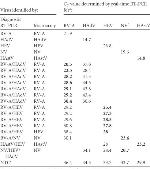
TABLE 2CTvalues for viral nucleic acid quantification in samples with coinfection
Virus identified by:
CTvalue determined by real-time RT-PCR
fora:
Diagnostic
RT-PCR Microarray RV-A HAdV HEV NVb HAstV
RV-A RV-A 21.9
HAdV HAdV 14.7
HEV HEV 23.8
NV NV 19.6
HAstV HAstV 14.8
RV-A/HAdV RV-A 20.5 37.6 RV-A/HAdV RV-A 22.5 28.4 RV-A/HAdV RV-A 28.2 41.3 RV-A/HAdV RV-A 28.6 44.5 RV-A/HAdV RV-A 29.1 43.8 RV-A/HAdV RV-A 29.2 43.4 RV-A/HAdV RV-A 30.4 30.6
RV-A/HEV RV-A 29.2 25.4
RV-A/HEV RV-A 29.2 27.3
RV-A/HEV RV-A 29.6 28.5
RV-A/HEV RV-A 30.8 27.8
RV-A/HEV HEV 38.4 28
RV-A/NV NV 30.1 23.6
HAstV/HEV HAstV 28 23.2
NV/HEV/ HAdV
NV 34.1 28.4 20.7
NTCc 36.4 44.5 33.7 33.7 29.9
aRV-A, rotavirus group A; HAdV, human adenovirus; HEV, human enterovirus; NV,
Norwalk virus; HAstV, human astrovirus. Single-infection samples were used as positive controls. LowerCTvalues are shown in bold.
b
NV is detected at the genus level as calicivirus.
cNTC, nontemplate control.
on May 16, 2020 by guest
http://jcm.asm.org/
with the amplified labeled DNA, regardless of its origin; in other
words, they were found to be “sticky,” and they were excluded
from further analysis. No common characteristic was found
be-tween these probes that could account for their nonspecific
bind-ing behavior.
One of the critical parameters in virus detection is the
sensitiv-ity of the assay. There are several factors that can affect the
sensi-tivity. In the case of a microarray, sample nucleic acids are
gener-ally processed by random-primed amplification prior to
hybridization to ensure amplification of a wide variety of viruses.
The product of random PCR could be lower than that of specific
PCR, decreasing the sensitivity of the assay, as all genetic material
is amplified, diluting the positive signal (
76
). The limit of
detec-tion for three viruses with different genome types (dsDNA,
dsRNA, and ssRNA
⫹
) was established at 10
3virus particles,
sug-gesting that the nature of the genome does not affect the sensitivity
of the assay. Moreover, testing the sensitivity of the microarray
with purified RV-A RNA, we observed that addition of 50 ng of
cellular RNA as a nonspecific diluting RNA decreased the
sensi-tivity of detection 10-fold. To try to solve the sensisensi-tivity problem
in complex clinical samples, agent-specific primers have been
in-cluded in previous studies, together with random primers during
amplification of the genetic material (
14
,
15
), with the
disadvan-tage of narrowing the scope of targets for the microarray assay.
We subsequently analyzed a group of clinical samples collected
from children with diarrhea. Initially, the clinical samples were
screened by SDS-PAGE, which led to the identification of 34
RV-A-positive samples, while the microarray presented in this work
identified 44, suggesting that the microarray platform has a higher
sensitivity than traditional methods. A similar sensitivity was
ob-tained by RT-PCR, as 42 samples were found to be RV-A positive.
Even though our results indicate that the limit of detection of
purified virus (1
⫻
10
3viral particles) is similar to that reached
with PCR assays (
8
), the microarray had a higher number of
pos-itive results when clinical samples were tested, possibly due to the
natural genetic variation in primer binding regions of viruses
found in sample viruses.
Although multiplexed assays are being developed, their use in
routine testing is not generally implemented, and most studies use
single-pathogen tests. When RT-PCR screening for the most
com-mon viruses is performed, the percentage of clinical samples
with-out a virus identified remains around 30 to 50% (
13
,
77
,
78
), while
the microarray presented in this study detected a virus in 92% of
the samples. This high detection rate could have been influenced
by the time of sampling, since winter is a high season for viral
gastroenteritis in the region and no preselection for pathogens was
performed. An additional advantage of the microarray test
com-pared to a set of different RT-PCR assays is the capacity to identify
viruses that are not commonly tested for, such as those previously
associated with diarrhea (like HPeV) and those of unclear clinical
significance in GI disease (HBoV and TTV). In this work we found
a wide range of circulating atypical viruses among children,
simi-larly as observed in other studies (
79
,
80
), and their continuous
surveillance should be considered. To our knowledge, this is first
report of HPeV, HBoV, and TTV in Mexican children.
As a consequence of the limited number of virus species
rou-tinely tested, the prevalence of coinfections is a poorly explored
issue. Usually, when a panel of up to 5 viruses is used, coinfection
rates of between 4 and 18% are observed, with the most common
combination being RV-A/NV (
2
,
13
,
77
,
81
,
82
,
83
). More
re-cently, wide-ranging metagenomic studies have shown that mixed
infections are more common than previously thought (
4
,
80
),
even in healthy individuals (
79
). The analysis of the small set of
clinical samples in this work showed that 30% (23 out of 76)
contained more than 1 gastrointestinal virus. The identification of
individual viruses in coinfections presented some discrepancies
when comparing the results from microarray and RT-PCR tests.
Of seven samples with mixed infections identified by the
microar-ray, five were confirmed by RT-PCR, while in 16 mixed infections
identified by RT-PCR, a single virus was identified by the
microar-ray, suggesting that the microarray may be less sensitive than
RT-PCR for detection of mixed infections. To address this
inconsis-tency, real-time RT-PCR was implemented for the principal
combinations of viruses that were missed by the microarray. This
platform showed a certain advantage for detection of RV-A over
HAdV and HEV, as RV-A was identified even when the HEV
genome was present in larger amounts. HEV was identified by the
microarray in samples coinfected with RV-A only when RV-A
RNA was present in small amounts, close to negative-control
lev-els (
Table 2
). Preferential identification of RV-A by the microarray
could be due to the large amount of virus particles excreted during
the acute phase of infection and to the large number of probes
selected (42 oligonucleotides, compared to 5 and 17 probes for
HEV A and HEV C, respectively, and 17 probes for HAdV). On the
other hand, the two HEV samples positive by microarray that were
missed by RT-PCR correspond to mixed infections with HAstV/
TTV and SV/HpeV/TTV, respectively. Several attempts to identify
HEV in these samples by RT-PCR resulted in negative results, and
thus the possibility of a microarray false-positive result cannot be
discarded.
The number of virus species identified has increased
consider-ably in the last decade with the application of emerging genomic
technologies such as microarrays and unbiased next-generation
sequencing in studies of fatal or rare cases of disease in humans
and in wild and domestic animals (
25
,
56
,
84
,
85
,
86
). Adequate
tools that allow detection of well-known pathogenic viruses while
being capable of detecting the new or rare viruses in a single assay
will contribute useful epidemiological information about both
kinds of viruses. This microarray includes viruses of different host
origins in order to extend the range of use to veterinary studies.
The oligonucleotide probes selected should allow the
identifica-tion of target viruses despite the sequence variaidentifica-tions that will
oc-cur in the future; however, it will be important to update the
microarray design on a regular basis to maintain the capacity to
broadly detect pathogenic viruses and to include newly found viral
species.
Parallel detection of gastroenteric viruses beyond the most
common viruses should facilitate a better understanding of virus
etiology, as it increases the rate of positive cases, closing the
diag-nostic gap, and allows inspection for mixed infections where
sec-ondary viral agents could represent an important factor. Adding
data from case-control studies and inclusion of other host
param-eters, such as serological data, will help to provide evidence of
virus pathogenicity. Furthermore, adequate and comprehensive
epidemiological studies in wild and domestic animals should be
considered.
ACKNOWLEDGMENTS
The computational analysis was performed using the cluster of the Insti-tuto de Biotecnología, UNAM, with the assistance of M. C. Jerome
on May 16, 2020 by guest
http://jcm.asm.org/
leyen. We thank Paul Gaytán Colín, Eugenio López-Bustos, and Santiago Becerra Ramírez from Unidad de Síntesis y Secuenciación de DNA, Insti-tuto de Biotecnología, UNAM, for synthesis of the oligonucleotides used in RT-PCR assays.
This work was supported by grant S0008-111593 from CONACYT. Miguel A. Martínez and María de los Dolores Soto-del Río were supported by a scholarship from CONACYT-Mexico.
REFERENCES
1.WHO.2011. World health statistics 2011. World Health Organization, Geneva, Switzerland.
2.Simpson VR.2002. Wild animals as reservoirs of infectious diseases in the UK. Vet J163:128 –146.http://dx.doi.org/10.1053/tvjl.2001.0662. 3.Wilhelmi I, Roman E, Sanchez-Fauquier A.2003. Viruses causing
gas-troenteritis. Clin Microbiol Infect9:247–262.http://dx.doi.org/10.1046/j .1469-0691.2003.00560.x.
4.Rovida F, Campanini G, Sarasini A, Adzasehoun KM, Piralla A, Bal-danti F.2013. Comparison of immunologic and molecular assays for the diagnosis of gastrointestinal viral infections. Diagn Microbiol Infect Dis
75:110 –111.http://dx.doi.org/10.1016/j.diagmicrobio.2012.09.016. 5.Allard A, Albinsson B, Wadell G.2001. Rapid typing of human
adeno-viruses by a general PCR combined with restriction endonuclease analysis. J Clin Microbiol 39:498 –505. http://dx.doi.org/10.1128/JCM.39.2.498 -505.2001.
6.Farkas T, Zhong WM, Jing Y, Huang PW, Espinosa SM, Martinez N, Morrow AL, Ruiz-Palacios GM, Pickering LK, Jiang X.2004. Genetic diversity among sapoviruses. Arch Virol149:1309 –1323. http://dx.doi .org/10.1007/s00705-004-0296-9.
7.Royuela E, Negredo A, Sanchez-Fauquier A.2006. Development of a one step real-time RT-PCR method for sensitive detection of human astrovirus. J Virol Methods133:14 –19.http://dx.doi.org/10.1016/j.jviromet.2005.10.012. 8.Schwarz BA, Bange R, Vahlenkamp TW, Johne R, Muller H. 2002. Detection and quantitation of group A rotaviruses by competitive and real-time reverse transcription-polymerase chain reaction. J Virol Meth-ods105:277–285.http://dx.doi.org/10.1016/S0166-0934(02)00118-0. 9.Coupland LJ, McElarney I, Meader E, Cowley K, Alcock L, Naunton J,
Gray J.2013. Simultaneous detection of viral and bacterial enteric patho-gens using the Seeplex(R) Diarrhea ACE detection system. Epidemiol In-fect141:2111–2121.http://dx.doi.org/10.1017/S0950268812002622. 10. Khamrin P, Okame M, Thongprachum A, Nantachit N, Nishimura S,
Okitsu S, Maneekarn N, Ushijima H.2011. A single-tube multiplex PCR for rapid detection in feces of 10 viruses causing diarrhea. J Virol Methods
173:390 –393.http://dx.doi.org/10.1016/j.jviromet.2011.02.012. 11. Rohayem J, Berger S, Juretzek T, Herchenroder O, Mogel M, Poppe M,
Henker J, Rethwilm A.2004. A simple and rapid single-step multiplex RT-PCR to detect norovirus, astrovirus and adenovirus in clinical stool samples. J Virol Methods118:49 –59.http://dx.doi.org/10.1016/j.jviromet .2004.01.016.
12. Svraka S, van der Veer B, Duizer E, Dekkers J, Koopmans M, Vennema H.2009. Novel approach for detection of enteric viruses to enable syn-drome surveillance of acute viral gastroenteritis. J Clin Microbiol47:
1674 –1679.http://dx.doi.org/10.1128/JCM.00307-09.
13. van Maarseveen NM, Wessels E, de Brouwer CS, Vossen AC, Claas EC.2010. Diagnosis of viral gastroenteritis by simultaneous detection of adenovirus group F, astrovirus, rotavirus group A, norovirus geno-groups I and II, and sapovirus in two internally controlled multiplex real-time PCR assays. J Clin Virol49:205–210.http://dx.doi.org/10 .1016/j.jcv.2010.07.019.
14. Palacios G, Quan PL, Jabado OJ, Conlan S, Hirschberg DL, Liu Y, Zhai J, Renwick N, Hui J, Hegyi H, Grolla A, Strong JE, Towner JS, Geisbert TW, Jahrling PB, Buchen-Osmond C, Ellerbrok H, San-chez-Seco MP, Lussier Y, Formenty P, Nichol MS, Feldmann H, Briese T, Lipkin WI.2007. Panmicrobial oligonucleotide array for diagnosis of infectious diseases. Emerg Infect Dis13:73– 81.http://dx .doi.org/10.3201/eid1301.060837.
15. Quan PL, Palacios G, Jabado OJ, Conlan S, Hirschberg DL, Pozo F, Jack PJ, Cisterna D, Renwick N, Hui J, Drysdale A, Amos-Ritchie R, Baumeister E, Savy V, Langer KM, Rucht JA, Boyle DB, Garcia-Sastre A, Casas I, Perez-Brena P, Briese T, Lipkin WI. 2007. Detection of respiratory viruses and subtype identification of influenza A viruses by GreeneChipResp oligonucleotide microarray. J Clin Microbiol45:2359 – 2364.http://dx.doi.org/10.1128/JCM.00737-07.
16. Sengupta S, Onodera K, Lai A, Melcher U.2003. Molecular detection and identification of influenza viruses by oligonucleotide microarray hy-bridization. J Clin Microbiol 41:4542– 4550. http://dx.doi.org/10.1128 /JCM.41.10.4542-4550.2003.
17. Xiao-Ping K, Yong-Qiang L, Qing-Ge S, Hong L, Qing-Yu Z, Yin-Hui Y.2009. Development of a consensus microarray method for identifica-tion of some highly pathogenic viruses. J Med Virol81:1945–1950.http: //dx.doi.org/10.1002/jmv.21602.
18. Striebel HM, Birch-Hirschfeld E, Egerer R, Foldes-Papp Z, Tilz GP, Stelzner A.2004. Enhancing sensitivity of human herpes virus diagnosis with DNA microarrays using dendrimers. Exp Mol Pathol77:89 –97.http: //dx.doi.org/10.1016/j.yexmp.2004.05.004.
19. Zhaohui S, Wenling Z, Bao Z, Rong S, Wenli M.2004. Microarrays for the detection of HBV and HDV. J Biochem Mol Biol37:546 –551.http: //dx.doi.org/10.5483/BMBRep.2004.37.5.546.
20. Boriskin YS, Rice PS, Stabler RA, Hinds J, Al-Ghusein H, Vass K, Butcher PD.2004. DNA microarrays for virus detection in cases of central nervous system infection. J Clin Microbiol42:5811–5818.http://dx.doi .org/10.1128/JCM.42.12.5811-5818.2004.
21. Wang D, Coscoy L, Zylberberg M, Avila PC, Boushey HA, Ganem D, DeRisi JL.2002. Microarray-based detection and genotyping of viral pathogens. Proc Natl Acad Sci U S A99:15687–15692.http://dx.doi.org /10.1073/pnas.242579699.
22. Holtz LR, Finkbeiner SR, Zhao G, Kirkwood CD, Girones R, Pipas JM, Wang D.2009. Klassevirus 1, a previously undescribed member of the family Picornaviridae, is globally widespread. Virol J6:86.http://dx.doi .org/10.1186/1743-422X-6-86.
23. Jones MS, II, Harrach B, Ganac RD, Gozum MM, Dela Cruz WP,
Riedel B, Pan C, Delwart EL, Schnurr DP.2007. New adenovirus species found in a patient presenting with gastroenteritis. J Virol81:5978 –5984. http://dx.doi.org/10.1128/JVI.02650-06.
24. Kapoor A, Victoria J, Simmonds P, Wang C, Shafer RW, Nims R, Nielsen O, Delwart E.2008. A highly divergent picornavirus in a marine mammal. J Virol82:311–320.http://dx.doi.org/10.1128/JVI.01240-07. 25. Li L, Victoria J, Kapoor A, Blinkova O, Wang C, Babrzadeh F, Mason
CJ, Pandey P, Triki H, Bahri O, Oderinde BS, Baba MM, Bukbuk DN, Besses JM, Bartkus JM, Delwart EL.2009. A novel picornavirus associ-ated with gastroenteritis. J Virol83:12002–12006.http://dx.doi.org/10 .1128/JVI.01241-09.
26. Rockett RJ, Sloots TP, Bowes S, O’Neill N, Ye S, Robson J, Whiley DM, Lambert SB, Wang D, Nissen MD, Bialasiewicz S.2013. Detection of novel polyomaviruses, TSPyV, HPyV6, HPyV7, HPyV9 and MWPyV in feces, urine, blood, respiratory swabs and cerebrospinal fluid. PLoS One
8:e62764.http://dx.doi.org/10.1371/journal.pone.0062764.
27. Zarate S, Espinosa R, Romero P, Guerrero CA, Arias CF, Lopez S.2000. Integrin alpha2beta1 mediates the cell attachment of the rotavirus neur-aminidase-resistant variant nar3. Virology278:50 –54.http://dx.doi.org /10.1006/viro.2000.0660.
28. Li W, Godzik A.2006. Cd-hit: a fast program for clustering and compar-ing large sets of protein or nucleotide sequences. Bioinformatics22:1658 – 1659.http://dx.doi.org/10.1093/bioinformatics/btl158.
29. Paulin LF, Soto-Del Rio Mde L, Sanchez I, Hernandez J, Gutierrez-Rios RM, Lopez-Martinez I, Wong-Chew RM, Parissi-Crivelli A, Isa P, Lopez S, Arias CF.2014. PhyloFlu, a DNA microarray for determining the phylogenetic origin of influenza A virus gene segments and the genomic fingerprint of viral strains. J Clin Microbiol52:803– 813.http: //dx.doi.org/10.1128/JCM.03134-13.
30. He Z, Wu L, Fields MW, Zhou J.2005. Use of microarrays with different probe sizes for monitoring gene expression. Appl Environ Microbiol71:
5154 –5162.http://dx.doi.org/10.1128/AEM.71.9.5154-5162.2005. 31. Hughes TR, Mao M, Jones AR, Burchard J, Marton MJ, Shannon KW,
Lefkowitz SM, Ziman M, Schelter JM, Meyer MR, Kobayashi S, Davis C, Dai H, He YD, Stephaniants SB, Cavet G, Walker WL, West A, Cofrey E, Shoemaker DD, Stoughton R, Blanchard AP, Friend SH, Linsey PS.2001. Expression profiling using microarrays fabricated by an ink-jet oligonucleotide synthesizer. Nat Biotechnol19:342–347.http://dx .doi.org/10.1038/86730.
32. Wang HY, Malek RL, Kwitek AE, Greene AS, Luu TV, Behbahani B, Frank B, Quackenbush J, Lee NH.2003. Assessing unmodified 70-mer oligonucleotide probe performance on glass-slide microarrays. Genome Biol4:R5.http://dx.doi.org/10.1186/gb-2003-4-1-r5.
33. SantaLucia J, Jr.1998. A unified view of polymer, dumbbell, and
on May 16, 2020 by guest
http://jcm.asm.org/
nucleotide DNA nearest-neighbor thermodynamics. Proc Natl Acad Sci U S A.95:1460 –1465.http://dx.doi.org/10.1073/pnas.95.4.1460.
34. Urisman A, Fischer KF, Chiu CY, Kistler AL, Beck S, Wang D, DeRisi JL.2005. E-Predict: a computational strategy for species identification based on observed DNA microarray hybridization patterns. Genome Biol
6:R78.http://dx.doi.org/10.1186/gb-2005-6-9-r78.
35. Bohlander SK, Espinosa R, III, Le Beau MM, Rowley JD, Diaz MO.
1992. A method for the rapid sequence-independent amplification of mi-crodissected chromosomal material. Genomics13:1322–1324.http://dx .doi.org/10.1016/0888-7543(92)90057-Y.
36. Chen EC, Miller SA, DeRisi JL, Chiu CY.27 April 2011. Using a pan-viral microarray assay (Virochip) to screen clinical samples for viral pathogens. J Vis Exphttp://dx.doi.org/10.3791/2536.
37. Breitling R, Armengaud P, Amtmann A, Herzyk P.2004. Rank products: a simple, yet powerful, new method to detect differentially regulated genes in replicated microarray experiments. FEBS Lett573:83–92.http://dx.doi .org/10.1016/j.febslet.2004.07.055.
38. Strimmer K.2008. fdrtool: a versatile R package for estimating local and tail area-based false discovery rates. Bioinformatics24:1461–1462.http: //dx.doi.org/10.1093/bioinformatics/btn209.
39. R Core Team.2013. R: a language and environment for statistical com-puting. R Foundation for Statistical Computing, Vienna, Austria. 40. Teo J, Di Pietro P, San Biagio F, Capozzoli M, Deng YM, Barr I,
Caldwell N, Ong KL, Sato M, Tan R, Lin R. 2011. VereFlu: an
integrated multiplex RT-PCR and microarray assay for rapid detection and identification of human influenza A and B viruses using lab-on-chip technology. Arch Virol156:1371–1378.http://dx.doi.org/10.1007 /s00705-011-0999-7.
41. Rotbart HA.1990. Enzymatic RNA amplification of the enteroviruses. J Clin Microbiol28:438 – 442.
42. Allander T, Tammi MT, Eriksson M, Bjerkner A, Tiveljung-Lindell A, Andersson B.2005. Cloning of a human parvovirus by molecular screen-ing of respiratory tract samples. Proc Natl Acad Sci U S A102:12891– 12896.http://dx.doi.org/10.1073/pnas.0504666102.
43. Nielsen AC, Gyhrs ML, Nielsen LP, Pedersen C, Bottiger B. 2013. Gastroenteritis and the novel picornaviruses aichi virus, cosavirus, saffold virus, and salivirus in young children. J Clin Virol57:239 –242.http://dx .doi.org/10.1016/j.jcv.2013.03.015.
44. Greninger AL, Runckel C, Chiu CY, Haggerty T, Parsonnet J, Ganem D, DeRisi JL.2009. The complete genome of klassevirus—a novel picornavirus in pediatric stool. Virol J6:82.http://dx.doi.org/10.1186 /1743-422X-6-82.
45. Biagini P, Uch R, Belhouchet M, Attoui H, Cantaloube JF, Brisbarre N, de Micco P.2007. Circular genomes related to anelloviruses identified in human and animal samples by using a combined rolling-circle amplifica-tion/sequence-independent single primer amplification approach. J Gen Virol88:2696 –2701.http://dx.doi.org/10.1099/vir.0.83071-0.
46. Okamoto H, Takahashi M, Nishizawa T, Tawara A, Fukai K, Mura-matsu U, Naito Y, Yoshikawa A.2002. Genomic characterization of TT viruses (TTVs) in pigs, cats and dogs and their relatedness with species-specific TTVs in primates and tupaias. J Gen Virol83:1291–1297. 47. Chu DK, Poon LL, Guan Y, Peiris JS. 2008. Novel astroviruses in
insectivorous bats. J Virol82:9107–9114.http://dx.doi.org/10.1128/JVI .00857-08.
48. Khamrin P, Maneekarn N, Peerakome S, Okitsu S, Mizuguchi M, Ushijima H.2008. Bovine kobuviruses from cattle with diarrhea. Emerg Infect Dis14:985–986.http://dx.doi.org/10.3201/eid1406.070784. 49. Atmar RL, Opekun AR, Gilger MA, Estes MK, Crawford SE, Neill
FH, Graham DY.2008. Norwalk virus shedding after experimental human infection. Emerg Infect Dis14:1553–1557.http://dx.doi.org/10 .3201/eid1410.080117.
50. Lee N, Chan MC, Wong B, Choi KW, Sin W, Lui G, Chan PK, Lai RW, Cockram CS, Sung JJ, Leung WK.2007. Fecal viral concentration and diarrhea in norovirus gastroenteritis. Emerg Infect Dis13:1399 –1401. http://dx.doi.org/10.3201/eid1309.061535.
51. Ramani S, Sankaran P, Arumugam R, Sarkar R, Banerjee I, Mohanty I, Jana AK, Kuruvilla KA, Kang G.2010. Comparison of viral load and duration of virus shedding in symptomatic and asymptomatic neonatal rotavirus infections. J Med Virol82:1803–1807.http://dx.doi.org/10.1002 /jmv.21872.
52. Dennehy PH.2011. Viral gastroenteritis in children. Pediatr Infect Dis J
30:63– 64.http://dx.doi.org/10.1097/INF.0b013e3182059102.
53. Breitbart M, Hewson I, Felts B, Mahaffy JM, Nulton J, Salamon P,
Rohwer F.2003. Metagenomic analyses of an uncultured viral commu-nity from human feces. J Bacteriol185:6220 – 6223.http://dx.doi.org/10 .1128/JB.185.20.6220-6223.2003.
54. Finkbeiner SR, Allred AF, Tarr PI, Klein EJ, Kirkwood CD, Wang D.
2008. Metagenomic analysis of human diarrhea: viral detection and dis-covery. PLoS Pathog4:e1000011.http://dx.doi.org/10.1371/journal.ppat .1000011.
55. Reyes A, Haynes M, Hanson N, Angly FE, Heath AC, Rohwer F,
Gordon JI.2010. Viruses in the faecal microbiota of monozygotic twins and their mothers. Nature466:334 –338.http://dx.doi.org/10 .1038/nature09199.
56. Victoria JG, Kapoor A, Li L, Blinkova O, Slikas B, Wang C, Naeem A, Zaidi S, Delwart E.2009. Metagenomic analyses of viruses in stool sam-ples from children with acute flaccid paralysis. J Virol83:4642– 4651.http: //dx.doi.org/10.1128/JVI.02301-08.
57. Zhang T, Breitbart M, Lee WH, Run JQ, Wei CL, Soh SW, Hibberd ML, Liu ET, Rohwer F, Ruan Y.2006. RNA viral community in human feces: prevalence of plant pathogenic viruses. PLoS Biol4:e3.http://dx.doi.org /10.1371/journal.pbio.0040003.
58. Lin B, Blaney KM, Malanoski AP, Ligler AG, Schnur JM, Metzgar D, Russell KL, Stenger DA.2007. Using a resequencing microarray as a multiple respiratory pathogen detection assay. J Clin Microbiol45:443– 452.http://dx.doi.org/10.1128/JCM.01870-06.
59. Brinkman NE, Fout GS.2009. Development and evaluation of a generic tag array to detect and genotype noroviruses in water. J Virol Methods
156:8 –18.http://dx.doi.org/10.1016/j.jviromet.2008.03.010.
60. Brown DW, Gunning KB, Henry DM, Awdeh ZL, Brinker JP, Tzipori S, Herrmann JE.2008. A DNA oligonucleotide microarray for detecting human astrovirus serotypes. J Virol Methods147:86 –92.http://dx.doi.org /10.1016/j.jviromet.2007.07.028.
61. Chizhikov V, Wagner M, Ivshina A, Hoshino Y, Kapikian AZ, Chuma-kov K.2002. Detection and genotyping of human group A rotaviruses by oligonucleotide microarray hybridization. J Clin Microbiol40:2398 – 2407.http://dx.doi.org/10.1128/JCM.40.7.2398-2407.2002.
62. Lovmar L, Fock C, Espinoza F, Bucardo F, Syvanen AC, Bondeson K.
2003. Microarrays for genotyping human group a rotavirus by multiplex capture and type-specific primer extension. J Clin Microbiol41:5153– 5158.http://dx.doi.org/10.1128/JCM.41.11.5153-5158.2003.
63. Vinje J, Koopmans MP.2000. Simultaneous detection and genotyping of “Norwalk-like viruses” by oligonucleotide array in a reverse line blot hy-bridization format. J Clin Microbiol38:2595–2601.
64. Ayodeji M, Kulka M, Jackson SA, Patel I, Mammel M, Cebula TA, Goswami BB.2009. A microarray based approach for the identification of common foodborne viruses. Open Virol J3:7–20.http://dx.doi.org/10 .2174/1874357900903010007.
65. Chen H, Mammel M, Kulka M, Patel I, Jackson S, Goswami BB.
2011. Detection and identification of common food-borne viruses with a tiling microarray. Open Virol J5:52–59.http://dx.doi.org/10.2174 /1874357901105010052.
66. Chen Q, Li J, Deng Z, Xiong W, Wang Q, Hu YQ.2010. Comprehensive detection and identification of seven animal coronaviruses and human respiratory coronavirus 229E with a microarray hybridization assay. In-tervirology53:95–104.http://dx.doi.org/10.1159/000264199.
67. Honma S, Chizhikov V, Santos N, Tatsumi M, Timenetsky Mdo C, Linhares AC, Mascarenhas JD, Ushijima H, Armah GE, Gentsch JR, Hoshino Y.2007. Development and validation of DNA microarray for genotyping group A rotavirus VP4 (P[4], P[6], P[8], P[9], and P[14]) and VP7 (G1 to G6, G8 to G10, and G12) genes. J Clin Microbiol45:2641–2648 http://dx.doi.org/10.1128/JCM.00736-07.
68. Jaaskelainen A J, Piiparinen H, Lappalainen M, Koskiniemi M, Vaheri A.2006. Multiplex-PCR and oligonucleotide microarray for detection of eight different herpesviruses from clinical specimens. J Clin Virol37:83– 90.http://dx.doi.org/10.1016/j.jcv.2006.05.010.
69. Kim JM, Kim SY, Park YB, Kim HJ, Min BS, Cho JC, Yang JM, Cho YH, Ko G.2012. Simultaneous detection of major enteric viruses using a combimatrix microarray. J Microbiol50:970 –977.http://dx.doi.org/10 .1007/s12275-012-2228-9.
70. Leblanc N, Gantelius J, Schwenk JM, Stahl K, Blomberg J, Andersson-Svahn H, Belak S.2009. Development of a magnetic bead microarray for simultaneous and simple detection of four pestiviruses. J Virol Methods
155:1–9.http://dx.doi.org/10.1016/j.jviromet.2008.04.010.
71. Lin B, Vora GJ, Thach D, Walter E, Metzgar D, Tibbetts C, Stenger DA.
2004. Use of oligonucleotide microarrays for rapid detection and