0095-1137/10/$12.00 doi:10.1128/JCM.00916-09
Copyright © 2010, American Society for Microbiology. All Rights Reserved.
In Situ Molecular Diagnosis and Histopathological Characterization of
Enteroadherent
Enterococcus hirae
Infection in
Pre-Weaning-Age Kittens
䌤
Jodi L. Nicklas,
1Peter Moisan,
2Maria R. Stone,
1and Jody L. Gookin
1*
Department of Clinical Sciences, College of Veterinary Medicine, North Carolina State University, Raleigh, North Carolina 27606,1
and Rollins Animal Disease Diagnostic Laboratory, Raleigh, North Carolina 276992
Received 8 May 2009/Returned for modification 9 February 2010/Accepted 22 May 2010
The bacterial causes of diarrhea can be frustrating to identify, and it is likely that many remain undiagnosed. The pathogenic potential of certain bacteria becomes less ambiguous when they are observed to intimately associate with intestinal epithelial cells. In the present study we sought to retrospectively characterize the
clinical,in situmolecular, and histopathological features of enteroadherent bacteria in seven unrelated kittens
that were presumptively diagnosed with enteropathogenicEscherichia coli(EPEC) on the basis of postmortem
light microscopic and, in some cases, microbiological examination. Characterization of the enteroadherent
bacteria in each case was performed by Gram staining, in situ hybridization using fluorescence-labeled
oligonucleotide probes, PCR amplification of species-specific gene sequences, and ultrastructural imaging applied to formalin-fixed paraffin-embedded sections of intestinal tissue. In only two kittens was EPEC
infection confirmed. In the remaining five kittens, enteroadherent bacteria were identified asEnterococcusspp.
The enterococci were further identified as Enterococcus hirae on the basis of PCR amplification of DNA
extracted from the formalin-fixed, paraffin-embedded tissue and amplified by using species-specific primers.
Transmission electron microscopy of representative lesions fromE. coli- andEnterococcusspp.-infected kittens
revealed coccobacilli adherent to intestinal epithelial cells without effacement of microvilli or cup-and-pedestal formation. Enterococci were not observed, nor were DNA sequences amplified from intestinal tissue obtained from age-matched kittens euthanized for reasons unrelated to intestinal disease. These studies suggest that
E. hiraemay be a common cause of enteroadherent bacterial infection in pre-weaning-age kittens and should be considered in the differential diagnosis of bacterial disease in this population.
Diarrhea is a frequent clinical sign at the time of death or euthanasia of kittens, particularly those residing in shelters or rescue facilities. Enteritis is second only to the specific diag-nosis of feline parvovirus infection as the most common cause of kitten mortality identified by histopathology-based studies (2). Aside from viral, protozoal, and helminthic causes of enteritis, the bacterial culprits of diarrhea are particularly problematic to identify. The intestinal tract harbors a diverse population of bacteria that play critical roles in nutrient assimi-lation, mucosal immunity, and colonization resistance. Recog-nition of pathogenic bacteria within this population is ham-pered by our limited knowledge of normal bacterial diversity, the challenge of distinguishing commensal from pathogenic bacteria, the frequent presence of pathogenic bacteria in clin-ically normal animals, and the ability of commensal bacteria to become pathogenic in genetically susceptible individuals or under abnormal environmental conditions. The pathogenic po-tential of certain bacteria becomes less ambiguous when they are observed to intimately adhere to intestinal epithelial cells. There are few reports of enteroadherent bacterial infection in cats. Most reported cases involve the cultivation from feces
ofEscherichia colithat are molecularly characterized as
con-taining the gene coding for intimin (eae) (10, 11, 16). Bacterial expression of intimin and the translocated intimin receptor (tir) mediates adherence of enteropathogenic and some en-terohemorrhagic strains ofE. colito the intestinal epithelium. This attachment is accompanied by effacement of the epithelial microvilli and the rearrangement of actin to form membranous projections beneath the bacteria resembling a cup and pedes-tal. Attaching and effacing lesions have been described in only two cats; however, images of the lesions were not provided (18).
As a prelude to prospective studies examining the clinical importance of enteropathogenic E. coli(EPEC) infection in kittens, we sought to retrospectively characterize the clinical,in situmolecular, and histopathological features of enteroadher-ent bacteria in seven unrelated kittens that were presumptively diagnosed with EPEC on the basis of light microscopic and, in some cases, microbiological examination. Our unexpected discovery that most of these kittens were infected with en-teroadherentEnterococcus hiraeand notE. colihas impor-tant implications for future selection and interpretation of microbiological tests in these cases.
MATERIALS AND METHODS
Intestinal samples.Formalin-fixed, paraffin-embedded intestinal tissue from seven unrelated kittens submitted to a state animal disease diagnostic laboratory for necropsy examination from 2004 to 2008 were retrospectively obtained. In each case, light microscopic examination of the small intestine revealed mild-to-moderate, subacute-to-acute, necrotizing enterocolitis with extensive coloniza-tion of the small intestinal epithelium by adherent coccobacilli. On the basis of
* Corresponding author. Mailing address: North Carolina State Uni-versity, College of Veterinary Medicine, 4700 Hillsborough Street, Ra-leigh, NC 27606. Phone: (919) 513-6295. Fax: (919) 513-6538. E-mail: Jody_Gookin@ncsu.edu.
䌤Published ahead of print on 2 June 2010.
2814
on May 16, 2020 by guest
http://jcm.asm.org/
these findings a diagnosis of attaching and effacingE. coliinfection was pre-sumed. The kittens ranged in age from 3 to 10 weeks (median age, 6 weeks) and were each being housed in shelter or foster facilities. Clinical signs prior to death or euthanasia included diarrhea and lethargy (four kittens), upper respiratory tract infection (one kitten), sudden death (one kitten), and unknown (one kit-ten). Aerobic bacterial culture of feces was performed at the time of necropsy for four kittens, three of which were positive forE. coli. Genomic DNA extracted from selectedE. colicolonies, was assayed by multiplex PCR for the presence of genes encoding intimin (eae),Shigatoxins (Stx1 and Stx2), heat-stable toxins (STa and STb), heat-labile toxin (LT), or cytotoxic necrotizing factors (CNF-1 and CNF-2) as previously described (9, 23). Two kittens were identified with eae-positive nonhemolyticE. coli, one kitten with CNF-1 and CNF-2-positive beta-hemolyticE. coli, and one kitten was culture-negative forE. coli. Based on the presumption that enterococci were normal flora, neither their presence nor their identity was reported.
For comparative purposes, formalin-fixed, paraffin-embedded intestinal tissues were prospectively obtained from an equal number of age-matched control kittens, euthanized by a local animal control facility for reasons unrelated to intestinal tract disease.
FISH.Formalin-fixed, paraffin-embedded tissue samples were sectioned at a thickness of 4m and mounted on poly-L-lysine-coated slides. The tissue sec-tions were deparaffinized, rehydrated, and air dried prior to hybridization with fluorescencein situhybridization (FISH) probes at a working strength of 5 ng/l (14). The universal bacterial probe Eub338 (14), labeled at the 3⬘end with 6-FAM, was used to identify eubacteria. For subsequent analyses, specific probes directed againstE. coli/Shigella(14) orEnterococcusspp. (Enc221, 5⬘-Cy3-CAC CGCGGGTCCATCCATCA-3⬘) were simultaneously applied. The genus-spe-cificEnterococcussp. probe Enc221 has been demonstrated to have an analytical specificity of 100% when tested against 14 differentEnterococcusspecies and 19 nonenterococcal reference strains (25). Probe specificity was additionally evalu-ated by using a sense probe non-Enc221 (5⬘-6FAM-TGATGGATGGACCCGC GGTG-3⬘) and by including positive and negative control slides in each assay. Positive control slides included formalin-fixed, paraffin-embedded intestinal tis-sue from a pig and dog with culture and multiplex PCR-confirmed eae-positive, enteropathogenicE. coliinfection and a pig with intestinalEnterococcus durans
infection. For the hybridization of Eub338 and Enc221 probes to enterococci, optimal permeabilization was achieved by washing the slides in permeabilization buffer (100 mM Tris-HCl [pH 7.5], 50 mM EDTA) followed by treatment with lysozyme (L6876 [Sigma Chemical Company, St. Louis, MO]; 10 mg/ml in per-meabilization buffer) for 30 min at 37°C in a humidified chamber. Slides were subsequently washed in diethylpyrocarbonate-treated deionized water, air dried, and hybridized as previously described (14).
DNA extraction from formalin-fixed, paraffin-embedded intestinal tissue. In-testinal tissue (⬃80 mg), corresponding in location to the site of microscopically observed enteroadherent bacteria, was excised from the paraffin block of each infected and all control kittens. Between each tissue block, an equivalent amount of paraffin was excised from a block devoid of tissue to assess for the possibility of cross-contamination between samples. The excised tissue samples were added to 200l of ATL buffer (Qiagen, Valencia, CA) and microwaved at 5-s intervals until the paraffin liquefied. The solution was centrifuged (150⫻gfor 10 min), followed by removal of the paraffin ring by using a sterile pipette tip. The remaining solution was vortexed for 5 min (vortex adaptor; MoBio, Carlsbad, CA) in the presence of ZR BashingBeads (Zymo Research, Orange, CA), treated with 100l of lysozyme (40 mg/ml; Sigma), and incubated for 7 h at 37°C. The solution was subsequently treated with proteinase K (20l) and incubated overnight at 56°C, followed by the addition of 200l of AL buffer (Qiagen) and incubation at 55°C for 30 min and at 95°C for 15 min. The remaining protocol, beginning with the addition of 200l of ethanol, was performed as described for the DNeasy blood and tissue kit (Qiagen). Extractions performed concurrently with the feline samples included paraffin wax spiked within vitro-culturedE. hirae
ATCC 8043 (positive control) and extraction reagents alone (negative control). DNA extraction fromE. hiraeandE. duransculture isolates.E. hirae(ATCC 8043) andE. durans(ATCC 6056) type strains were purchased from the Amer-ican Type Culture Collection and then reconstituted and cultured for 24 h at 37°C in brain heart infusion medium. Bacteria were pelleted from 1 ml of broth culture and pretreated with 180l of lysozyme (20 mg/ml in PCR-grade water) at 37°C for 30 min. DNA was extracted by using a commercially available kit (DNeasy blood and tissue kit; Qiagen) in accordance with recommendations for Gram-positive bacteria.
Identification ofE. hiraeandE. durans by PCR amplification of species-specific muramidase gene sequences.All samples of DNA extracted from par-affin-embedded intestinal tissue, as well as paraffin blocks devoid of tissue (neg-ative controls), were first subjected to PCR amplification of an⬃400-bp gene
sequence of feline GAPDH (glyceraldehyde-3-phosphate dehydrogenase) DNA as previously described (12). Subsequent PCR was performed using the species-specific primer pairsmur-2ed(E. durans) andmur-2(E. hirae), which were synthesized (IDT Technologies, Coralville, IA) in accordance with published sequence data (1). Reaction conditions formur-2edandmur-2gene amplification were as follows: a 100-l reaction volume of PCR buffer II (Perkin-Elmer, Foster City, CA) containing 2 U of AmpliTaq Gold DNA polymerase, 2 mM MgCl2, 70 pmol of each primer, 200M each deoxynucleotide triphosphate, and 10l of DNA template. DNA amplification was performed in a Bio-Rad iCycler at the following temperature profiles: initial denaturation at 95°C for 5 min; denatur-ation at 94°C for 1 min, annealing at 55°C for 2 min, and extension at 72°C for 3 min for 50 cycles; followed by a final extension for 10 min at 72°C. Amplicons were visualized by UV illumination after electrophoresis of 10l of the reaction solution in a 1.5% agarose gel containing ethidium bromide. Reaction products were purified (QIAquick PCR purification kit; Qiagen) and sequenced by a commercial laboratory (Davis Sequencing, Davis, CA).
Transmission electron microscopy.Intestinal tissue (⬃80 mg), corresponding in location to the site of microscopically observed enteroadherent bacteria, was cut from the original paraffin block of three kittens. Excess paraffin was trimmed away, and the remaining tissue deparaffinized overnight in xylene. The tissue samples were rehydrated in a graded series of alcohol (100%, twice for 30 min each time; 95%, twice for 15 min each time; 75%, once for 15 min; 50%, once for 15 min), transferred to 0.1 M phosphate buffer (pH 7.2 to 7.4), and fixed in 1% osmium tetroxide for 1 h. Tissue samples were then dehydrated in a graded series of alcohol (50%, once for 15 min; 75%, once for 15 min; 95%, twice for 15 min each time; 100%, twice for 30 min each time), fixed in acetone (2⫻10-min) and embedded in Spurr resin. Ultrathin sections were placed on 200-mesh copper grids, post-stained with uranyl acetate and lead citrate, and examined with a FEI/Philips EM 208S transmission electron microscope.
RESULTS
Light microscopic findings in kittens with enteroadherent
bacterial infection and in control kittens.In the small
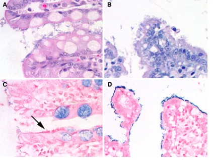
intes-tines of all seven kittens extensive colonies of coccobacilli were present in diplococcal and palisading formations over the lu-minal surface (Fig. 1). In one kitten the colon was also in-volved. The organisms were tightly adherent to enterocytes of the villous tips, as well as to necrotic enterocytes within the luminal detritus. Affected cells were shrunken, hypereosino-philic, and often contained pyknotic nuclei. Within the super-ficial and deep lamina propria only occasional neutrophils and small numbers of lymphocytes were observed. In three kittens concurrent intestinal pathogens were identified, including
Tritrichomonas foetus, panleukopenia virus, and adenovirus (1
kitten each). Enteroadherent bacteria were not observed in intestinal tissue samples obtained from seven age-matched control kittens euthanized for reasons unrelated to gastroin-testinal disease.
FISH using eubacterial andE. coli/Shigella-specific
oligonu-cleotide probes.Because each kitten had been presumptively
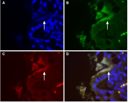
diagnosed with enteroadherentE. coliinfection on the basis of light microscopy, in situ hybridization was initially per-formed using individual and simultaneously applied eubac-terial (Eub338) and E. coli/Shigella-specific oligonucleotide probes. Hybridization of both probes to the enteroadherent bacteria was observed for only two kittens (Fig. 2). Identical results were obtained with intestinal tissue from a dog and pig diagnosed with eae-positiveE. coliinfection. For the remain-ing five kittens, neither probe hybridized to the enteroadherent bacteria.E. coliwas also not observed by FISH in small intes-tinal tissues from age-matched control kittens, with the excep-tion of one case in which a fewE. coliorganisms were observed in the intestinal lumen.
on May 16, 2020 by guest
http://jcm.asm.org/
Characterization of enteroadherent bacteria by tissue Gram
staining.To determine whether failure of probes to hybridize
to enteroadherent bacteria in the remaining five kittens could be attributed to the presence of Gram-positive organisms, in-testinal tissue from all kittens were subjected to Gram staining. Enteroadherent bacteria in the two kittens for which positive hybridization with eubacterial andE. coli/Shigellaprobes were observed stained negative by Gram staining. In the remaining five kittens in which neither probe hybridized, enteroadherent bacteria stained Gram positive (Fig. 1). Only small numbers of Gram-negative rods and rare Gram-positive coccobacilli were observed in the small intestinal lumens of control kittens.
FISH using eubacterial and Enterococcus-specific
oligonu-cleotide probes.To determine the genus of Gram-positive
bac-teria responsible for enteroadherent infection in the remaining five kittens,in situhybridization was performed using individ-ual and simultaneously applied eubacterial (Eub338) and
En-terococcus sp.-specific oligonucleotide probes after
pretreat-ment of tissue sections with lysozyme. Hybridization of both eubacterial andEnterococcusprobes were observed in the five kittens with enteroadherent Gram-positive bacterial infection (Fig. 3) and in intestinal tissue from a pig with E. durans
infection. In the two kittens with enteroadherent
Gram-nega-tive E. coli infection, hybridization of only the eubacterial
probe was observed. Positive hybridization was not observed
when Gram-positive bacteria were probed with a negative con-trol, fluorescence-labeled sense oligonucleotide for the
Entero-coccusprobe or the E. coli/Shigellaprobe. Small numbers of
enterococci were observed in the small intestinal lumens of two age-matched control kittens.
Identification of enteroadherent bacteria asE. hirae.Both
E. hiraeand E. duranshave been variably cultured from the
feces of puppies, calves, foals, piglets, and suckling rats having diarrhea associated with intestinal epithelial colonization by Gram-positive bacteria (3, 4, 6–8, 17, 20, 22, 24). In none of these cases however, were the enteroadherent bacteria defin-itively identified as enterococci. To unambiguously determine the identity of the enteroadherent organisms in the kittens examined in the present study, infected sections of intestine were selectively excised from each paraffin block for DNA extraction and PCR amplification using E. durans- and E.
hirae-specific primers. Robust amplification of a 400-bp
[image:3.585.82.505.69.386.2]frag-ment of the feline GAPDH gene from all blocks containing tissue demonstrated that quality DNA was extracted from each sample. Subsequent PCR performed on DNA extracted from intestinal tissues of four of the five kittens with enteroadherent enterococci resulted in amplification of a 521-bp product cor-responding in size to the target sequence of theE. hirae muram-idase gene. Sequence analysis of the PCR amplicons revealed 99% sequence identity to that of E. hirae (accession no.
FIG. 1. Light microscopic appearance of the small intestinal epithelium from a kitten with enteroadherentE. coli(A) and enteroadherent Enterococcus(B) infection after staining with hematoxylin and eosin. Note the similarity in the histological appearance of the bacteria. After Gram staining of the tissue sections, bacteria from a cat withE. colistain negative (C; arrow), whereas those from the cat withEnterococcus
stain positive (D).
on May 16, 2020 by guest
http://jcm.asm.org/
M77639). Amplification of products corresponding in size to the target sequence of theE. duransmuramidase gene homo-logue (177 bp) was not observed for any samples. Further, neitherE. hiraeorE. durans gene sequences were amplified from paraffin blocks serving as extraction controls, the paraffin block of a kitten infected with enteroadherentE. coli, or those of the control kittens in which enteroadherent bacteria were not identified.
Transmission electron microscopy.To characterize the
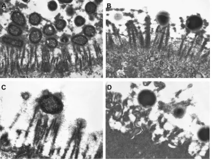
ul-trastructural relationship ofE. hiraewith the intestinal epithe-lium and compare these findings to those of enteroadherentE. coli, representative lesions fromE. coli- andE. hirae-infected kittens and anE. coli-infected pig were excised from paraffin-embedded intestinal tissue, processed, and examined by trans-mission electron microscopy. The ultrastructural relationship between intestinal epithelial cells and enteroadherentE. colior
E. hiraewere similar (Fig. 4). In both infections bacteria were
observed to intimately attach to the brush border of the mi-crovillus tips. In the case of E. hirae, this attachment was associated with fine electron-dense filaments, which radiated from the surface of the cell wall to microvilli and adjacent
bacteria. In neither case was classic cup-and-pedestal forma-tion or effacement of the microvillus architecture observed.
DISCUSSION
Enterococci are motile, Gram-positive bacteria that reside in the feces of most healthy humans and animals, where they are considered to be members of the normal intestinal flora. In fact, recent analysis of microbial diversity of the feline intestine using 16S rRNA gene sequence analysis identified
Entero-coccus spp. as one of the predominant bacterial species
isolated from the jejuna, colons, and feces of healthy young-adult cats (19). Gram-positive cocci, includingEnterococcus
[image:4.585.79.503.69.405.2]spp., are rarely implicated as a cause of diarrhea. Therefore, it is not surprising that efforts were not taken to specifically culture or identify enterococci from feces of these kittens at the time of necropsy. Particular members of the enterococci, however, have been sporadically implicated as a cause of diar-rhea in suckling animals of several host species, including pig-lets, calves, foals, rats, puppies, and a kitten (3, 4, 6–8, 17, 20, 22, 24). In these cases, light microscopic examination of the
FIG. 2. Fluorescencein situhybridization (FISH) of the small intestinal epithelium from a kitten with enteroadherentE. coliinfection. (A) The nuclear counterstain, DAPI (4⬘,6⬘-diamidino-2-phenylindole), enables visualization of intestinal epithelial and lamina propria nuclei. Nucleic acid within the enteroadherent bacteria (arrow) can be seen in the lumen along the junction of two intestinal villi. (B) FISH using the eubacterial probe Eub338 labeled with FAM (green fluorescence) identifies the bacteria as eubacteria. (C) FISH using anE. coli/Shigella-specific probe labeled with Cy3 (red fluorescence) identifies the eubacteria asE. coli/Shigella. (D) Merged image demonstrating the simultaneous hybridization (light yellow) of enteroadherent bacteria with eubacterial andE. coli/Shigella-specific oligonucleotide probes.
on May 16, 2020 by guest
http://jcm.asm.org/
small intestine revealed the adhesion of Gram-positive cocci to the apical surface of enterocytes in association with cul-tivation from feces ofE. duransorE. hirae. However, in no case were the adherent bacteria specifically identified as
Enterococcusspp.
Identification of the enteroadherent bacteria as enterococci in the kittens described here was unexpected. In each case,E. coliinfection was presumed on the basis of light microscopic examination of intestinal tissue, and in three cases this was supported by culture from feces of a necrotoxigenic or attach-ing and effacattach-ing strain ofE. coli. In fact, the light microscopic appearance of E. coli versusEnterococcus infection in these kittens was virtually indistinguishable. It is unclear why a Gram
stain was not initially considered in the diagnostic approach to these cases, other than the fact that enteroadherent Enterococ-cusspp. have only rarely been reported in cats (13, 17). Due to our false presumption that the adherent bacteria wereE. coli, we did not consider an alternate identity for the organisms until they failed to hybridize to the eubacterium-specific oligo-nucleotide probe Eub338 by FISH. Treatment of tissue sec-tions with the Gram-positive cell wall lytic enzyme, lysozyme, was required for successful hybridization of oligonucleotide probes to the enterococci. By using FISH, our studies unam-biguously demonstrated that the adherent bacteria belong to the genusEnterococcus. Further, their species was identified as
E. hiraeon the basis of PCR amplification of DNA extracted
directly from the site of intestinal lesions. This approach is superior to the historical identification of enteroadherent bac-teria on the basis of fecal culture, particularly becauseE. hirae
is second only to E. faecalisas the most common species of
Enterococcusisolated from the feces of clinically normal cats
(5). Given its high prevalence in normal cats, the use of fecal culture for diagnosis of clinical disease caused byE. hiraeis likely to be problematic. Likewise, presumptive identification of enteroadherent bacteria on the basis of positive culture results forE. colican also be misleading because a necrotoxi-genic and attaching and effacing strain ofE. coliwas cultured from the feces of two cats with enteroadherentE. hirae infec-tion in the present study.
The most characteristic feature of enteroadherent entero-coccal infection was an extensive colonization of small intesti-nal villous epithelial cells by Gram-positive bacteria. The abil-ity to adhere to the intestinal epithelium is reported to be a virulence attribute of only some strains of enterococci,
includ-ingE. hirae,E. durans,E. villorum, andE. faecium(7, 8, 13, 24).
Under stressful growth conditions enterococci have been dem-onstrated to produce binding proteins that mediate adhesion to the intestinal epithelium (21). Ultrastructurally, this adhe-sion was characterized by the presence of fine filaments, a finding consistent with fimbriae, that radiated from the surface of the cocci to the microvillus brush border, as well to adjacent bacteria (4, 6, 20). There was no evidence in the present or prior cases that adherence of enterococci results in microvillus effacement or cup and pedestal formation, as is characteristic of attaching and effacing E. coli (4, 6, 17, 20). Enterococci appear to be sufficient to mediate diarrhea in suckling foals, gnotobiotic piglets, and rat pups experimentally infected with
E. hiraecultured from the feces of animals with
enteroadher-ent bacteria and diarrhea (6–8, 22). However, the mechanisms by which enteroadherent enterococci cause diarrhea remain unclear. Villous architecture is invariably preserved, subepi-thelial inflammation is minimal in infected animals (4, 6–8, 13, 15, 17), and enterotoxin secretion has not been demonstrated (6, 22). Infected animals have significantly decreased small intestinal lactase and alkaline phosphatase activities, suggest-ing that the diarrhea may be malabsorptive (22). Little to no research aimed at further clarifying the pathogenesis of en-terococcal diarrhea has been performed in the last 2 decades. Whether E. hirae was the primary or contributing cause of diarrhea, death, or euthanasia in the kittens reported here is unknown. The invasive potential of E. hirae is suggested by prior reports wherein cats with enteroadherent infection and diarrhea succumbed to acute death or terminal bacteremia and
FIG. 3. FISH of small intestinal villous epithelium from kittens with enteroadherentEnterococcusinfection. Merged images demon-strate the simultaneous hybridization of enteroadherent bacteria with DAPI (blue) eubacterial (green) andEnterococcus-specific oligonucle-otide probes (red), which generate an orange fluorescence.
on May 16, 2020 by guest
http://jcm.asm.org/
septic shock (13, 17). In dog pups with enteroadherentE. hirae
infection, cocci were observed within intestinal epithelial cell lysosomes (4, 6). Commonalities between the kittens reported here and prior descriptions of enteroadherent Enterococcus
infection suggest that young, suckling animals are predisposed to conditions capable of promoting pathogenicity of these normally commensal bacteria (3, 4, 6–8, 17, 20, 22, 24). These conditions could include immaturity of immune func-tion or failure of passive transfer, impaired colonizafunc-tion resistance, stressful housing conditions, concurrent infec-tious disease as identified in several of the cats reported here, or antibiotic administration.
Although the prevalence of enteroadherentE. hirae infec-tion in pre-weaning-age kittens is unknown, kitten mortality in association with diarrhea is common in shelter and rescue facilities, and necropsy examinations are seldom requested. Based on results of the present study, it appears thatE. hirae
may be a common cause of enteroadherent bacterial infection in kittens and easily misdiagnosed asE. coliif based on light microscopic examination alone. Consequently, we advocate the routine use of a Gram stain as the first discriminatory test to diagnosis of enteroadherent bacterial infection in kittens.
ACKNOWLEDGMENTS
This study was supported by a grant from the Winn Feline Founda-tion.
We thank Stacy Robinson and Jennifer Haugland for case material and Kenny Simpson for advice with this project. Transmission electron microscopy was performed by the Laboratory for Advanced Electron and Light Optical Methods, College of Veterinary Medicine, North Carolina State University.
REFERENCES
1.Arias, C. A., B. Robredo, K. V. Singh, C. Torres, D. Panesso, and B. E. Murray.2006. Rapid identification ofEnterococcus hiraeandEnterococcus duransby PCR and detection of a homologue of theE. hirae mur-2gene in
E. durans. J. Clin. Microbiol.44:1567–1570.
2.Cave, T. A., H. Thompson, S. W. Reid, D. R. Hodgson, and D. D. Addie.2002. Kitten mortality in the United Kingdom: a retrospective analysis of 274 histopathological examinations (1986 to 2000). Vet. Rec.151:497–501. 3.Cheon, D. S., and C. Chae.1996. Outbreak of diarrhea associated with
Enterococcus duransin piglets. J. Vet. Diagn. Invest.8:123–124.
4.Collins, J. E., M. E. Bergeland, C. J. Lindeman, and J. R. Duimstra.1988.
Enterococcus(Streptococcus)duransadherence in the small intestine of a diarrheic pup. Vet. Pathol.25:396–398.
5.Devriese, L. A., J. I. Cruz Colque, P. De Herdt, and F. Haesebrouck.1992. Identification and composition of the tonsillar and anal enterococcal and streptococcal flora of dogs and cats. J. Appl. Bacteriol.73:421–425. 6.Devriese, L. A., and F. Haesebrouck.1991.Enterococcus hiraein different
animal species. Vet. Rec.129:391–392.
7.Etheridge, M. E., and S. L. Vonderfecht.1992. Diarrhea caused by a slow-growingEnterococcus-like agent in neonatal rats. Lab. Anim. Sci.42:548– 550.
8.Etheridge, M. E., R. H. Yolken, and S. L. Vonderfecht.1988.Enterococcus hiraeimplicated as a cause of diarrhea in suckling rats. J. Clin. Microbiol. 26:1741–1744.
9.Franck, S. M., B. T. Bosworth, and H. W. Moon.1998. Multiplex PCR for enterotoxigenic, attaching and effacing, and Shiga toxin-producing Esche-richia colistrains from calves. J. Clin. Microbiol.36:1795–1797.
10.Goffaux, F., B. China, L. Janssen, and J. Mainil.2000. Genotypic
charac-FIG. 4. Transmission electron micrographs of feline small intestinal epithelium from kittens naturally infected with enteropathogenicE. coli(A and C) or enteroadherentE. hirae(B and D). Interactions between the bacteria and the microvillus brush border of the epithelial cells are similar
in both infections.
on May 16, 2020 by guest
http://jcm.asm.org/
[image:6.585.83.502.69.387.2]terization of enteropathogenicEscherichia coli(EPEC) isolated in Belgium from dogs and cats. Res. Microbiol.151:865–871.
11.Goffaux, F., B. China, L. Janssen, V. Pirson, and J. Mainil.1999. The locus for enterocyte effacement (LEE) of enteropathogenic Escherichia coli
(EPEC) from dogs and cats. Adv. Exp. Med. Biol.473:129–136.
12.Gray, S. G., S. A. Hunter, M. R. Stone, and J. L. Gookin.Assessment of reproductive tract disease in cats at risk forTritrichomonas foetusinfection. Am. J. Vet. Res.71:76–81.
13.Helie, P., and R. Higgins.1999. Diarrhea associated withEnterococcus fae-ciumin an adult cat. J. Vet. Diagn. Invest.11:457–458.
14.Janeczko, S., D. Atwater, E. Bogel, A. Greiter-Wilke, A. Gerold, M. Baum-gart, H. Bender, P. L. McDonough, S. P. McDonough, R. E. Goldstein, and K. W. Simpson.2008. The relationship of mucosal bacteria to duodenal histopathology, cytokine mRNA, and clinical disease activity in cats with inflammatory bowel disease. Vet. Microbiol.128:178–193.
15.Jergens, A. E., F. M. Moore, J. C. Prueter, and P. J. Yankauskas.1991. Adherent gram-positive cocci on the intestinal villi of two dogs with gastro-intestinal disease. J. Am. Vet. Med. Assoc.198:1950–1952.
16.Krause, G., S. Zimmermann, and L. Beutin.2005. Investigation of domestic animals and pets as a reservoir for intimin (eae) gene-positiveEscherichia colitypes. Vet. Microbiol.106:87–95.
17.Lapointe, J. M., R. Higgins, N. Barrette, and S. Milette.2000.Enterococcus hiraeenteropathy with ascending cholangitis and pancreatitis in a kitten. Vet. Pathol.37:282–284.
18.Pospischil, A., J. G. Mainil, G. Baljer, and H. W. Moon.1987. Attaching and effacing bacteria in the intestines of calves and cats with diarrhea. Vet. Pathol.24:330–334.
19.Ritchie, L. E., J. M. Steiner, and J. S. Suchodolski.2008. Assessment of microbial diversity along the feline intestinal tract using 16S rRNA gene analysis. FEMS Microbiol. Ecol.66:590–598.
20.Rogers, D. G., D. H. Zeman, and E. D. Erickson.1992. Diarrhea associated withEnterococcus duransin calves. J. Vet. Diagn. Invest.4:471–472. 21.Styriak, I., A. Laukova, V. Strompfova, and A. Ljungh. 2004. Mode of
binding of fibrinogen, fibronectin and iron-binding proteins by animal en-terococci. Vet. Res. Commun.28:587–598.
22.Tzipori, S., J. Hayes, L. Sims, and M. Withers.1984.Streptococcus durans: an unexpected enteropathogen of foals. J. Infect. Dis.150:589–593. 23.Van Bost, S., E. Jacquemin, E. Oswald, and J. Mainil.2003. Multiplex PCRs
for identification of necrotoxigenicEscherichia coli. J. Clin. Microbiol.41: 4480–4482.
24.Vancanneyt, M., C. Snauwaert, I. Cleenwerck, M. Baele, P. Descheemaeker, H. Goossens, B. Pot, P. Vandamme, J. Swings, F. Haesebrouck, and L. A. Devriese.2001.Enterococcus villorumsp. nov., an enteroadherent bacterium associated with diarrhoea in piglets. Int. J. Syst. Evol. Microbiol.51:393–400. 25.Wellinghausen, N., M. Bartel, A. Essig, and S. Poppert.2007. Rapid iden-tification of clinically relevantEnterococcusspecies by fluorescence in situ hybridization. J. Clin. Microbiol.45:3424–3426.