0095-1137/10/$12.00
doi:10.1128/JCM.01061-10
Copyright © 2010, American Society for Microbiology. All Rights Reserved.
Prevalence and Diversity of
Borrelia
Species in Ticks That Have
Bitten Humans in Sweden
䌤
Peter Wilhelmsson,
1Linda Fryland,
2Stefan Bo
¨rjesson,
1† Johan Nordgren,
1Sven Bergstro
¨m,
4Jan Ernerudh,
2Pia Forsberg,
3* and Per-Eric Lindgren
1,5Division of Medical Microbiology, Department of Clinical and Experimental Medicine, Linko
¨ping University, Linko
¨ping, Sweden
1;
Division of Clinical Immunology, Department of Clinical and Experimental Medicine, Linko
¨ping University, Linko
¨ping,
Sweden
2; Division of Infectious Diseases, Department of Clinical and Experimental Medicine, Linko
¨ping University,
Linko
¨ping, Sweden
3; Department of Molecular Biology, Umeå University, Umeå, Sweden
4; and
Department of Microbiology, Ryhov County Hospital, Jo
¨nko
¨ping, Sweden
5Received 26 May 2010/Returned for modification 28 July 2010/Accepted 3 September 2010
Members of the genus
Borrelia
are among the most common infectious agents causing tick-borne disease in
humans worldwide. Here, we developed a Light Upon eXtension (LUX) real-time PCR assay that can detect and
quantify
Borrelia
species in ticks that have fed on humans, and we applied the assay to 399 such ticks.
Borrelia
PCR-positive ticks were identified to species level by sequencing the products of conventional PCR performed
using
Borrelia
group-specific primers. There was a 19% prevalence of
Borrelia
spp. in the detached ticks, and
the number of spirochetes per
Borrelia
PCR-positive tick ranged from 2.0
ⴛ
10
2to 4.9
ⴛ
10
5, with a median
of 7.8
ⴛ
10
3spirochetes. Adult ticks had a significantly larger number of spirochetes, with a median of 8.4
ⴛ
10
4compared to the median of nymphs of 4.4
ⴛ
10
4. Adult ticks also exhibited a higher prevalence of
Borrelia
(33%) than nymphs (14%). Among the identified species,
Borrelia afzelii
was found to predominate (61%) and
was followed by
B
.
garinii
(23%),
B
.
valaisiana
(13%),
B
.
burgdorferi
sensu stricto (1%),
B
.
lusitaniae
(1%), and
B
.
miyamotoi
-like (1%). Also, 3% of the ticks were coinfected with multiple strains of
B. afzelii
. Notably, this is
the first report of
B. lusitaniae
being detected in ticks in Sweden. Our LUX real-time PCR assay proved to be
more sensitive than a corresponding TaqMan assay. In conclusion, the novel LUX real-time PCR method is a
rapid and sensitive tool for detection and quantification of
Borrelia
spp. in ticks.
Lyme borreliosis (LB) is the most common tick-borne
dis-ease in humans in Europe (26), and it is caused by spirochetes
belonging to the
Borrelia burgdorferi
sensu lato complex. That
group comprises the species
B. burgdorferi
sensu stricto,
B.
afzelii
, and
B. garinii
, which are usually transmitted by the
vector
Ixodes ricinus
. Furthermore, there have been reports of
B. valaisiana
,
B. lusitaniae
, and
B. spielmanii
being detected in
samples of human skin and cerebrospinal fluid (5, 7, 30), which
suggests that those three species can also give rise to LB. It is
often hard to distinguish the clinical symptoms of LB from
those of other diseases (10), and hence, it can be difficult to
establish a correct diagnosis, especially if the patient is unable
to recall having a tick bite.
Today, diagnosis is based mainly on serological tests,
al-though some PCR-based approaches, such as the TaqMan
real-time PCR assay (3, 12), have been developed to detect
Borrelia
species in clinical samples. Even if real-time PCR is
not yet considered to be a routine method in clinical practice,
it can nonetheless provide valuable information about
Borrelia
infections, with regard to species type and the number of
spi-rochetes present. Additional major advantages of PCR in this
context are its simplicity, sensitivity, robustness, and speed.
Other assays besides the TaqMan assay include a method
based on SYBR green dye chemistry (37) and another using
Light Upon eXtension (LUX) (Invitrogen Corporation).
Com-pared to the SYBR green real-time PCR assay, the LUX assay
offers the benefit of using a self-quenched primer with a
hair-pin loop structure, which makes it more specific; that is, it
entails less unspecific binding and primer-dimer formation.
Furthermore, the fluorophore is attached to the hairpin loop in
the LUX setup, and thus, in contrast to the TaqMan assay, this
PCR technique does not need an internal probe and is
there-fore a better choice if broader specificity is required. The LUX
assay also has the capacity for melting curve analysis, which
offers the possibility of discriminating between PCR products
with different base pair compositions (23) and thereby
reveal-ing false-positive samples.
Ixodes ricinus
has been found in 23 of the 25 provinces in
Sweden (9), but it is most common in the southern and central
parts of the country and along the northeastern coast (14).
Various investigators have described the prevalence and
diver-sity of
Borrelia
in ticks collected in the field in Sweden (4, 8, 9,
14), and to date, five species of these bacteria have been
re-corded:
B. afzelii
,
B. garinii
,
B. valaisiana
,
B. burgdorferi
sensu
stricto, and also one that is closely related to
B. miyamotoi
,
which is known to be associated with relapsing fever.
Accord-ing to the cited studies, the prevalence of
Borrelia
spp. in
Sweden varies between 3% and 23%. However, detection was
not achieved by real-time PCR in those investigations, and
thus, no attempts were made to quantify the
Borrelia
spiro-chetes in the ticks. To our knowledge, no quantification of
* Corresponding author. Mailing address: Department of Clinical
and Experimental Medicine, Linko
¨ping University, SE-581 85 Linko
¨-ping, Sweden. Phone: 46 101031398. Fax: 46 101034764. E-mail: pia
.forsberg@liu.se.
† Present address: Department of Animal Health and Antimicrobial
Strategies, National Veterinary Institute (SVA), Uppsala, Sweden.
䌤
Published ahead of print on 15 September 2010.
4169
on May 16, 2020 by guest
http://jcm.asm.org/
Downloaded from
on May 16, 2020 by guest
http://jcm.asm.org/
Downloaded from
on May 16, 2020 by guest
http://jcm.asm.org/
Borrelia
spirochetes in ticks detached from humans has ever
been performed.
Our aim was to study the prevalence of
Borrelia
and to
quantify
Borrelia
cells in ticks that had fed on humans, and we
developed a LUX real-time PCR assay for that purpose. In
addition, we examined possible geographical differences in
prevalence, and we also studied the temporal and spatial
dis-tribution of
Borrelia
species.
MATERIALS AND METHODS
Study sites and collection of ticks.The ticks analyzed in the present study were also used in an investigation focused on the clinical outcome in the humans involved (L. Fryland, P. Wilhelmsson, P.-E. Lindgren, D. Nyman, C. Ekerfelt, and P. Forsberg, submitted for publication), and more detailed information about collection of the specimens is to be published by the group conducting the latter investigation. In short, we used a total of 399 ticks that had been attached to humans in nine areas in O¨ stergo¨tland County, Sweden, between June 2007 and January 2008. The specimens were obtained from eight primary health care centers (PHCs) located in the towns/communities of Ekholmen, Johannelund, Linghem, Kisa, Ska¨rblacka, So¨derko¨ping, Valdemarsvik, and Åtvidaberg, and some were also acquired from the Department of Infectious Diseases at Linko ¨-ping University Hospital. The ticks were available at those facilities because people had been asked to bring detached ticks to their local PHCs. The subjects also completed a health questionnaire and provided a blood sample during the initial visit made to donate the ticks. A second blood sample was obtained 3 months later, and both samples were analyzed for anti-Borreliaantibodies to determine seroconversion or increase in antibody titer. The ticks that people provided were kept in plastic tubes at room temperature and were transported to the Division of Medical Microbiology, Linko¨ping University, within 3 days. They were photographed to determine species type and developmental stage, based on size and color of the dorsal shield. This study was approved by the Ethics Committee of the Faculty of Medicine, Linko¨ping University (no. M132-06).
DNA extraction from ticks.The ticks were washed in 70% ethanol and then in phosphate-buffered saline (PBS), and they were subsequently sectioned longitu-dinally into two equal parts using a sterile scalpel. One half of each tick was subjected to DNA extraction using a DNeasy blood and tissue kit (Qiagen, Hilden, Germany) and the supplementary protocol designated “purification of total DNA from ticks,” according to the manufacturer’s instructions, which gave 50l of DNA in the supplied elution buffer. The DNA concentration in each sample was determined using a spectrophotometer (NanoDrop ND-1000; Thermo Fisher Scientific, Wilmington, DE). The extracted DNA was stored at ⫺20°C pending further analysis.
Reference bacterial strains and samples used to develop the real-time PCR assay.A panel comprising DNA from 16 bacterial species, three human blood samples, and one human skin surface sample was used to develop a real-time PCR assay as described below. DNA from one strain of each of the following referenceBorreliaspecies served as positive controls:B. burgdorferisensu stricto B31 ATCC 35210,B. afzeliiACA-1 (2),B. gariniiIP90 (18),B. valaisianaVS116 (35),B. japonicaH014 (16),B. duttonii1120 (obtained from the strain collection of Guy Baranton, Institut Pasteur),B. hispanicaCR1 (obtained from the strain collection of Guy Baranton, Institut Pasteur),B. persica(obtained from the strain collection of Eduard Korenberg, Gamaleya Research Institute, Moscow, Russia), B. coriaceae(obtained from the strain collection of Alan G. Barbour, UC Irvine), B. anserina(obtained from the strain collection of Alan G. Barbour, UC Irvine), andB. turicatae(obtained from the strain collection of Alan G. Barbour, UC Irvine). These strains were cultivated for 12 days at 35°C in 8 ml of Barbour-Stoenner-Kelly (BSK) medium supplemented with 9% rabbit serum (Sigma-Aldrich Sweden, Stockholm, Sweden) and then harvested by centrifugation at 8,000⫻gfor 10 min at 20°C. DNA was extracted from the bacterial pellets using a DNeasy blood and tissue kit (Qiagen) according to the instructions of the manufacturer. DNA from one strain of each of the five bacterial species that can be members of human skin flora (i.e.,Escherichia coliC-1467,Staphylococcus aureusATCC 3359,Staphylococcus epidermidisCCUG 21989,Streptococcus pyo-genesCCUG 33061, andPropionibacterium acnesCCUG 1794) was used as a negative control. These strains were cultivated on blood agar plates at 37°C overnight. One colony of each strain was then transferred to LB medium and incubated overnight at 37°C, after which bacterial DNA was isolated using a DNeasy blood and tissue kit (Qiagen) according to the manufacturer’s protocol. DNA from human blood and skin surface samples was used as additional neg-ative controls. The human blood and skin surface samples were collected from
staff at the Division of Medical Microbiology, Linko¨ping University. A skin surface sample was taken by gently scraping a scalpel on the lower arm. An aliquot (200l) of each blood sample and 5 mg of the skin surface sample were used for DNA extraction, which was also done with the DNeasy blood and tissue kit (Qiagen) according to the manufacturer’s protocol.
Design ofBorreliaprimers for real-time PCR and for conventional PCR.All sequences of the 16S rRNA gene available for different strains ofBorreliaspp. in GenBank were obtained from the National Center for Biotechnology Informa-tion (www.ncbi.nlm.nih.gov), and these sequences were aligned using BioEdit software (Tom Hall, Ibis Therapeutics, Carlsbad, CA). The forward primer B16S_FL, 5⬘-GAC TCG TCA AGA CTG ACG CTA AGT C-3⬘, and reverse primer B16S_R, 5⬘-GCA CAC TTA ACA CGT TAG CTT CGG TAC TAA C-3⬘, were designed to target a conserved, 131-bp-longBorrelia-specific region of the 16S rRNA gene (bases in bold at the 5⬘end of the B16S_FL primer corre-spond to additional bases added to create the hairpin loop structure). According to the BLAST search (1), the designed primers matched 100% with the se-quences of strains of the following species:B.burgdorferisensu stricto,B.garinii, B.afzelii,B.valaisiana,B.lusitaniae,B.spielmanii,B.andersonii,B.hispanica, B.miyamotoi,B.turdi,B.parkeri,B.crocidurae,B.tanukii,B.duttonii,B.hermsii, B.theileri,B.persica,B.anserina,B.turicatae,B.turcica,B.japonica,B.coriaceae, B.recurrentis, andB.lonestari. The LUX primer pair was designed and evaluated using OligoAnalyzer 3.0 (Integrated DNA Technologies, Coralville, IA). The forward primer B16S_FL was labeled with the reporter dye 6-carboxyfluorescein (FAM) at the first thymine base from the 3⬘ end (Invitrogen Corporation, Paisley, United Kingdom).
PCR primers for species identification were based on the 5S-23S rRNA inter-genic spacer (IGS). We used the same set of primers as that reported by Postic et al. (28): 5⬘-CTG CGA GTT CGC GGG AGA-3⬘and 5⬘-TCC TAG GCA TTC ACC ATA-3⬘, which amplify a genetically diverse region within the IGS in a conventional PCR assay. To increase the sensitivity of the assay, we applied a nested PCR approach with an additional set of primers designed to target the PCR product obtained from the first amplification: B5S-23S_Fn, 5⬘-GAG TTC GCG GGA GAG TAA G-3⬘, and B5S-23S_Rn, 5⬘-TAG GCA TTC ACC ATA GAC TCT T-3⬘. According to the BLAST search, the designed primers for the 5S-23S IGS matched 100% with sequences of strains belonging to the following Borreliaspecies, all of which are present in Europe (26, 29):B.burgdorferisensu stricto,B.afzelii,B.garinii,B.valaisiana,B. spielmanii, andB.lusitaniae. How-ever, it is not known whether the primers targeting the 5S-23S rRNA IGS can detect theB. miyamotoi-like species, which has previously been found in Sweden (8). All tick samples positive forBorreliain the LUX real-time PCR assay, which failed to produce PCR products with primers targeting the 5S-23S IGS, were instead analyzed with primers targeting the 16S-23S IGS (4): F, 5⬘-GTA TGT TTA GTG AGG GGG GTG-3⬘; R, 5⬘-GGA TCA TAG CTC AGG TGG TTA G-3⬘; Fn, 5⬘-AGG GGG GTG AAG TCG TAA CAA G-3⬘; and Rn, 5⬘-GTC TGA TAA ACC TGA GGT CGG A-3⬘. These primers were employed to detect theB. miyamotoi-like spirochete (4), and, according to the BLAST search, they matched 100% with sequences belonging to the following species:B. miyamotoi, B. burgdorferisensu stricto,B. afzelii,B. garinii,B. recurrentis,B. duttonii,B. turicatae,B. hermsii, andB. japonica.
Optimization of primers designed for detection and quantification ofBorrelia spp. by conventional PCR.Optimization and evaluation of assay specificity were performed using the designed primers B16S_FL (without a fluorophore) and B16S_R in a conventional PCR assay with DNA templates from the reference panel, as described above. Different annealing temperatures (55 to 60°C) were tested. The reaction mixture in the optimized assay (final volume of 50l) contained 5l of 10⫻PCR buffer (Amersham Biosciences, Uppsala, Sweden), 1l of deoxynucleoside triphosphate (dNTP) (10 mM), 1l of each primer (10 M), 0.25l ofTaqDNA polymerase (5 Ul⫺1
; Amersham Biosciences), 5l of template DNA (2 to 4 ngl⫺1), and 36.75l of RNase-free water. The
amplification program comprised 95°C for 2 min, followed by 95°C for 15 s, 58°C for 30 s, 72°C for 30 s in 40 cycles, and finally 72°C for 7 min. The reactions were performed in a PTC-100TM programmable thermal controller (M. J. Research, Inc., Waltham, MA), and products were analyzed by agarose gel electrophoresis. Conventional PCR assays used for species identification.A nested PCR assay was performed to amplify the 5S-23S rRNA IGS for species identification. Specificity of the assay was determined using the same reference panel as that employed to develop the LUX assay described above. The reaction mixture (final volume of 50l) contained the following: 5l of 10⫻PCR buffer (Amersham Biosciences), 1l of dNTP (10 mM), 1l each of the primers targeting the 5S-23S IGS (28) (10M), 0.38l of high-fidelity polymerase (3.5 Ul⫺1
; Amersham Biosciences), 5l of template DNA (2 to 4 ngl⫺1
), and 36.62l of RNase-free water. The amplification program comprised 95°C for 5 min, fol-lowed by 95°C for 15 s, 57°C for 30 s, 39 cycles of 72°C for 30 s, and finally 72°C
on May 16, 2020 by guest
http://jcm.asm.org/
for 7 min. An aliquot (5l) of the PCR product obtained in this assay was added to the second PCR mixture, which was prepared using the same volumes, con-centrations, and amplification program as those used for the first mixture, except with a different primer pair (B5S-23S_Fn and B5S-23S_Rn), and the number of cycles was increased to 42. The nested PCR assay used to amplify the 16S-23S rRNA IGS forB. miyamotoi-like identification was performed as described by Bunikis et al. (4).
All reactions were conducted in a PTC-100TM programmable thermal con-troller (M. J. Research, Inc., Waltham, MA), and PCR products were analyzed by agarose gel electrophoresis.
LUX real-time PCR assay. Each PCR amplification was carried out in a 96-well reaction plate (Applied Biosystems, Warrington, United Kingdom), us-ing a 20-l aliquot of reaction mixture containing the following: 10l of Plati-num qPCR SuperMix uracil-D-glycosylase (UDG) (Invitrogen), 0.04l of Rox reference (Invitrogen), 0.4l of LUX B16S_FL primer (10 M), 0.4l of B16S_R primer (10M; Invitrogen), 4.16l RNase-free water, and 5l of template DNA. Thereafter, the plate was centrifuged at 900⫻gfor 5 min.
The reactions were performed on an ABI PRISM 7500 fast real-time PCR system (Applied Biosystems). The reaction mixture was preheated at 50°C for 2 min (carry-over prevention step, activation of the enzyme UDG) and 95°C for 2 min (denaturation of UDG and activation of PlatinumTaqDNA polymerase) and then subjected to 45 cycles of 95°C for 15 s, 58°C for 30 s, and 72°C for 30 s. Immediately after completion of PCR, melting curve analyses were performed by heating to 95°C for 15 s, followed by cooling to 60°C for 1 min, and subsequent heating to 95°C at 0.8°C min⫺1
with continuous fluorescence recording. The real-time PCR and melting curve results were analyzed using Sequence Detec-tion Software version 1.3.1 (Applied Biosystems).
Determination of the detection/quantification limit and efficiency of the LUX real-time PCR assay.A 10-fold serial dilution ranging from 101to 107gene
copies of theB. burgdorferiB31 was used as a standard to determine the detec-tion limit and efficiency of the real-time PCR assay. The gene copy numbers were calculated by converting the concentration of total double-stranded DNA (dsDNA) ofB. burgdorferiB31 (measured spectrophotometrically) to the num-ber of genome copies based on the molecular weight of the genome. According to Ornstein and Barbour (25), there is a mean of 10 genomes perB. burgdorferi B31 cell and one 16S rRNA gene copy per genome. Therefore, calculations included the assumption that allBorreliaspecies have 10 16S rRNA gene copies per cell.
To check for possible inhibition, we used five ticks in different developmental stages (i.e., five larvae, five nymphs, and five adults), which were collected in the field and kindly provided by Jan Landin, Department of Physics, Chemistry and Biology, Linko¨ping University. These specimens were processed as described above.B. burgdorferiB31 cells cultivated in BSK medium were washed with PBS and counted in a phase-contrast microscope. One half of each tick was spiked with a known number ofB. burgdorferiB31 cells, and the other half served as a negative control forBorrelia. A serial dilution was prepared in PBS to represent Borreliaconcentrations of 104
, 103
, 102
, 101
, and 100
spirochetes per tick sample and then incubated for 30 min at 37°C. The tick samples were subsequently used for DNA extraction as described above. Real-time PCR amplification was per-formed using LUX primer pairs targeting the 16S rRNA gene to verify the efficiency and the quantification limit of the assay.
Validation of the LUX real-time PCR assay.Considering the aspects of sen-sitivity and specificity, we compared the LUX assay with the TaqMan-based real-time PCR method reported by Ornstein and Barbour (25). The primer pair and probe in the TaqMan assay are designed to target the same 136-bp region of the 16S rRNA gene as the primer pair designed for the LUX assay. We also applied an internal control to all extracted tick samples to check for PCR inhibition and thereby prevent false-negative results. A modified real-time PCR assay for the mitochondrial tick housekeeping gene 16SIxodesDNA was run as previously described by Schwaiger and Cassinotti (32). The same primers (F-16sIxodes and R-(F-16sIxodes), but no TaqMan probe, were employed in a SYBR green assay. The PCR amplification was carried out in 96-well reaction plates (Applied Biosystems), and the reaction mixture (20l) contained 10l of FastStart Universal SYBR green Master (ROX) (Roche, Mannheim, Germany), 0.4l of each primer (10M) (Sigma-Aldrich Sweden AB, Stockholm, Sweden), 7.2l of RNase-free water, and 2l of template DNA. The reactions were performed in an ABI Prism 7500 fast real-time PCR system (Applied Biosys-tems) using the same reaction conditions as those described by Schwaiger and Cassinotti (32). Melting analyses of all reactions were performed as reported above.
Nucleotide sequencing of the PCR products and species identification by sequence analysis.Macrogen, Inc. (Seoul, South Korea), performed nucleotide sequencing of the PCR products that we obtained from primers targeting the
following: the 16S rRNA gene, 5S-23S rRNA IGS, 16S-23S rRNA IGS, and the 16SIxodesDNA. The sequencing reactions were based on BigDye chemistry.
In addition to the 5S-23S IGS sequences acquired in this study, IGS sequences fromBorreliaspp. in GenBank were used in the phylogenetic analysis (n⫽41). By including a representative selection of IGS sequences fromBorreliaspp. that are common in and around Europe (i.e.,B. afzelii,B. garinii,B. valaisiana,B. burgdorferisensu stricto, andB. lusitaniae), we were able to identify the species found in our investigation. Sequence alignment was performed using Clustal W2 (European Bioinformatics Institute, Cambridge, United Kingdom). Phylogenetic analyses were conducted usingMEGAversion 4 (17, 31), and the phylogenetic tree was constructed by applying neighbor-joining and Kimura-2-parameter methods with pairwise deletion. The significance of the relationship was ascer-tained by bootstrap analysis (500 replicates).
Cloning of PCR products of 5S-23S IGS from ticks carrying more than one Borreliastrain.PCR products containing differentBorreliasequences, deter-mined as dual peaks in the sequences analysis, were separated by cloning of the PCR products from amplification of the 5S-23S IGS. The PCR products were purified using a GeneJET PCR purification kit (Fermentas, Glen Burnie, MD) according to the manufacturer’s protocol. For bacterial transformation and clon-ing procedures, a TransformAid bacterial transformation kit and a CloneJET PCR cloning kit (both from Fermentas) were used as stipulated in the protocols provided by the manufacturer. DNA was extracted from transformants with plasmids containing PCR products as inserts and purified using a GeneJET plasmid miniprep kit (Fermentas) according to the manufacturer’s instructions. Sequencing of the inserted PCR products was performed by Macrogen, Inc. (Seoul, South Korea).
Statistical analysis.Fisher’s exact test and chi-square test were applied to compare the distribution ofBorreliaPCR-positive ticks andBorreliaspecies among the different PHCs (i.e., geographic sampling locations). The Mann-Whitney test was used to compare adults and nymphs, as well as different months, regarding the number ofBorreliaspirochetes per tick. Median values and 95% confidence intervals were determined. Statistical analyses were per-formed, and graphs were drawn using GraphPad Prism version 5.00 for Windows (GraphPad Software, San Diego, CA). AllPvalues of⬍0.05 were considered significant.
Nucleotide sequence accession number.Sequences obtained in this investiga-tion have been deposited in GenBank with accession numbers HM173532 to HM173598.
RESULTS
Development of a broad-range and sensitive LUX real-time
PCR assay.
We designed a LUX real-time PCR primer pair to
target a highly conserved 131-bp region of the 16S rRNA gene.
Without a fluorophore and at a primer annealing temperature
of 58°C, this pair could detect all of the tested
Borrelia
refer-ence strains, as indicated by conventional PCR followed by
sequence analysis of the PCR products (data not shown). As
expected, when analyzing the DNA samples that served as
negative controls, human blood, human skin surface,
E. coli
,
S.
aureus
,
S. epidermidis
,
S. pyogenes
, and
P. acnes
, no PCR
prod-ucts were detected.
Five independent LUX real-time PCR runs, including a
10-fold serial dilution of gene copies from
B. burgdorferi
B31,
were performed, exhibiting a dynamic range in the interval of
10
1to 10
7gene copies per reaction. The slope as a mean of the
standard curves was
⫺
3.64
⫾
0.08 (
r
2⫽
0.99). The melting
temperature of the PCR products was 80.3
⫾
0.2°C. Using the
known copy numbers of reference DNA, the lower
quantifica-tion limit was 10
1gene copies per PCR. However, it was
pos-sible to detect, but not to accurately quantify, fewer than 10
1gene copies. Ten gene copies of the 16S rRNA is equivalent to
the number of copies that exists in one
Borrelia
cell (25). The
lower quantification limit was similar in the PCR assay using
the
Borrelia
spiked tick samples; thus, no inhibition of the PCR
amplification was detected. The assay did not show any
unspe-cific binding or primer-dimer formation. The primers targeting
on May 16, 2020 by guest
http://jcm.asm.org/
the 5S-23S IGS were able to amplify PCR products from all the
Borrelia
strains used as references.
Borrelia in every fifth tick detected with a novel, sensitive
LUX real-time PCR assay.
All 399 ticks that we analyzed were
identified as
I. ricinus
; 101 (25.3%) were adult females, 296
(74.2%) were nymphs, and two (0.5%) were in the larval stage
(Table 1). The LUX-based real-time PCR assay showed the
presence of
Borrelia
spp. in 75 ticks (19%; Table 1), whereas
the TaqMan assay detected
Borrelia
in only 72 of those 75 and
in no other ticks (data not shown). No obvious seasonal trend
in the number of
Borrelia
-positive ticks was detected during the
collection period.
The SYBR green real-time PCR assay detected the
tick-specific extraction control 16S
Ixodes
DNA in all samples (data
not shown). The threshold cycle (
C
T) value range for this DNA
was 11 to 23 (median of 16) for adult ticks and 14 to 26
(median of 18) for nymph ticks.
Higher number of
Borrelia
cells in adults than in nymphs.
According to the LUX real-time PCR assay, the number of
Borrelia
cells per
Borrelia
PCR-positive tick ranged from 2.0
⫻
10
2to 4.9
⫻
10
5(Fig. 1), with a median of 7.8
⫻
10
3. The
number of
Borrelia
cells in adults ranged from 6.0
⫻
10
2to
4.9
⫻
10
5and in nymphs from 2.0
⫻
10
2to 7.0
⫻
10
4. The
number of
Borrelia
cells was significantly higher in adult ticks
than in nymphs (median per tick of 8.4
⫻
10
3versus 4.4
⫻
10
3).
Furthermore, the prevalence of
Borrelia
was greater in adult
ticks than in nymphs, and no
Borrelia
was detected in larvae.
The prevalence varied from 11% to 33% in the ticks obtained
at the PHCs in O
¨ stergo¨tland County (data not shown).
Com-parison of the PCR-positive ticks from all collection sites
in-dicated that the prevalence of
Borrelia
was significantly lower
in those from one PHC (Kisa) than in those from the other
PHCs. However, no seasonal trend in
Borrelia
cell number was
observed over the collection period. Moreover, no significant
seasonal difference could be detected for the number of
de-velopmental stages of the ticks provided over the study period.
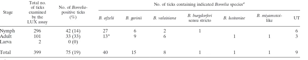
Six different
Borrelia
species detected in the detached ticks.
It was possible to determine the
Borrelia
species in 66 of the 75
ticks that were positive for
Borrelia
in the LUX real-time PCR
(Table 1) using the primer pairs targeting the 5S-23S IGS and
16S-23S IGS regions. Six different species were recorded
(Ta-ble 1), among which
B
.
afzelii
(
n
⫽
40) predominated and was
followed by
B
.
garinii
(
n
⫽
15),
B
.
valaisiana
(
n
⫽
8),
B
.
burgdorferi
sensu stricto (
n
⫽
1),
B
.
lusitaniae
(
n
⫽
1), and
B.
miyamotoi
-like (
n
⫽
1). Notably,
B. lusitaniae
was identified for
[image:4.585.43.546.91.190.2]the first time in Sweden.
B. afzelii
dominated at both the adult
TABLE 1. Prevalence of
Borrelia
species in
I. ricinus
ticks that had been removed from humans and obtained at primary
health care centers in O
¨ stergo¨tland County, Sweden
Stage
Total no. of ticks examined
by the LUX assay
No. of Borrelia-positive ticks
(%)
No. of ticks containing indicatedBorreliaspeciesa
B. afzelii B. garinii B. valaisiana B. burgdorferi
sensu stricto B. lusitaniae
B.
miyamotoi-like UT
Nymph
296
42 (14)
27
6
2
1
6
Adult
101
33 (33)
13*
9
6
1
1
3
Larva
2
0 (0)
Total
399
75 (19)
40
15
8
1
1
1
9
aAbbreviations: UT, untypeable; *, includes the coinfected ticks.
FIG. 1. Total number of ticks, adult ticks (
●
), and nymphs (
⫻
) PCR positive for
Borrelia
plotted against the number of
Borrelia
cells per tick.
Horizontal lines indicate the median, with upper and lower quartiles.
on May 16, 2020 by guest
http://jcm.asm.org/
[image:4.585.135.450.507.705.2](39%) and the nymph (64%) stages. Considering the diversity
of
Borrelia
species in relation to the developmental stages of
the ticks, we found that
B. afzelii
occurred more often in
nymphs than in adults, whereas the opposite pattern was
ob-served for
B. garinii
. Three times more adult ticks (
n
⫽
6) than
nymphs (
n
⫽
2) were positive for
B. valaisiana
. Two adult ticks
were coinfected with different strains of
B. afzelii
(Table 1), and
both of those specimens were obtained at the same PHC
(Åtvidaberg).
B. afzelii
and
B. garinii
were also found in ticks
from all of the collection sites.
Nine LUX real-time PCR-positive samples contained
spe-cies that were not typeable, possibly because the primers
targeting the 5S-23S and 16S-23S IGS do not amplify these
Borrelia
sequences. However, both the LUX and TaqMan
real-time PCR assays successfully amplified the correct length of
PCR products from these nine samples, as confirmed by
elec-trophoresis. Nucleotide sequencing also verified that the LUX
PCR products originated from
Borrelia
.
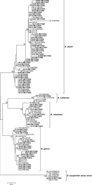
Neighbor-joining was used to construct a phylogenetic tree
based on the 5S-23S rRNA IGS sequences of
Borrelia
species
(Fig. 2). Sixty-seven sequences from the current study (i.e.,
from the coinfected ticks included) and 41 reference sequences
retrieved from GenBank were gathered into clusters. A cluster
represented a group of sequences within the same
Borrelia
species that displayed more than 93% sequence similarity, and
we found that some sequences within the same cluster showed
100% similarity, even though they had disparate origins (e.g.,
the ticks came from different sampling sites). The
B. afzelii
sequences of the two coinfected ticks included in Fig. 2 are
denoted At26A, At26B, At50A, and At50B.
DISCUSSION
Using our new LUX real-time PCR assay, we found that
19% of ticks removed from humans in O
¨ stergo¨tland County,
Sweden, were positive for
Borrelia
. This prevalence is similar to
that observed in a study conducted in the Netherlands (13)
showing that 20.4% of ticks detached from humans were
Bor-relia
positive, whereas it is twice as high as the proportion
detected in an investigation performed in Switzerland (24).
Another study, conducted in Texas (38), found only 1%
Bor-relia
prevalence in ticks removed from humans. In the latter
investigation, analyzed ticks were provided by individuals that
had been bitten in areas where the associated Lyme borreliosis
was considered to be nonendemic. Additionally, in the United
States, only three species of the
B. burgdorferi
sensu lato
com-plex have been described and only one of them is known to be
human pathogenic (36). It should be mentioned that
Borrelia
spirochetes were not quantified in these three studies, because
real-time PCR assays were not used. Furthermore,
Borrelia
species were not determined in the Swiss investigation.
Using indirect immunofluorescence to detect
Borrelia
in
field-collected ticks, Gustafson et al. (9) found positive
speci-mens in 20 of the 23 Swedish provinces where
I. ricinus
was
encountered, with an average prevalence of 10% in nymphs
and 15% in adults. However, the prevalence of
Borrelia
varied
greatly between the provinces, as exemplified by 0% and 13%
found in nymph and adult ticks, respectively, in O
¨ stergo¨tland.
It is plausible that the higher prevalences that we observed
were due to the increased occurrence of
Borrelia
in ticks since
1995. The
Borrelia
prevalence in adults that we recorded (33%)
is also three times higher than that noted by Fraenkel et al. (8)
in a study of field-collected adult ticks from the south and east
coasts of Sweden. This discrepancy might be the result of
dissimilarities in climate and ecosystem conditions, but it may
also be explained by the use of different PCR assays. When a
Borrelia
-positive tick bites a host, dramatic changes occur in
the expression pattern of the
Borrelia
population, seen as rapid
multiplication of the spirochetes in the tick midgut, leading to
the overall higher density of
Borrelia
cells in the tick (27). If the
PCR assay applied is not sensitive enough, a lower density of
Borrelia
spirochetes in field-collected ticks may give
false-neg-ative results.
In our study, the prevalence of
Borrelia
was significantly
higher in adult ticks than in nymphs. This was probably the
case because adult ticks require an extra blood meal from a
host that may be infected with the bacteria, an assumption that
is supported by the results of an investigation of field-collected
ticks conducted in Switzerland in 2004 (15). We observed
geo-graphical differences in
Borrelia
prevalence in O
¨ stergo¨tland
County, and these local disparities may be related to factors
such as the presence/density of reservoir hosts, forest structure,
and types of biotope.
Considering both adult and nymph ticks, we found that
B.
afzelii
was the dominating species in O
¨ stergo¨tland County,
followed by
B. garinii
,
B
,
valaisiana
,
B. burgdorferi
sensu stricto,
B. lusitaniae
, and
B. miyamotoi
-like (Table 1). Furthermore,
B.
afzelii
and
B. garinii
were identified at all collecting sites. Those
two species have also been described to predominate among
ticks detached from humans in the Netherlands (13), and the
same pattern has been seen in field-collected ticks from the
south and east coasts of Sweden (8). Moreover,
B. afzelii
and
B.
garinii
are the most abundant
Borrelia
species in Europe (11).
The diversity of reservoir hosts is likely to have an impact on
the geographic distribution of
Borrelia
species. It is well known
that small mammals (e.g., rodents) frequently serve as
inter-mediate hosts for strains of
B. afzelii
and that strains of
B.
garinii
and
B. valaisiana
are associated with a variety of bird
species (19). The fact that we also identified
B. lusitaniae
for
the first time in Sweden may be important, because it is
pos-sible that some strains of this species give rise to human LB (6).
In addition, there is evidence that
B. lusitaniae
is becoming
established in the northern part of Europe (33).
In our investigation, two adult ticks coinfected with two
different strains of
B. afzelii
were obtained from the same PHC.
In comparison, other studies have shown various prevalences
of coinfections in field-collected ticks: 3% among adult ticks in
England (21), 4% in both adults and nymphs in Switzerland
(15), 14% in nymphs in the United States (34), 64% in nymphs
in Denmark (33), and 16% among adults and nymphs in
Slo-vakia and Poland (22). In the study conducted in SloSlo-vakia and
Poland, 5% of all the positive ticks were coinfected with
dif-ferent strains of one particular species (i.e.,
B. garinii
or
B.
valaisiana
). The discrepancies in the prevalences of
coinfec-tions between our investigation and other studies might be
explained by differences in the transmission pathway, that is,
whether there was host-tick or tick-tick (cofeeding)
transmis-sion (20). Notably, all the coinfected ticks that we found came
from the same area, and there was high sequence similarity
between the
Borrelia
strains that they carried (Fig. 2), which
on May 16, 2020 by guest
http://jcm.asm.org/
FIG. 2. Phylogenetic tree based on the 5S-23S rRNA intergenic spacer region of different
Borrelia
species, constructed by neighbor-joining
using Kimura 2-parameter and pairwise deletion with a bootstrap value of 500 replicates. Strains found in O
¨ stergo¨tland County, Sweden, are shown
in bold. Brackets denote
Borrelia
species clusters with more than 93% sequence similarity. The scale bar corresponds to the expected number of
substitutions per nucleotide site. The reliability of the tree was tested by 500 bootstrap replicate analyses; only values greater than 50% are shown.
The source of each reference sequence is indicated by an accession number preceded by a country code: CZ, Czech Republic; DE, Germany; FR,
France; GB, Great Britain; MA, Morocco; SK, Slovakia; CH, Switzerland; TR, Turkey; RU, Russia.
4174
on May 16, 2020 by guest
seems to suggest closely related transmission pathways (e.g.,
these strains may cocirculate among intermediate hosts in the
area).
The number of
Borrelia
cells ranged from 2.0
⫻
10
2to 4.9
⫻
10
5per tick in our study (Fig. 1), which agrees with the range
and medians found in field-collected nymph and adult ticks in
the northeastern United States (34). We also observed a
sig-nificantly higher number of
Borrelia
cells in adults than in
nymphs. Adults have a larger body volume than nymphs and
can thus be engorged with more host blood, which should allow
faster replication of
Borrelia
, resulting in the detection of
higher numbers of the spirochetes.
Our LUX real-time PCR assay was able to reveal a wide
variety of
Borrelia
species at a detection limit of less than 10
gene copies, which is equivalent to the number of copies that
exists in one
Borrelia
cell (25). Furthermore, compared to a
TaqMan real-time PCR assay (25), our method showed greater
sensitivity, which was seen as detection of more
Borrelia
-posi-tive ticks. Inasmuch as all these samples were determined to
species level, the possibility of false-positive results due to
carryover contamination of PCR products can probably be
excluded. We also noted that the mean amplification efficiency
was higher for the LUX assay than the results previously
re-ported for the TaqMan assay (25), which is important because
such efficiency is a crucial marker of the success of gene
quan-tification. In addition, again compared to the TaqMan assay
(25), our assay gave a lower standard deviation, as calculated
from a set of independent real-time PCR runs. Constant
am-plification efficiency is an important criterion for reliable
com-parison between samples and between real-time PCR runs, as
well as for assay reproducibility.
In summary, we found that approximately 20% of 399 ticks
that had fed on humans in O
¨ stergo¨tland, Sweden, were positive
for
Borrelia
. Six
Borrelia
species were detected, and
B.
lus-itaniae
was identified for the first time in Sweden. These
ob-servations suggest that the novel LUX real-time PCR assay
provides a rapid and sensitive tool for detection and
quantifi-cation of
Borrelia
in ticks. It is also plausible that this assay can
be a valuable tool in clinical diagnostics as a complement to
serological tests.
ACKNOWLEDGMENTS
We are grateful for the enthusiasm and support of staff members at
the primary health care centers in Ekholmen, Johannelund, Linghem,
Kisa, Ska
¨rblacka, So
¨derko
¨ping, Valdemarsvik, and Åtvidaberg and at
the Department of Infectious Diseases, University Hospital, Linko
¨p-ing, Sweden.
Patricia O
¨ dman is acknowledged for comments and linguistic
revi-sion of the manuscript. We also thank Liselott Lindvall and Mari-Anne
Åkeson for excellent specimen collection logistics.
This study was supported by the Medical Research Council of
Southeast Sweden and by ALF funds.
REFERENCES
1.Altschul, S. F., W. Gish, W. Miller, E. W. Myers, and D. J. Lipman.1990. Basic local alignment search tool. J. Mol. Biol.215:403–410.
2.Asbrink, E., A. Hovmark, and B. Hederstedt.1984. The spirochetal etiology of acrodermatitis chronica atrophicans Herxheimer. Acta Derm. Venereol. 64:506–512.
3.Babady, N. E., L. M. Sloan, E. A. Vetter, R. Patel, and M. J. Binnicker.2008. Percent positive rate of Lyme real-time polymerase chain reaction in blood, cerebrospinal fluid, synovial fluid, and tissue. Diagn. Microbiol. Infect. Dis. 62:464–466.
4.Bunikis, J., U. Garpmo, J. Tsao, J. Berglund, D. Fish, and A. G. Barbour.
2004. Sequence typing reveals extensive strain diversity of the Lyme borre-liosis agents Borrelia burgdorferi in North America and Borrelia afzelii in Europe. Microbiology150:1741–1755.
5.Collares-Pereira, M., S. Couceiro, I. Franca, K. Kurtenbach, S. M. Schafer, L. Vitorino, L. Goncalves, S. Baptista, M. L. Vieira, and C. Cunha.2004. First isolation of Borrelia lusitaniae from a human patient. J. Clin. Microbiol. 42:1316–1318.
6.da Franca, I., L. Santos, T. Mesquita, M. Collares-Pereira, S. Baptista, L. Vieira, I. Viana, E. Vale, and C. Prates.2005. Lyme borreliosis in Portugal caused by Borrelia lusitaniae? Clinical report on the first patient with a positive skin isolate. Wien. Klin. Wochenschr.117:429–432.
7.Fingerle, V., U. C. Schulte-Spechtel, E. Ruzic-Sabljic, S. Leonhard, H. Hofmann, K. Weber, K. Pfister, F. Strle, and B. Wilske.2008. Epidemi-ological aspects and molecular characterization of Borrelia burgdorferi s.l. from southern Germany with special respect to the new species Bor-relia spielmanii sp. nov. Int. J. Med. Microbiol.298:279–290.
8.Fraenkel, C. J., U. Garpmo, and J. Berglund.2002. Determination of novel Borrelia genospecies in Swedish Ixodes ricinus ticks. J. Clin. Microbiol. 40:3308–3312.
9.Gustafson, R., T. G. Jaenson, A. Gardulf, H. Mejlon, and B. Svenungsson. 1995. Prevalence of Borrelia burgdorferi sensu lato infection in Ixodes rici-nus in Sweden. Scand. J. Infect. Dis.27:597–601.
10.Hengge, U. R., A. Tannapfel, S. K. Tyring, R. Erbel, G. Arendt, and T. Ruzicka.2003. Lyme borreliosis. Lancet Infect. Dis.3:489–500.
11.Huba´lek, Z., and J. Halouzka.1997. Distribution of Borrelia burgdorferi sensu lato genomic groups in Europe, a review. Eur. J. Epidemiol.13:951– 957.
12.Ivacic, L., K. D. Reed, P. D. Mitchell, and N. Ghebranious.2007. A Light-Cycler TaqMan assay for detection of Borrelia burgdorferi sensu lato in clinical samples. Diagn. Microbiol. Infect. Dis.57:137–143.
13.Jacobs, J. J., G. T. Noordhoek, J. M. Brouwers, P. R. Wielinga, J. P. Jacobs, and A. H. Brandenburg.2008. [Small risk of developing Lyme borreliosis following a tick bite on Ameland: research in a general practice.] Ned. Tijdschr. Geneeskd.152:2022–2026. (In Dutch.)
14.Jaenson, T. G., L. Talleklint, L. Lundqvist, B. Olsen, J. Chirico, and H. Mejlon.1994. Geographical distribution, host associations, and vector roles of ticks (Acari: Ixodidae, Argasidae) in Sweden. J. Med. Entomol.31:240– 256.
15.Jouda, F., J. L. Perret, and L. Gern.2004. Density of questing Ixodes ricinus nymphs and adults infected by Borrelia burgdorferi sensu lato in Switzer-land: spatio-temporal pattern at a regional scale. Vector Borne Zoonotic Dis.4:23–32.
16.Kawabata, H., T. Masuzawa, and Y. Yanagihara.1993. Genomic analysis of Borrelia japonica sp. nov. isolated from Ixodes ovatus in Japan. Microbiol. Immunol.37:843–848.
17.Kimura, M.1980. A simple method for estimating evolutionary rates of base substitutions through comparative studies of nucleotide sequences. J. Mol. Evol.16:111–120.
18.Kriuchechnikov, V. N., E. I. Korenberg, S. V. Shcherbakov, V. Kovalevskii Iu, and M. L. Levin.1988. [Identification of Borrelia isolated in the USSR from Ixodes persulcatus Schulze ticks.] Zh. Mikrobiol. Epidemiol. Immuno-biol.12:41–44.
19.Kurtenbach, K., S. De Michelis, S. Etti, S. M. Schafer, H. S. Sewell, V. Brade, and P. Kraiczy.2002. Host association of Borrelia burgdorferi sensu lato—the key role of host complement. Trends Microbiol.10:74–79. 20.Kurtenbach, K., S. De Michelis, H. S. Sewell, S. Etti, S. M. Schafer, R. Hails,
M. Collares-Pereira, M. Santos-Reis, K. Hanincova, M. Labuda, A. Bor-mane, and M. Donaghy.2001. Distinct combinations of Borrelia burgdorferi sensu lato genospecies found in individual questing ticks from Europe. Appl. Environ. Microbiol.67:4926–4929.
21.Kurtenbach, K., M. Peacey, S. G. Rijpkema, A. N. Hoodless, P. A. Nuttall, and S. E. Randolph.1998. Differential transmission of the genospecies of Borrelia burgdorferi sensu lato by game birds and small rodents in England. Appl. Environ. Microbiol.64:1169–1174.
22.Lencakova, D., C. Hizo-Teufel, B. Petko, U. Schulte-Spechtel, M. Stanko, B. Wilske, and V. Fingerle.2006. Prevalence of Borrelia burgdorferi s.l. OspA types in Ixodes ricinus ticks from selected localities in Slovakia and Poland. Int. J. Med. Microbiol.296(Suppl. 1):108–118.
23.Lowe, B., H. A. Avila, F. R. Bloom, M. Gleeson, and W. Kusser.2003. Quantitation of gene expression in neural precursors by reverse-transcrip-tion polymerase chain reacreverse-transcrip-tion using self-quenched, fluorogenic primers. Anal. Biochem.315:95–105.
24.Nahimana, I., L. Gern, D. S. Blanc, G. Praz, P. Francioli, and O. Peter.2004. Risk of Borrelia burgdorferi infection in western Switzerland following a tick bite. Eur. J. Clin. Microbiol. Infect. Dis.23:603–608.
25.Ornstein, K., and A. G. Barbour.2006. A reverse transcriptase-polymerase chain reaction assay of Borrelia burgdorferi 16S rRNA for highly sensitive quantification of pathogen load in a vector. Vector Borne Zoonotic Dis. 6:103–112.
26.Piesman, J., and L. Gern. 2004. Lyme borreliosis in Europe and North America. Parasitology129(Suppl.):S191–S220.
27.Piesman, J., B. S. Schneider, and N. S. Zeidner.2001. Use of quantitative
on May 16, 2020 by guest
http://jcm.asm.org/
PCR to measure density of Borrelia burgdorferi in the midgut and salivary glands of feeding tick vectors. J. Clin. Microbiol.39:4145–4148.
28.Postic, D., M. V. Assous, P. A. Grimont, and G. Baranton.1994. Diversity of Borrelia burgdorferi sensu lato evidenced by restriction fragment length polymorphism of rrf (5S)-rrl (23S) intergenic spacer amplicons. Int. J. Syst. Bacteriol.44:743–752.
29.Richter, D., D. B. Schlee, R. Allgower, and F. R. Matuschka.2004. Rela-tionships of a novel Lyme disease spirochete, Borrelia spielmani sp. nov., with its hosts in Central Europe. Appl. Environ. Microbiol.70:6414–6419. 30.Rijpkema, S. G., D. J. Tazelaar, M. J. Molkenboer, G. T. Noordhoek, G.
Plantinga, L. M. Schouls, and J. F. Schellekens.1997. Detection of Borrelia afzelii, Borrelia burgdorferi sensu stricto, Borrelia garinii and group VS116 by PCR in skin biopsies of patients with erythema migrans and acroderma-titis chronica atrophicans. Clin. Microbiol. Infect.3:109–116.
31.Saitou, N., and M. Nei.1987. The neighbor-joining method: a new method for reconstructing phylogenetic trees. Mol. Biol. Evol.4:406–425. 32.Schwaiger, M., and P. Cassinotti. 2003. Development of a quantitative
real-time RT-PCR assay with internal control for the laboratory detection of tick borne encephalitis virus (TBEV) RNA. J. Clin. Virol.27:136–145.
33.Vennestrøm, J., H. Egholm, and P. M. Jensen.2008. Occurrence of multiple infections with different Borrelia burgdorferi genospecies in Danish Ixodes ricinus nymphs. Parasitol. Int.57:32–37.
34.Wang, G., D. Liveris, B. Brei, H. Wu, R. C. Falco, D. Fish, and I. Schwartz. 2003. Real-time PCR for simultaneous detection and quantification of Bor-relia burgdorferi in field-collected Ixodes scapularis ticks from the North-eastern United States. Appl. Environ. Microbiol.69:4561–4565.
35.Wang, G., A. P. van Dam, A. Le Fleche, D. Postic, O. Peter, G. Baranton, R. de Boer, L. Spanjaard, and J. Dankert.1997. Genetic and phenotypic anal-ysis of Borrelia valaisiana sp. nov. (Borrelia genomic groups VS116 and M19). Int. J. Syst. Bacteriol.47:926–932.
36.Wang, G., A. P. van Dam, I. Schwartz, and J. Dankert.1999. Molecular typing of Borrelia burgdorferi sensu lato: taxonomic, epidemiological, and clinical implications. Clin. Microbiol. Rev.12:633–653.
37.Wilhelm, J., and A. Pingoud.2003. Real-time polymerase chain reaction. Chembiochem4:1120–1128.
38.Williamson, P. C., P. M. Billingsley, G. J. Teltow, J. P. Seals, M. A. Turn-bough, and S. F. Atkinson.2010. Borrelia, Ehrlichia, and Rickettsia spp. in ticks removed from persons, Texas, U. S. A. Emerg. Infect. Dis.16:441–446.