Specimens from the United States
Alejandra Giraldo,aDeanna A. Sutton,bKittipan Samerpitak,c,dG. Sybren de Hoog,cNathan P. Wiederhold,bJosep Guarro,a Josepa Genéa
Unitat de Micologia, Facultat de Medicina i Ciències de la Salut and IISPV, Universitat Rovira i Virgili, Reus, Spaina
; Fungus Testing Laboratory, University of Texas Health Science Center, San Antonio, Texas, USAb
; CBS-KNAW Fungal Biodiversity Centre, Utrecht, the Netherlandsc
; Department of Microbiology, Faculty of Medicine, Khon Kaen University, Khon Kaen, Thailandd
Ochroconisis a dematiaceous fungus able to infect immunocompetent people. Recently, the taxonomy of the genus has been re-evaluated, and the most relevant species,Ochroconis gallopava, was transferred to the new genusVerruconis. Due to the impor-tant clinical implications of these fungi and based on the recent classification, it was of interest to know the spectra ofOchroconis andVerruconisspecies in clinical samples received in a reference laboratory in the United States. A set of 51 isolates was identi-fied morphologically and molecularly based on sequence analyses of the nuclear ribosomal RNA (nrRNA), actin, and-tubulin genes.Verruconis gallopavawas the most common species (68.6%), followed byOchroconis mirabilis(21.5%). One isolate of Ochroconis cordanaewas found, being reported for the first time in a clinical setting. The most common anatomical site of isola-tion was the lower respiratory tract (58.8%), followed by superficial and deep tissues at similar frequencies (21.6 and 19.6%, re-spectively). Interestingly, three new species were found, which areOchroconis olivaceaandOchroconis ramosafrom clinical specimens andOchroconis icarusof an environmental origin. Thein vitroantifungal susceptibilities of eight antifungal drugs against theOchroconisisolates revealed that terbinafine and micafungin were the most active drugs.
O
chroconisis a dematiaceous anamorphic genus described byde Hoog and von Arx (1) to accommodate species with slow
to moderate growth, brown to olivaceous colonies, brownish con-idiophores, and septate, dark-pigmented, and rough-walled conidia, which are produced by sympodial conidiogenesis and
liberated rhexolytically (1–3). The species of the genus have a
cos-mopolitan distribution and are isolated from different sources,
i.e., soil, decaying vegetable material (4,5), indoor and outdoor
environments (6), cave rocks, and Paleolithic paintings (7,8). Due
to the thermotolerance of some species, it is common to find them
in thermal soils (9–12), hot spring effluents (11,13), sewage from
nuclear power plants, coal waste piles (14–16), and broiler-house
litters (17,18). Some species have been reported to be
opportu-nistic pathogens in humans, producing localized infections in the brain and lungs, as well as subcutaneous and systemic infections,
sometimes with fatal outcomes (13,19–23). These organisms also
cause infections in birds (18,24–27), cats (28,29), and dogs (30).
The type species of the genus,Ochroconis constricta, was initially
described by Abbott (31) as a species ofScolecobasidium. However,
Samerpitak et al. (32) reviewed these fungi, andScolecobasidium
was abandoned, since the original material of the type species,
Scolecobasidium terreum(strain CBS 203.27), was found to be of
doubtful identity. In the same study, the thermophilic species with light to dark brown and verrucose to coarsely ornamented
conidia, such asOchroconis gallopava,Ochroconis calidifluminalis,
andOchroconis verrucosa, were transferred to the new genus
Ver-ruconis. The mesophilic species with subhyaline, smooth-walled
to verruculose conidia and commonly associated with infections
in cold-blooded animals were retained inOchroconis(32). Both
VerruconisandOchroconiswere located within the
Sympoventuri-aceaefamily in the recently described orderVenturiales(
Dothideo-mycetes) (33).
Due to the clinical relevance of these fungi and the recent tax-onomical studies involving them, it was of interest to assess the
spectra of the species ofVerruconisandOchroconisin clinical
sam-ples received by a reference center in the United States. Because
little is known about the antifungal susceptibility ofOchroconis,
we have determined thein vitroactivity to the clinically available
antifungal drugs against theOchroconisspecies identified in the
present study.
MATERIALS AND METHODS
Fungal isolates.Fifty-one clinical isolates (Table 1) received at the Fungus Testing Laboratory at the University of Texas Health Science Center (UTHSC) at San Antonio, TX, were investigated in this study. Several type and reference strains provided by the CBS-KNAW Fungal Biodiversity Centre (Utrecht, the Netherlands) were also included in the study.
Phenotypic studies.Morphological characterization of the isolates was done on oatmeal agar (OA) (30 g of filtered oat flakes after 1 h of simmering, 20 g of agar, distilled water to final volume of 1,000 ml), potato carrot agar (PCA) (20 g each of filtered potatoes and carrots, 20 g of agar, distilled water to final volume of 1,000 ml), 2% malt extract agar (MEA 2%) (10 g of malt extract, 20 g of agar, distilled water to final volume of 1,000 ml), and potato dextrose agar (PDA) (Pronadisa, Madrid, Spain). The cultures were incubated at 25°C in the dark and examined after 4 weeks. The colony diameters were measured after 14 days of growth, and colony colors were determined using the color charts of Kornerup and Wanscher (34). In addition, the ability of the isolates to grow at 4, 15, 30, 32, 33, 35, 37, 40, and 42°C was tested on PDA.
Micro-Received15 July 2014 Returned for modification15 August 2014 Accepted10 September 2014
Published ahead of print17 September 2014
Editor:D. W. Warnock
Address correspondence to Josepa Gené, josepa.gene@urv.cat.
Copyright © 2014, American Society for Microbiology. All Rights Reserved.
doi:10.1128/JCM.02027-14
on May 16, 2020 by guest
http://jcm.asm.org/
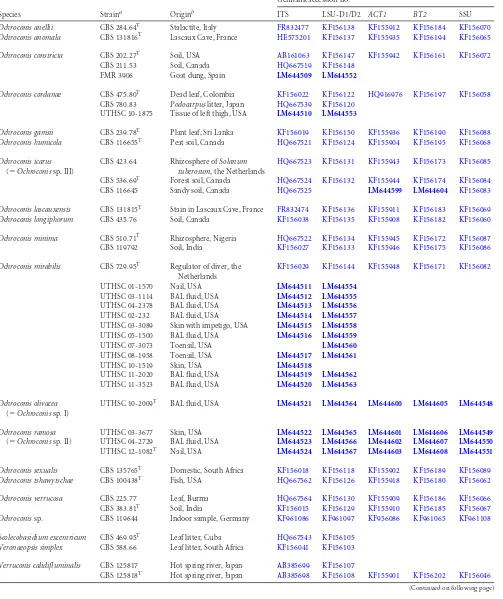
TABLE 1Strains included in the study
Species Straina Originb
GenBank accession no.c
ITS LSU-D1/D2 ACT1 BT2 SSU
Ochroconis anellii CBS 284.64T Stalactite, Italy FR832477 KF156138 KF155912 KF156184 KF156070
Ochroconis anomala CBS 131816T Lascaux Cave, France HE575201 KF156137 KF155935 KF156194 KF156065
Ochroconis constricta CBS 202.27T Soil, USA AB161063 KF156147 KF155942 KF156161 KF156072
CBS 211.53 Soil, Canada HQ667519 KF156148
FMR 3906 Goat dung, Spain LM644509 LM644552
Ochroconis cordanae CBS 475.80T Dead leaf, Colombia KF156022 KF156122 HQ916976 KF156197 KF156058
CBS 780.83 Podocarpuslitter, Japan HQ667539 KF156120
UTHSC 10-1875 Tissue of left thigh, USA LM644510 LM644553
Ochroconis gamsii CBS 239.78T Plant leaf, Sri Lanka KF156019 KF156150 KF155936 KF156190 KF156088
Ochroconis humicola CBS 116655T Peat soil, Canada HQ667521 KF156124 KF155904 KF156195 KF156068
Ochroconis icarus
(⫽Ochroconissp. III)
CBS 423.64 Rhizosphere ofSolanum tuberosum, the Netherlands
HQ667523 KF156131 KF155943 KF156173 KF156085
CBS 536.69T Forest soil, Canada HQ667524 KF156132 KF155944 KF156174 KF156084
CBS 116645 Sandy soil, Canada HQ667525 LM644599 LM644604 KF156083
Ochroconis lascauxensis CBS 131815T Stain in Lascaux Cave, France FR832474 KF156136 KF155911 KF156183 KF156069
Ochroconis longiphorum CBS 435.76 Soil, Canada KF156038 KF156135 KF155908 KF156182 KF156060
Ochroconis minima CBS 510.71T Rhizosphere, Nigeria HQ667522 KF156134 KF155945 KF156172 KF156087
CBS 119792 Soil, India KF156027 KF156133 KF155946 KF156175 KF156086
Ochroconis mirabilis CBS 729.95T Regulator of diver, the
Netherlands
KF156029 KF156144 KF155948 KF156171 KF156082
UTHSC 01-1570 Nail, USA LM644511 LM644554
UTHSC 03-1114 BAL fluid, USA LM644512 LM644555
UTHSC 04-2378 BAL fluid, USA LM644513 LM644556
UTHSC 02-232 BAL fluid, USA LM644514 LM644557
UTHSC 03-3089 Skin with impetigo, USA LM644515 LM644558
UTHSC 05-1500 BAL fluid, USA LM644516 LM644559
UTHSC 07-3073 Toenail, USA LM644560
UTHSC 08-1958 Toenail, USA LM644517 LM644561
UTHSC 10-1519 Skin, USA LM644518
UTHSC 11-2020 BAL fluid, USA LM644519 LM644562
UTHSC 11-3523 BAL fluid, USA LM644520 LM644563
Ochroconis olivacea
(⫽Ochroconissp. I)
UTHSC 10-2009T BAL fluid, USA LM644521 LM644564 LM644600 LM644605 LM644548
Ochroconis ramosa
(⫽Ochroconissp. II)
UTHSC 03-3677 Skin, USA LM644522 LM644565 LM644601 LM644606 LM644549
UTHSC 04-2729 BAL fluid, USA LM644523 LM644566 LM644602 LM644607 LM644550
UTHSC 12-1082T Nail, USA LM644524 LM644567 LM644603 LM644608 LM644551
Ochroconis sexualis CBS 135765T Domestic, South Africa KF156018 KF156118 KF155902 KF156189 KF156089
Ochroconis tshawytschae CBS 100438T Fish, USA HQ667562 KF156126 KF155918 KF156180 KF156062
Ochroconis verrucosa CBS 225.77 Leaf, Burma HQ667564 KF156130 KF155909 KF156186 KF156066
CBS 383.81T Soil, India KF156015 KF156129 KF155910 KF156185 KF156067
Ochroconissp. CBS 119644 Indoor sample, Germany KF961086 KF961097 KF956086 KF961065 KF961108
Scolecobasidium excentricum CBS 469.95T Leaf litter, Cuba HQ667543 KF156105
Veronaeopsis simplex CBS 588.66 Leaf litter, South Africa KF156041 KF156103
Verruconis calidifluminalis CBS 125817 Hot spring river, Japan AB385699 KF156107
CBS 125818T Hot spring river, Japan AB385698 KF156108 KF155901 KF156202 KF156046
(Continued on following page)
on May 16, 2020 by guest
http://jcm.asm.org/
scopic features were examined and measured by making direct wet mounts with 85% lactic acid or by slide cultures on OA and PCA using the light microscope Olympus CH-2 (Olympus Corporation, Tokyo, Japan). Photomicrographs were made with a Zeiss Axio Imager M1 light micro-scope (Zeiss, Oberkochen, Germany), using Nomarski differential inter-ference contrast. The 95% confidence intervals were derived from 50 obser-vations (⫻1,000 magnification), with the extremes given in parentheses. The ranges of the dimensions of other characters are given in the descriptions of new taxa.
DNA extraction, amplification, and sequencing.The isolates were grown on YES agar (20 g of yeast extract, 150 g of sucrose, 20 g of agar, distilled water to final volume of 1,000 ml) for 10 days at 25°C. DNA extraction was done by using FastDNA kit protocol (MP Biomedicals, Solon, OH), with the homogenization step done with a FastPrep FP120 cell disrupter (Thermo Savant, Holbrook, NY). The 18S nuclear small
subunit (nuSSU), the internal transcribed spacer regions (ITS), including the 5.8S small subunit gene, and the D1/D2 domains of the 28S nuclear large subunit (nuLSU) were amplified with the primer pairs NS1/NS4, ITS5/ITS4, and NL1/NL4b or LR0R/LR5, respectively (35–37). The frag-ments of the actin (ACT1) and-tubulin (BT2) genes were amplified using the primers ACT-512F/ACT-783R and Bt1a/Bt1b, respectively (38,
[image:3.585.43.545.81.501.2]39). The amplified fragments were purified and sequenced at Macrogen Corp. Europe (Amsterdam, the Netherlands) with a 3730XL DNA ana-lyzer (Applied Biosystems, Foster City, CA). The sequencing was per-formed with the same primers used for amplification to ensure good-quality sequences over the total length of the amplicon. Consensus sequences were obtained using SeqMan version 7.0.0 (DNAStar, Madi-son, WI). Some ITS, D1/D2,ACT1, andBT2sequences corresponding to several species ofOchroconisorVerruconiswere retrieved from GenBank (32,33) and included in the phylogenetic study (Table 1).
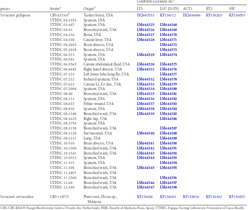
TABLE 1(Continued)
Species Straina Originb
GenBank accession no.c
ITS LSU-D1/D2 ACT1 BT2 SSU
Verruconis gallopava CBS 437.64T Turkey brain, USA HQ667553 KF156112 HQ916989 KF156203 KF156053
UTHSC 04-1355 Sputum, USA
UTHSC 03-447 Sputum, USA LM644525 LM644568
UTHSC 04-43 Bronchial wash, USA LM644526 LM644569
UTHSC 04-236 Brain, USA LM644527 LM644570
UTHSC 04-539 Canine liver, USA LM644528 LM644571
UTHSC 04-2693 Brain abscess, USA LM644572
UTHSC 05-1018 Brain abscess, USA LM644573
UTHSC 06-513 Sputum, USA LM644529 LM644574
UTHSC 06-541 Sputum, USA
UTHSC 06-3565 Canine abdominal fluid, USA LM644530 LM644575
UTHSC 06-4445 Right hand abscess, USA LM644531 LM644576
UTHSC 07-153 Left lower lobe lung Bx, USA LM644577
UTHSC 07-212 Induced sputum, USA LM644532 LM644578
UTHSC 07-623 Canine L2-L3 disc, USA LM644533 LM644579
UTHSC 07-2994 Sputum, USA LM644534 LM644580
UTHSC 08-40 Bronchial wash, USA LM644535 LM644581
UTHSC 08-112 Sputum, USA LM644536 LM644582
UTHSC 08-657 Feline wound, USA LM644537 LM644583
UTHSC 08-810 Sputum, USA LM644538 LM644584
UTHSC 08-1340 Bronchial wash, USA LM644539 LM644585
UTHSC 08-1625 Right hip, USA LM644586
UTHSC 08-1756 Sputum, USA
UTHSC 08-3158 Bronchial wash, USA LM644587
UTHSC 09-1229 Ear/mastoid, USA LM644540 LM644588
UTHSC 09-3111 Lung, USA LM644589
UTHSC 10-510 Brain abscess, USA LM644541 LM644590
UTHSC 10-1509 Bronchial wash, USA LM644542 LM644591
UTHSC 10-1541 Bronchial wash, USA LM644543 LM644592
UTHSC 10-3013 Sputum, USA LM644544 LM644593
UTHSC 11-315 Sputum, USA LM644594
UTHSC 11-509 Bronchial wash, USA LM644545 LM644595
UTHSC 11-2401 Bronchial wash, USA
UTHSC 11-2569 Bronchial wash, USA LM644596
UTHSC 12-69 Bronchial wash, USA LM644546 LM644597
UTHSC 12-549 Bronchial wash, USA LM644547 LM644598
Verruconis verruculosa CBS 119775 Plant root,Heveasp.,
Malaysia
KF156041 KF156103 KF155919 KF156193 KF156055
a
CBS, CBS-KNAW Fungal Biodiversity Centre, Utrecht, the Netherlands; FMR, Faculty of Medicine Reus, Spain; UTHSC, Fungus Testing Laboratory, University of Texas Health Science Center, San Antonio, TX;T, type strain.
b
BAL, bronchoalveolar lavage; Bx, biopsy.
cThe accession numbers of sequences newly determined in this study are indicated in bold type. ITS, internal transcribed spacer regions of the nuclear ribosomal DNA (nrDNA)
and intervening 5.8S nrDNA; LSU, large subunit of the nrDNA;ACT1, partial actin gene;BT2,-tubulin gene; SSU, small subunit of the nrDNA.
on May 16, 2020 by guest
http://jcm.asm.org/
Phylogenetic analysis. Multiple sequence alignments using the Clustal W and MUSCLE applications (40,41) were made in MEGA ver-sion 5.05 (42), in which the best substitution models were searched for each locus and for the combined data set, and maximum likelihood (ML) and maximum parsimony (MP) analyses were also performed. For ML, gaps and missing data were treated as a partial deletion with a site coverage cutoff 95%, and nearest-neighbor interchange (NNI) was used as the heu-ristic method. Internal branch support was assessed by a search of 1,000 bootstrapped sets of data. A bootstrap support (BS) ofⱖ70 was consid-ered significant. The phylogenetic distance values between the isolates were estimated with Kimura 2-parameter as a nucleotide substitution model under MEGA version 5.05. For the multilocus analysis, a phyloge-netic analysis using a Markov chain Monte Carlo (MCMC) algorithm was done with MrBayes version 3.1.2 (43) on the CIPRES portal (http://www .phylo.org), with two simultaneous runs for 10 million generations, with a sampling frequency of 1,000 trees. A burn-in tree sample of 10% was discarded. Bayesian posterior probabilities (PP) were obtained from the 50% majority-rule consensus of trees. A PP value ofⱖ0.95 was considered significant. The congruencies of the sequence data sets for the separate loci were determined using tree topologies of 70% reciprocal neighbor-join-ing (NJ) bootstrap trees with maximum likelihood distances, which were compared visually to identify conflicts between the partitions (44).
Scole-cobasidium excentricumstrain CBS 469.95 andVeronaeopsis simplexstrain
CBS 588.66 were used as outgroups.
Antifungal susceptibility.Thein vitroactivities of amphotericin B (AMB), itraconazole (ITC), posaconazole (PSC), voriconazole (VRC), anidulafungin (AFG), caspofungin (CFG), micafungin (MFG), and terbi-nafine (TBF) were determined for all the clinical isolates ofOchroconis, according to the methods outlined in the CLSI document M38-A2 (46) but with an incubation temperature of 30°C. The minimal effective con-centration (MEC) was determined at 48 h for the echinocandins, and the MICs at 48 h and 72 h were determined for the remaining drugs. The MIC was defined as the lowest concentration exhibiting 100% visual inhibition of growth for AMB, ITC, PSC, and VRC and an 80% reduction in growth for TBF.Paecilomyces variotiistrain ATCC MYA-3630 andAspergillus
fumigatusstrain ATCC MYA-3626 were used as quality control strains.
Statistical analyses of the data were done using the Kruskal-Wallis test in GraphPad Prism version 6.0 for Windows (GraphPad Software, San Di-ego, CA, USA). Statistical significance was defined as aPvalue ofⱕ0.05 (two-tailed).
Nucleotide sequence accession numbers.All novel DNA sequences were deposited in GenBank under accession numbers shown in bold type inTable 1, and taxonomic novelties were deposited in MycoBank (http: //www.MycoBank.org) (45).
RESULTS
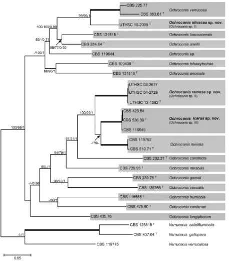
Phylogenetic analysis.The D1/D2 analysis (Fig. 1) showed that 68.6% and 31.4% of the isolates studied were distributed in two
main groups corresponding toOchroconisandVerruconis,
respec-tively. TheVerruconisisolates grouped with high support in the
same clade as the ex-type strain ofV. gallopava(CBS 437.64).
Eleven of the clinical isolates ofOchroconiswere nested with the
ex-type strain ofO. mirabilis(CBS 729.95) and one with the
ex-type strain ofO. cordanae(CBS 475.80). Three clinical isolates
(UTHSC 03-3677, UTHSC 12-1082, and UTHSC 04-2729),
mor-phologically similar toOchroconis minima, constituted a clade
that was phylogenetically distant from the type strain of that spe-cies. Although the isolate UTHSC 10-2009 clearly showed the
dif-ferential features ofOchroconis, it did not group with any of the
species of that genus included in this study, including the
refer-ence strain CBS 536.69, which was received asO. minima.
With the aim of confirming and clarifying the data obtained in the D1/D2 analysis, a multilocus analysis of five concatenated gene
regions (nuSSU, ITS, nuLSU,ACT1, andBT2) was performed,
including the type and some reference strains of the currently
accepted Ochroconis species. The concatenated sequence
con-sisted of 4,188 bp (Fig. 2), of which 1,000 were parsimony
infor-mative (51 nuSSU, 127 LSU, 432 ITS, 175ACT1, and 215BT2) and
revealed the formation of 17 lineages, 13 of them corresponding to
knownOchroconisspecies and three representing putative new
species (Ochroconissp. I,Ochroconissp. II, andOchroconissp. III).
Ochroconissp. I (strain UTHSC 10-2009) was located in the same
clade as that of two reference strains ofOchroconis verrucosa,
in-cluding the ex-type strain (CBS 383.81), although with significant
phylogenetic distance. The clade corresponding toOchroconissp.
II consisted of three clinical isolates (UTHSC 03-3677, UTHSC
12-1082, and UTHSC 04-2729), whileOchroconissp. III included
three environmental reference strains all previously identified as
O. minima(CBS 423.64, CBS 536.69, and CBS 116645). The clades
representingOchroconissp. II andOchroconissp. III were
phyloge-netically related (6.5% phylogenetic distance in the combined data
set) although distinct from the sequences ofO. minimatype strain
CBS 510.71 (5.7% and 7.0% phylogenetic distances with
Ochroco-nissp. III andOchroconissp. II, respectively).
Most of the clinical isolates included in this study were of re-spiratory origin (58.8%), mainly obtained from bronchoalveolar lavage (BAL) fluid and sputum samples, followed by superficial
tissue samples (21.6%), principally from the nails and skin (Table
2). The remaining 19.6% of the isolates were from miscellaneous
deep tissue or sterile fluid specimens, with most of them collected
from the lungs and brain.V. gallopavawas recovered from a wide
range of clinical specimens, being isolated from all the deep tissues and in equal numbers from sputum and bronchial wash fluids.
The isolates ofOchroconisspp. were exclusively recovered from
respiratory samples (BAL fluid), skin, and nails.
Phenotypic studies.Most of the isolates belonging to theV.
gallopavaclade showed the typical phenotypic characteristics
de-scribed for the species, i.e., colonies on PDA at 25°C with moder-ate growth (up to 58 mm diameter after 14 days), brownish gray (5E2) with a diffusible brown pigment, poorly differentiated idiogenous cells, and pale brown clavate 1-septum conidia
con-stricted at the septum that were (7)10 to 21m long by 2.5 to 4.5
m wide. All the strains showed optimum growth at 42°C (up to
80 mm at 14 days). Some strains showed atypical features not previously reported for the species. Isolate UTHSC 07-623 pro-duced yellowish white (4A2) colonies on all media tested and hy-aline conidia. The isolates UTHSC 07-212 and UTHSC 12-549 showed slow growth at 25°C (17 to 20 mm and 7 to 10 mm in 14 days, respectively), and their colonies were radially striate, with a lobulate edge, and moist, with a cerebriform aspect, respectively. Several isolates, apart from the clavate conidia, produced some ellipsoidal conidia with an apiculated base (UTHSC 03-447, UTHSC 04-43, UTHSC 04-236, UTHSC 06-513, UTHSC 07-153, UTHSC 08-112, UTHSC 08-810, UTHSC 10-510, UTHSC
11-509, and UTHSC 12-549). The isolates that grouped in theO.
mirabilisclade produced slow-growing (20 to 32 mm diameter in
14 days), olive brown (4D4), and velvety colonies on PDA and dark brown (5F8) and dry colonies on OA and PCA; the conidio-phores were cylindrical, thick walled, and with denticles distrib-uted sympodially along the conidiophore; the conidia were cylin-drical or ellipsoidal 1-septum, slightly constricted at the septum, and pale brown with rugose walls. The isolate UTHSC 10-1875,
clustered in theO. cordanaeclade, displayed long (up to 40m),
brown, and simple conidiophores, some of them with several
on May 16, 2020 by guest
http://jcm.asm.org/
FIG 1Maximum-likelihood (ML) tree constructed with sequences of the D1/D2 domains of the 28S rRNA gene. Bootstrap support (BS) values of⬎70% are shown at the nodes.V. simplexCBS 588.66 andS. excentricumCBS 469.95 were used as outgroup taxa.T, ex-type strains.
on May 16, 2020 by guest
http://jcm.asm.org/
[image:5.585.74.511.63.693.2]septa, and were thick walled, with denticles on the apical region; the conidia were ellipsoidal, slightly constricted at the septum,
smooth or finely verruculose, and 7 to 13m by 3 to 3.5m.
Ochroconissp. I (isolate UTHSC 10-2009) was mainly
character-ized by the production of long conidiophores (up to 60m) and
verrucose broadly ellipsoidal conidia, sometimes slightly
apicu-lated at the base (Fig. 3CandF). BothOchroconissp. II (Fig. 4) and
Ochroconissp. III (Fig. 5) showed phenotypic characteristics
sim-ilar to those ofO. minimabut with some differences, i.e., in
Ochro-conissp. II, the conidia had a rough surface (Fig. 4FandG), the
conidiophores reached up to 20m long, the chlamydospores
were smaller (3 to 3.5m by 3 to 3.5m), and the maximum and
minimum temperatures for growth were 35°C and 15°C,
respec-tively; inOchroconissp. III, the conidia were slightly smaller, with
FIG 2Bayesian tree from a concatenated data set, including the five regions nuSSU, ITS, nuLSU,ACT1, andBT2. Bootstrap support obtained with maximum-likelihood (left) and maximum parsimony (middle) of⬎70% and Bayesian posterior probability (right) values of⬎0.95 are shown at the nodes.T, ex-type
strains. Full supported branches are depicted as thick lines.
on May 16, 2020 by guest
http://jcm.asm.org/
[image:6.585.62.523.64.586.2]a narrower lower cell (up to 2.5m wide), and the maximum and minimum temperatures for growth were 33°C and 4°C, respec-tively.
Antifungal susceptibility.The results of thein vitroactivities
of the antifungal drugs tested are summarized inTable 3. No
sta-tistical difference in antifungal activities were observed among the
Ochroconisspp. studied. Terbinafine was the most active drug
against all the species tested, followed by MFG, with MICs and
MECs of 0.02g/ml and 0.25g/ml, respectively. AMB showed
poorin vitroactivity against all the isolates tested, with geometric
mean (GM) MICs and MIC90s of 24.5g/ml and 32g/ml,
re-spectively. Similarly, the three azoles showed very little activity, with elevated MICs.
TAXONOMY
Based on the mentioned phylogenetic data, which correlated with
the phenotypic features observed, we concluded thatOchroconis
sp. I,Ochroconissp. II, andOchroconissp. III are different from the
taxa currently accepted in this genus and are therefore described here as new.
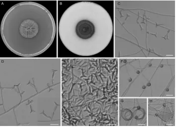
Ochroconis icarusSamerpitak, Giraldo, Guarro, & de Hoog, sp.
nov., MycoBank accession no. MB809376 (Fig. 5). Etymology: the
conidia look like the mythological figure Icarus, son of Daedalus,
who made wings to reach the sun. Diagnosis: it differs fromO.
minimaby the production of smaller conidia with a narrower
lower cell and a maximum growth temperature of 33°C and from
O. ramosamainly by having longer denticles and smooth-walled
conidia, with a wider upper cell.
Colonies on OA and PDA at 25°C attaining 16 to 20 mm and 17 to 24 mm diameter after 14 days, respectively; chocolate brown (6F4), flat, slightly curled, velvety at center, membranous at pe-riphery. Colonies on PCA at 25°C attaining 21 to 24 mm diameter after 14 days, dark brown (7F4), flat, membranous becoming vel-vety. On MEA 2% at 25°C, reaching 13 to 17 mm diameter after 14 days, yellowish brown (5E5), flat, woolly at center. Vegetative
hy-phae septate, pale brown, smooth and thin walled, 1 to 2m wide;
anastomosing and coiled hyphae usually present. Conidiophores poorly differentiated, arising laterally from vegetative hyphae,
flexuose, clavate, or cylindrical, 15 to 20m by 1.5 to 2m, pale
brown, thin and smooth walled, bearing one or more denticles in the apical region; denticles cylindrical, subhyaline to pale brown,
up to 2m long. Conidia abundant on OA, moderate on MEA
2%, and scarce on PCA, mostly two celled, trilobate, T or Y
shaped, 8 to 12m long, lower cell 1.5 to 2.5m wide, upper cell
up to 4 to 8m wide, pale brown, smooth and thin walled,
re-leased by rhexolytic secession. Chlamydospores abundant on PCA, moderate on OA and MEA 2%, growing directly on veg-etative hyphae, terminal or lateral, sessile, solitary, unicellular,
globose to slightly subglobose, 4 to 5m in diameter, brown,
smooth and thick walled. Sexual morph not observed. Cardinal temperatures for growth: optimum 24 to 27°C, maximum 33°C, minimum 4°C.
Specimens examined: Canada, Ontario, from forest soil, 1969,
G. L. Barron (holotype CBS H-21643; cultures ex-type CBS 536.69⫽
MUCL 15054⫽OAC 10212). From sandy soil, 1963, G. L. Barron
(CBS 116645⫽ATCC 16074⫽MUCL 102118⫽OAC 10094). The
Netherlands, Wageningen, from rhizosphere ofSolanum tuberosum,
1964, J. H. van Emden (CBS 423.64⫽MUCL 10610).
Ochroconis olivacea Giraldo, Gené, Deanna A. Sutton, &
Guarro, sp. nov., MycoBank accession no. MB809377 (Fig. 3).
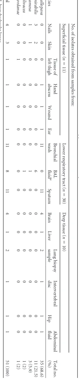
TABLE 2 Anatomical sources of isolates of Verruconis and Ochroconis spp. from clinical samples Species No. of isolates obtained from samples from: Total no. (%) Superficial tissue ( n ⫽ 11) Lower respiratory tract ( n ⫽ 30) Deep tissue ( n ⫽ 10) Nails Skin Tissue of left thigh Hand abscess Wound Ear Bronchial wash BAL fluid a Sputum Brain Liver Lung biopsy sample Intervertebral disc Hip Abdominal fluid V. gallopava 0 0 0 1 1 1 11 0 11 4 1 2 1 1 1 35 (68.6) O. mirabilis 3 2 0 0 0 0 0 6 0 0 0 0 0 0 0 11 (21.5) O. ramosa 1 1 0 0 0 0 0 1 0 0 0 0 0 0 0 3 (5.9) O. olivacea 0 0 0 0 0 0 0 1 0 0 0 0 0 0 0 1 (2) O. cordanae 0 0 1 0 0 0 0 0 0 0 0 0 0 0 0 1 (2) Total 4 3 1 1 1 1 11 8 11 4 1 2 1 1 1 51 (100) a BAL, bronchoalveolar lavage.
on May 16, 2020 by guest
http://jcm.asm.org/
[image:7.585.92.225.79.725.2]Etymology: referring to the colony color. Diagnosis: it differs from
Ochroconis humicola mainly by having slower growth, shorter
conidiophores, and verrucose conidia, and fromO. verrucosaby
the production of solitary two-celled conidia.
Colonies on OA at 25°C attaining 23 to 24 mm diameter after 14 days, from olive (1F4) to olive brown (4E5), flat, felty. Colonies on PCA at 25°C reaching 18 to 23 mm after 14 days, olive (2F8),
flat, woolly at center, becoming felty toward the periphery. Colo-nies on MEA 2% and PDA attaining 15 to 17 mm and 22 to 23 mm diameter in 14 days, respectively, olive brown (4E6), radially folded, velvety. Vegetative hyphae septate, pale brown, smooth
and thin walled, 1.5 to 2m wide. Conidiophores differentiated,
arising directly from vegetative hyphae, erect, straight or slightly
bent, simple, with 0 to 2 septa, cylindrical, (14)21 to 42(60)m by
FIG 3Ochroconis olivaceasp. nov. UTHSC 10-2009. (A and B) Colonies on OA and PCA, respectively, at 25°C after 14 days. (C, D, and F) Simple conidiophores arising directly from vegetative hyphae and conidia. (E) Young conidium growing on the apex of a conidiophore. Scale bars⫽10m.
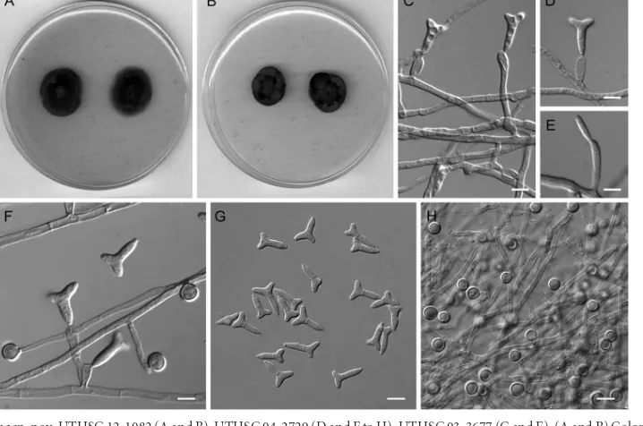
FIG 4Ochroconis ramosasp. nov. UTHSC 12-1082 (A and B), UTHSC 04-2729 (D and F to H), UTHSC 03-3677 (C and E). (A and B) Colonies on OA and PCA, respectively, at 25°C after 14 days. (C to F) Conidiophores producing trilobate conidia. (G) Trilobate conidia. (H) Chlamydospores growing directly on vegetative hyphae. Scale bars⫽10m.
on May 16, 2020 by guest
http://jcm.asm.org/
[image:8.585.114.473.66.309.2] [image:8.585.114.473.453.691.2]2 to 3m, brown, thick and smooth walled, producing conidia sympodially on long open denticles; denticles cylindrical, pale
brown, up to 1m long. Conidia abundant on OA and PCA,
absent on PDA and MEA 2%, mostly two celled, cylindrical or
broadly ellipsoidal, 6 to 9.5 m by 1.9 to 4.5m, sometimes
slightly apiculate at the base and constricted at the septum, pale brown, verrucose and thick walled, released by rhexolytic seces-sion. Chlamydospores and sexual morph not observed. Cardinal temperatures for growth: optimum 20 to 25°C, maximum 35°C, minimum 15°C.
Specimen examined: USA, Utah, from bronchoalveolar lavage fluid, 2010, D. A. Sutton (CBS H-21779 holotype; cultures ex-type
CBS 137170⫽FMR 12509⫽UTHSC 10-2009).
Ochroconis ramosaGiraldo, Gené, Deanna A. Sutton & Guarro,
sp. nov., MycoBank accession no. MB809378 (Fig. 4). Etymology:
referring to branched conidia. Diagnosis: it differs fromO.
min-imaby the production of smaller and narrower conidia and a
maximum growth temperature of 35°C and fromO. icarusmainly
by having shorter denticles and rough-walled conidia with a nar-rower upper cell.
[image:9.585.112.475.66.323.2]Colonies on OA at 25°C attaining 22 to 24 mm diameter in 14 days, chocolate brown (6F4), flat, felty at center, membranous toward the periphery. Colonies on PCA at 25°C reaching 17 to 20 mm after 14 days, brownish black (6H8), flat, woolly at center, membranous toward the periphery. Colonies on MEA 2% and PDA attaining 13 to 16 mm and 20 to 24 mm diameter in 14 days, FIG 5Ochroconis icarussp. nov. CBS 536.69. (A and B) Colonies on MEA 2% and OA, respectively, at 25°C after 21 days. (C and D) Conidiophores bearing trilobate conidia. (E) Trilobate conidia. (F to H) Chlamydospores and coiled hyphae. Scale bars⫽10m.
TABLE 3Results ofin vitroantifungal susceptibility testing of the 16 clinical isolates ofOchroconisspp. included in the study
Species
(no. of isolates tested)
GM or MIC dataa
MIC or MEC (g/ml) forb:
AMB ITC PSC VRC AFG CFG MFG TBF
O. mirabilis(11) GM 28.36 7.00 18.23 11.09 3.93 7.90 0.22 0.03
MIC range 8–32 1–32 0.5–32 2–32 0.015–32 1–32 0.06–0.5 0.015–0.125
MIC90 32 32 32 32 4 4 0.25 0.02
O. cordanae(1) GM 16 1 2 4 0.03 2 0.125 0.015
O. olivacea(1) GM 32 1 1 4 0.03 1 0.125 0.015
O. ramosa(3) GM 10.66 1 11.08 2.1 0.051 1 0.16 0.015
MIC range 8–16 1 0.25–32 0.5–4 0.015–0.125 1 0.125–0.25 0.015
Overall (16) GM 24.5 5.13 14.80 8.53 2.72 5.67 0.2 0.02
MIC range 8–32 1–32 0.25–32 0.5–32 0.02–32 1–32 0.06–0.50 0.02–0.13
MIC90 32 2 32 16 4 8 0.25 0.02
aGM, geometric mean. b
MEC, minimal effective concentration; AMB, amphotericin B; ITC, itraconazole; PSC, posaconazole; VRC, voriconazole; AFG, anidulafungin; CFG, caspofungin; MFG, micafungin; TBF, terbinafine.
on May 16, 2020 by guest
http://jcm.asm.org/
[image:9.585.41.546.545.697.2]respectively, olive (3F5) to olive brown (4E4 to F4), slightly raised, velvety. Vegetative hyphae septate, pale brown, smooth and thin
walled, 1.5 to 2m wide. Conidiophores differentiated arising
directly from vegetative hyphae, erect, straight, simple, clavate or
cylindrical with beaked apex, 15 to 20m by 1.5 to 2m, pale
brown, thin and smooth walled, bearing one or more denticles in the apical region; denticles cylindrical, subhyaline to pale brown,
up to 0.8m long. Conidia abundant on OA and PCA, absent on
PDA and MEA 2%, mostly two celled, trilobate, T or Y shaped,
(7)8 to 10(12)m long, lower cell 1.5 to 2.5m wide, upper cell 3
to 6m wide, pale brown, rough and thin walled, released by
rhexolytic secession. Chlamydospores abundant on PCA and OA, absent on MEA 2%, growing directly on vegetative hyphae, lateral,
sessile, solitary, unicellular, globose or subglobose, 3 to 3.5m by
3 to 3.5m, brown, smooth and thick walled. Sexual morph not
observed. Cardinal temperatures for growth: optimum 20 to 25°C, maximum 35°C, minimum 15°C.
Specimens examined: USA, California, from human nail, 2012, D. A. Sutton (CBS H-21780 holotype; cultures ex-type CBS
137173⫽FMR 12514⫽UTHSC 12-1082). Pennsylvania, from
human skin, 2003, D. A. Sutton (CBS 137171⫽FMR 12512⫽
UTHSC 03-3677). Utah, from bronchoalveolar lavage fluid, 2004,
D. A. Sutton (CBS 137172⫽FMR 12513⫽UTHSC 04-2729).
DISCUSSION
In this study, we determined the distributions ofVerruconisand
Ochroconisspecies in a set of clinical specimens from human and
animal origin from the United States, based on molecular and
phenotypic analyses.V. gallopavawas the most common species,
found mainly on respiratory samples (BAL fluid and sputum), followed by deep-tissue samples (brain and others). These
find-ings agree with those of previous studies, in whichV. gallopava
frequently involved the lung, producing either cavitary or non-cavitary lung lesions. This fungus also shows special neurotropism
in warm-blooded animals (21,22). AlthoughV. gallopavais
com-monly known as producing brain abscesses and encephalitis in birds and other animals, several reports document it as an etio-logic agent in human disease. Most of these reports occur in im-munocompromised patients, with organ transplantation being
the most common underlying condition (13,21,22,48). A few
cases in immunocompetent patients with pulmonary
manifesta-tions have also been described. In these cases,V. gallopavawas
isolated from BAL fluid and lobectomy samples (49–51).
Some of ourV. gallopavaisolates showed atypical
morpholo-gies, such as yellowish white or moist colonies, slow growth at 25°C, and ellipsoidal conidia with an apiculate base. However, the molecular analysis demonstrated that they in fact belong to this
species. The morphological variability inV. gallopava has also
been reported in other studies (13,28). Yarita et al. (13) analyzed
four isolates from hot springs in Japan, which showed differences in the shape and size of the conidia, being slender or thicker and shorter than those of the type species; however, their D1/D2
re-gions were 99.7% identical with the those of the type strain ofV.
gallopava. The isolates studied by Dixon and Salkin (28) also
pro-duced a similar variation in conidial size.
V. gallopavahas been described as a thermotolerant species
(22,32,52), and all the isolates studied here grew well at 42°C. This
explains its ability to survive in warm environments, like thermal soils or hot springs, and to infect warm-blooded animals, poultry,
and other birds, in addition to humans. Thein vitro
thermotoler-ance is a useful physiological feature for identifying this species
(13,20).
In the present study, we report for the first time an albino
isolate ofV. gallopava (UTHSC 07-623), which was recovered
from a canine intervertebral disc. This is an unusual phenomenon
that occurs in a few other fungi, such asNeoscytalidium hyalinum,
Aspergillus flavus,Ophiostoma floccosum,Ophiostoma piceae, and
Ophiostoma pluriannulatum (53–55), which in general are
in-volved almost exclusively in skin and nail disease (56, 57). The
melanin plays an important role in fungal pathogenesis, and its absence often generates less virulent isolates than with pigmented
ones (47,58–60). Although we could not demonstrate that this
isolate was the etiological agent of the infection, the presence of albino isolates recovered from deep tissues might suggest that this fungus has an additional mechanism of pathogenicity, apart from that of melanin. However, additional isolates should be studied to demonstrate this hypothesis.
O. mirabiliswas the most common species recovered from
su-perficial lesions, but it was also isolated from BAL fluid samples. This species is a waterborne fungus usually isolated from moist places in bathrooms and rarely from soil and plant material. How-ever, several isolates of this species have also been recovered from clinical samples, producing mild cutaneous infections (skin,
fin-gers, and toenails) in humans and fishes (32). It has been
sug-gested that the bathroom-associated fungi can penetrate the skin and the nails during showering, when these barriers are weakened
(6,61).
The species O. constricta and O. humicola, occasionally
re-ported from superficial infections in humans (20,21,61), were not
represented in our set of isolates. However, most of the isolates
identified here asO. mirabiliswere received as eitherO. constricta
orO. humicola. Only subtle morphological features allow the
dis-tinction between these three species:O. constrictahas poorly
dif-ferentiated conidiophores and markedly verruculose conidia,
with a conspicuous constriction at the septum,O. mirabilishas
differentiated cylindrical conidiophores (up to 20 m) and
conidia slightly constricted at the septum, andO. humicolahas
rapid growth, longer conidiophores than the other two species, and cylindrical conidia with a smooth or slightly rugose wall.
We obtained a single isolate ofO. cordanaefrom the tissue of a
left thigh, which was previously identified asO. humicola.O.
cor-danaecan be differentiated by its more slowly growing colonies,
shorter conidiophores, and smaller conidia, which are mostly
el-lipsoidal.O. cordanaewas recently described by Samerpitak et al.
(32) as a cosmopolitan species commonly inhabiting living leaves,
sometimes found on decaying vegetal material, and less frequently from ant nests, but it has never been obtained from clinical sam-ples. Therefore, this is the first report of this species in the clinical setting. Because only one isolate was obtained from a superficial tissue sample and because of its mesophilic abilities, the human-pathogenic role of this species is still doubtful.
Based on our multilocus sequence analysis and detailed
phe-notypic study, three new species are proposed here, i.e.,O. icarus
from environmental sources andO. ramosaandO. olivaceafrom
clinical specimens. BothO. icarusandO. ramosaare
morpholog-ically similar and phylogenetmorpholog-ically related toO. minima(Fig. 2).
However, they can be easily differentiated by their maximum
tem-peratures for growth (37°C forO. minima, 35°C forO. ramosa,
and 33°C forO. icarus). Additionally, inO. minima, the conidia
are longer than those of the other two species (up to 13.5m), in
on May 16, 2020 by guest
http://jcm.asm.org/
O. ramosa, the conidia have a rugose wall, and inO. icarus, the
lower cell of the conidia is narrower than that ofO. minima
(1.5-to 2.5-m wide forO. icarusversus up to 4.5m forO. minima).
In the phylogenetic analyses,O. olivaceawas placed close toO.
verrucosa(Fig. 2). Both species produce verrucose conidia, but in
O. verrucosa, they are cylindrical or fusiform, mostly four celled,
and sometimes arranged in acropetal, branched, or unbranched
chains (32). In contrast, inO. olivacea, the conidia are cylindrical
or ellipsoidal, mostly two celled, and not arranged in chains.
Mor-phologically,O. olivaceais similar toOchroconis gamsiiandO.
humicolain its production of one-celled conidia and erected long
cylindrical conidiophores. However, inO. gamsii, the conidia are
broadly fusiform and unilaterally flattened, and the
conidio-phores are darker, while inO. humicola, the conidia are broadly
cylindrical and finely echinulate and the conidiophores are longer
(up to 300m) (2,62).Ochroconis macrozamiae, a recently
de-scribed species onMacrozamialeaf litter, also resemblesO.
oliva-cea, but, in addition to having broadly fusiform conidia, it is
phy-logenetically close toO. gamsii(63).
Fewin vitroantifungal susceptibility studies are available for
Ochroconisspecies, as members of this genus are rarely involved in
human disease. The most relevant species in the clinical setting,O.
gallopava, was recently transferred toVerruconis. Recently,
Seyed-mousavi et al. (64) evaluated the antifungal susceptibilities of
nu-merous strains ofVerruconisandOchroconisspp. from clinical and
environmental origins against eight antifungal drugs, using the
broth microdilution test. In that study, the isolates ofV. gallopava
showed low MICs for AFG, PSC, ITC, AMB, CFG, and VRC, while 5-flucytosine and fluconazole showed no activity. In contrast,
only AFG, PSC, and CFG showed goodin vitroactivity againstO.
mirabilis, the most frequently isolated Ochroconisspecies from
clinical sources in that study (64). These results are in
disagree-ment with our data, for which these drugs demonstrated high MICs. TBF and MFG, which were not tested by Seyedmousavi et
al. (64), were the only drugs with some activity againstO.
mirabi-lis. These differences in the data between those of Seyedmousavi et
al. (64) and our study might be explained by the different origins
of theOchroconisisolates tested (Europe versus the United States)
but also by differences in the procedure, i.e., the incubation tem-perature (25°C versus 30°C) and the methodology or criteria used to read the endpoint.
There are few clinical cases reported in the literature regarding
Ochroconisinfections. Mancini and McGinnis (65) described a
case of pulmonary abscess byO. constrictain a heart transplant
recipient. The patient was successfully treated with systemic AMB, resulting in the resolution of clinical symptoms and a cavitary
lesion. Recently, Ge et al. (23) reported a human case of
subcuta-neous phaeohyphomycosis in an immunocompetent patient due
toOchroconis tshawytschae. Several short courses of treatment
with ITC or TBF were begun, but no cure was obtained. A
subcutaneous infection byO. humicolain a cat was reported by
VanSteenhouse et al. (66). The fungus was recovered from a
gran-ulomatous lesion, and although no antifungal test was performed, the cat was successfully treated with ketoconazole.
Despite the fact thatV. gallopavais the species most frequently
implicated in the clinical setting, the repeated isolation of several
species ofOchroconisin the clinical setting never before reported
suggests their potential pathogenic role and deserves further re-search.
ACKNOWLEDGMENTS
This study was supported by the Spanish Ministerio de Economía y Com-petitividad, grant CGL 2011-27185.
We thank the Ph.D. student M. Sandoval-Denis for his assistance with the antifungal susceptibility tests.
REFERENCES
1.de Hoog GS, von Arx JA.1973. Revision ofScolecobasidiumand
Pleuro-phragmium. Kavaka1:55– 60.
2.Ellis MB.1971. Dematiaceous hyphomycetes. Commonwealth Mycolog-ical Institute, Kew, Surrey, England.
3.Ellis MB.1976. More dematiaceous hyphomycetes. Commonwealth My-cological Institute, Kew, Surrey, England.
4.Dwivedi RS.1959. Occurrence of the genusScolecobasidiumAbbott in India. Curr. Sci. Bangalore28:374 –375.
5.Barron GI, Busch LV.1962. Studies on the soil hyphomycete
Scolecoba-sidium. Can. J. Bot.40:77– 84.http://dx.doi.org/10.1139/b62-009.
6.Lian X, de Hoog GS.2010. Indoor wet cells harbour melanized agents of cutaneous infection. Med. Mycol.48:622– 628.http://dx.doi.org/10.3109 /13693780903405774.
7.Nováková A.2009. Microscopic fungi isolated from the Domica Cave system (Slovak Karst National Park, Slovakia). A review. Int. J. Speleol.
38:71– 82.http://dx.doi.org/10.5038/1827-806X.38.1.8.
8.Martin-Sanchez PM, Nováková A, Bastian F, Alabouvette C, Saiz-Jimenez C.2012. Two new species of the genusOchroconis,O. lascauxensis
andO. anomalaisolated from black stains in Lascaux Cave, France. Fungal
Biol.116:574 –589.http://dx.doi.org/10.1016/j.funbio.2012.02.006. 9.Evans HC.1971. Thermophilous fungi of coal spoil tips. I. Taxonomy.
Trans. Br. Mycol. Soc. 57:241–254. http://dx.doi.org/10.1016/S0007 -1536(71)80006-2.
10. Evans HC.1971. Thermophilous fungi of coal spoil tips. II. Occurrence, distribution and temperature relationships. Trans. Br. Mycol. Soc.57:
255–266.
11. Tansey MR, Brock TD.1973.Dactylaria gallopava, a cause of avian en-cephalitis, in hot spring effluents, thermal soils and self-heated coal waste piles. Nature242:202–203.http://dx.doi.org/10.1038/242202a0. 12. Redman RS, Litvenseva A, Sheehan KB, Henson JM, Rodriguez RJ.
1999. Fungi from geothermal soils in Yellowstone National Park. Appl. Environ. Microbiol.65:5193–5197.
13. Yarita K, Sano A, Murata Y, Takayama A, Takahashi Y, Takahashi H, Yaguchi T, Ohori A, Kamei K, Miyaji M, Nishimura K.2007. Pathoge-nicity ofOchroconis gallopavaisolated from hot spring in Japan and a review of published reports. Mycopathologia164:135–147.http://dx.doi .org/10.1007/s11046-007-9034-7.
14. Tansey MR, Fliermans CB, Kern CD.1979. Aerosol dissemination of veterinary pathogenic and human opportunistic thermophilic and ther-motolerant fungi from thermal effluents of nuclear production reactors. Mycopathologia69:91–115.http://dx.doi.org/10.1007/BF00428608. 15. Rippon JW, Gerhold R, Heath M.1980. Thermophilic and thermotolerant
fungi isolated from the thermal effluent of nuclear power generating reactors: dispersal of human opportunistic and veterinary pathogenic fungi. Myco-pathologia70:169 –179.http://dx.doi.org/10.1007/BF00443028.
16. Kralovic SM, Rhodes JC.1995. Phaeohyphomycosis caused byDactylaria (human dactylariosis): report of a case with review of the literature. J. Infect.31:107–113.http://dx.doi.org/10.1016/S0163-4453(95)92060-9. 17. Waldrip DW, Padhye AA, Ajello L, Ajello M.1974. Isolation of
Dacty-laria gallopavafrom broiler-house litter. Avian Dis.18:445– 451.http://dx
.doi.org/10.2307/1589112.
18. Randall CJ, Owen DM.1981. Encephalitis in broiler chickens caused by a hyphomycete resemblingDactylaria gallopava. Avian Pathol.10:31– 41.
http://dx.doi.org/10.1080/03079458108418456.
19. Wong JS, Schousboe MI, Metcalf SS, Endre ZH, Hegarty JM, Maze MJ, Keith ER, Seaward LM, Podmore RG.2010.Ochroconis gallopava peri-tonitis in a cardiac transplant patient on continuous ambulatory perito-neal dialysis. Transpl. Infect. Dis.12:455– 458.http://dx.doi.org/10.1111/j .1399-3062.2010.00523.x.
20. de Hoog GS, Guarro J, Gené J, Figueras MJ.2011. Atlas of clinical fungi. CD-ROM version 3.1. CBS-KNAW Fungal Biodiversity Centre, Utrecht, the Netherlands.
21. Meriden Z, Marr KA, Lederman HM, Illei PB, Villa K, Riedel S, Carroll KC, Zhang SX.2012.Ochroconis gallopavainfection in a patient with
on May 16, 2020 by guest
http://jcm.asm.org/
chronic granulomatous disease: case report and review of the literature. Med. Mycol.50:883– 889.http://dx.doi.org/10.3109/13693786.2012.681075. 22. Qureshi ZA, Kwak EJ, Nguyen MH, Silveira FP. 2012.Ochroconis
gallopava: a dematiaceous mold causing infections in transplant recip-ients. Clin. Transplant.26:E17–E23.http://dx.doi.org/10.1111/j.1399 -0012.2011.01528.x.
23. Ge YP, Lv Gx, Shen YN, Li M, Deng SW, De Hoog S, Samerpitak K, Liu WD.2012. First report of subcutaneous phaeohyphomycosis caused by
Ochroconis tshawytschaein an immunocompetent patient. Med. Mycol.
50:637– 640.http://dx.doi.org/10.3109/13693786.2011.653834. 24. Blalock HG, Georg LK, Derieux WT.1973. Encephalitis in Turkey poults
due toDactylaria(Diplorhinotrichum)gallopava, a case report and its ex-perimental reproduction. Avian Dis. 17:197–204. http://dx.doi.org/10 .2307/1588939.
25. Shane SM, Markovits J, Snider TG, Harrington KS.1985. Encephalitis attributed to dactylariosis in Japanese quail chicks (Coturnix coturnix
ja-ponica). Avian Dis.29:822– 828.http://dx.doi.org/10.2307/1590673.
26. Karesh WB, Russell R, Gribble D.1987.Dactylaria gallopavaencephalitis in two grey-winged trumpeters (Psophia crepitans). Avian Dis.31:685– 688.http://dx.doi.org/10.2307/1590762.
27. Salkin IF, Dixon DM, Kemna ME, Danneman PJ, Griffith JW.1990. Fatal encephalitis caused byDactylaria constrictavar.gallopavain a snowy owl chick (Nyctea sandiaca). J. Clin. Microbiol.28:2845–2847.
28. Dixon DM, Salkin I.1986. Morphologic and physiologic studies of three dematiaceous pathogens. J. Clin. Microbiol.24:12–15.
29. Padhye AA, Amster RL, Browning M, Ewing EP.1994. Fatal encephalitis caused byOchroconis gallopavumin a domestic cat (Felis domesticus). J. Med. Vet. Mycol.32:141–145.http://dx.doi.org/10.1080/02681219480000191. 30. Singh K, Flood J, Welsh RD, Wyckoff JH, Snider TA, Sutton DA.2006.
Fatal systemic phaeohyphomycosis caused byOchroconis gallopavumin a dog (Canis familaris). Vet. Pathol.43:988 –992.http://dx.doi.org/10.1354 /vp.43-6-988.
31. Abbott EV.1927.Scolecobasidium, a new genus of soil fungi. Mycologia
19:29 –31.http://dx.doi.org/10.2307/3753662.
32. Samerpitak K, Van der Linde E, Choi HJ, Gerrits van den Ende AHG, Machouart M, Gueidan C, de Hoog GS.2014. Taxonomy ofOchroconis, genus including opportunistic pathogens on humans and animals. Fungal Divers.65:89 –126.http://dx.doi.org/10.1007/s13225-013-0253-6. 33. Machouart M, Samerpitak K, de Hoog GS, Gueidan C.2014. A
multi-gene phylogeny reveals thatOchroconisbelongs to the family
Sympoven-turiaceae(Venturiales,Dothideomycetes). Fungal Divers.65:77– 88.http:
//dx.doi.org/10.1007/s13225-013-0252-7.
34. Kornerup A, Wanscher JH.1978. Methuen handbook of colour, 3rd ed. Methuen, London, England.
35. Vilgalys R, Hester M.1990. Rapid genetic identification and mapping of enzymatically amplified ribosomal DNA from severalCryptococcus spe-cies. J. Bacteriol.172:4238 – 4246.
36. White TJ, Bruns T, Lee S, Taylor JW.1990. Amplification and direct sequencing of fungal ribosomal RNA genes for phylogenetics, p 315–322. InInnis MA, Gelfand DH, Sninsky JJ, White TJ (ed), PCR protocols: a guide to the methods and applications. Academic Press, New York, NY. 37. O’Donnell K.1993.Fusariumand its near relatives, p 225–233.In
Reyn-olds DR, Taylor JW (ed), The fungal holomorph: mitotic, meiotic and pleomorphic speciation in fungal systematics. CABI, Wallingford, United Kingdom.
38. Glass NL, Donaldson GC.1995. Development of primer sets designed for use with the PCR to amplify conserved genes from filamentous ascomy-cetes. Appl. Environ. Microbiol.61:1323–1330.
39. Carbone L, Kohn LM. 1999. A method for designing primer sets for speciation in filamentous ascomycetes. Mycologia91:553–556.http://dx .doi.org/10.2307/3761358.
40. Thompson JD, Higgins DG, Gibson TJ.1994. CLUSTAL W: improving the sensitivity of progressive multiple sequence alignment through se-quence weighting, position-specific gap penalties and weight matrix choice. Nucleic Acids Res.22:4673– 4680.http://dx.doi.org/10.1093/nar /22.22.4673.
41. Edgar RC.2004. MUSCLE: multiple sequence alignment with high accu-racy and high throughput. Nucleic Acids Res.32:1792–1797.http://dx.doi .org/10.1093/nar/gkh340.
42. Tamura K, Peterson D, Peterson N, Stecher G, Nei M, Kumar S.2011. MEGA 5: molecular evolutionary genetics analysis using maximum like-lihood, evolutionary distance and maximum parsimony methods. Mol. Biol. Evol.28:2731–2739.http://dx.doi.org/10.1093/molbev/msr121.
43. Ronquist F, Huelsenbeck JP.2003. MrBayes 3: Bayesian phylogenetic inference under mixed models. Bioinformatics19:1572–1574.http://dx .doi.org/10.1093/bioinformatics/btg180.
44. Gueidan C, Roux C, Lutzoni F.2007. Using multigene phylogeny anal-ysis to assess generic delineation and character evolution inVerrucariaceae (Verrucariales,Ascomycota). Mycol. Res.111:1145–1168.http://dx.doi.org /10.1016/j.mycres.2007.08.010.
45. Crous PW, Gams W, Stalpers JA, Robert V, Stegehuis G.2004. Myco-Bank: an online initiative to launch mycology into the 21st century. Stud. Mycol.50:19 –22.
46. Clinical and Laboratory Standards Institute.2008. Reference method for broth dilution antifungal susceptibility testing of filamentous fungi. Ap-proved standard, 2nd ed. CLSI document M38-A2. Clinical and Labora-tory Standards Institute, Wayne, PA.
47. Urán ME, Nosanchuk JD, Restrepo A, Hamilton AJ, Gómez BL, Cano LE.2011. Detection of antibodies againstParacoccidioides brasiliensis mel-anin inin vitroandin vivostudies during infection. Clin. Vaccine Immu-nol.18:1680 –1688.http://dx.doi.org/10.1128/CVI.05099-11.
48. Shoham S, Pic-Aluas L, Taylor J, Cortez K, Rinaldi MG, Shea Y, Walsh TJ.2008. Transplant-associatedOchroconis gallopavainfections. Transpl. Infect. Dis.10:442– 448.http://dx.doi.org/10.1111/j.1399-3062 .2008.00327.x.
49. Odell JA, Alvarez S, Cvitkovich DG, Cortese DA, McComb BL.2000. Multiple lung abscesses due toOchroconis gallopavum, a dematiaceous fungus, in a nonimmunocompromised wood pulp worker. Chest118:
1503–1505.http://dx.doi.org/10.1378/chest.118.5.1503.
50. Bravo LO, Jr, Ngamy V.2004.Ochroconis gallopavumandMycobacterium
avium intracellularein an immunocompetent patient. Chest126:975S.
http://dx.doi.org/10.1378/chest.126.4_MeetingAbstracts.975S.
51. Hollingsworth JW, Shofer S, Zaas A. 2007. Successful treatment of
Ochroconis gallopavuminfection in an immunocompetent host. Infection
35:367–369.http://dx.doi.org/10.1007/s15010-007-6054-7.
52. Yarita K, Sano A, Samerpitak K, Kamei K, de Hoog GS, Nishimura K.
2010.Ochroconis calidifluminalis, a sibling of the neurotropic pathogenO.
gallopava, isolated from hot spring. Mycopathologia170:21–30.http://dx
.doi.org/10.1007/s11046-010-9292-7.
53. Held BW, Thwaites JM, Farrell RL, Blanchette RA.2003. Albino strains
ofOphiostomaspecies for biological control of sapstaining fungi.
Holz-forschung.57:237–242.http://dx.doi.org/10.1515/HF.2003.036. 54. Brandt ME, Gade L, McCloskey CB, Balajee A.2009. AtypicalAspergillus
flavusisolates associated with chronic azole therapy. J. Clin. Microbiol.
47:3372–3375.http://dx.doi.org/10.1128/JCM.00671-09.
55. Phillips AJ, Alves A, Abdollahzadeh J, Slippers B, Wingfield MJ, Groe-newald JZ, Crous PW.2013. TheBotryosphaeriaceae: genera and species known from culture. Stud. Mycol.76:51–167.http://dx.doi.org/10.3114 /sim0021.
56. Sigler L, Summerbell RC, Poole L, Wieden M, Sutton DA, Rinaldi MG, Aguirre M, Estes GW, Galgiani JN.1997. InvasiveNattrassia mangiferae infections: case report, literature review, and therapeutic and taxonomic appraisal. J. Clin. Microbiol.35:433– 440.
57. Sriaroon C, Vincent AL, Silapunt S, Chandler A, Houston SH, Greene JN.2008. Successful treatment of subcutaneousScytalidium
hyalinuminfection with voriconazole and topical terbinafine in a cardiac
transplant patient. Transplantation 85:780 –782. http://dx.doi.org/10 .1097/TP.0b013e3181664ecb.
58. Gómez BL, Nosanchuk JD.2003. Melanin and fungi. Curr. Opin. Infect. Dis.16:91–96.http://dx.doi.org/10.1097/00001432-200304000-00005. 59. Langfelder K, Streibel M, Jahn B, Haase G, Brakhage AA.2003.
Bio-synthesis of fungal melanins and their importance for human pathogenic fungi. Fungal Genet. Biol.38:143–158.http://dx.doi.org/10.1016/S1087 -1845(02)00526-1.
60. Madrid H, Ruíz-Cendoya M, Cano J, Stchigel A, Orofino R, Guarro J.
2009. Genotyping andin vitroantifungal susceptibility ofNeoscytalidium
dimidiatumisolates from different origins. Int. J. Antimicrob. Agents34:
351–354.http://dx.doi.org/10.1016/j.ijantimicag.2009.05.006.
61. Saunte DM, Tarazooie B, Arendrup MC, de Hoog GS. 2012. Black yeast-like fungi in skin and nail: it probably matters. Mycoses55:161–167.
http://dx.doi.org/10.1111/j.1439-0507.2011.02055.x.
62. de Hoog GS.1985. Taxonomy of theDactylariacomplex, IV.Dactylaria,
Neta,SubulisporaandScolecobasidium. Stud. Mycol.26:1– 60.
63. Crous PW, Shivas RG, Quaedvlieg W, van der Bank M, Zhang Y, Summerell BA, Guarro J, Wingfield MJ, Wood AR, Alfenas AC, Braun U, Cano-Lira JF, García D, Marin-Felix Y, Alvarado P, Andrade JP,
on May 16, 2020 by guest
http://jcm.asm.org/
Armengol J, Assefa A, den Breeÿen A, Camele I, Cheewangkoon R, De Souza JT, Duong TA, Esteve-Raventós F, Fournier J, Frisullo S, García-Jiménez J, Gardiennet A, Gené J, Hernández-Restrepo M, Hirooka Y, Hospenthal DR, King A, Lechat C, Lombard L, Mang SM, Marbach PAS, Marincowitz S, Montaño-Mata NJ, Moreno G, Perez CA, Pérez Sierra AM, Robertson JL, Roux J, Rubio E, Schumacher RK, Stchigel AM, Sutton DA, Tan YP, Thompson EH, et al. 2014. Fungal planet description sheets: 214 –280. Persoonia32:184 –306.http://dx.doi.org/10 .3767/003158514X682395.
64. Seyedmousavi S, Samerpitak K, Rijs AJ, Melchers WJ, Mouton JW,
Verweij PE, de Hoog GS.2014. Antifungal susceptibility patterns of opportunistic fungi in the generaVerruconisandOchroconis. Antimi-crob. Agents Chemother.58:3285–3292.http://dx.doi.org/10.1128/AAC .00002-14.
65. Mancini MC, McGinnis MR. 1992.Dactylaria infection of a human being: pulmonary disease in a heart transplant recipient. J. Heart Lung Transplant.11:827– 830.
66. VanSteenhouse JL, Padhye AA, Ajello L.1988. Subcutaneous phaeohy-phomycosis caused byScolecobasidium humicolain a cat. Mycopathologia
102:123–127.http://dx.doi.org/10.1007/BF00437449.