Copyright © 2001, American Society for Microbiology. All Rights Reserved.
Evaluation of Mycobacteria Growth Indicator Tube for Direct
and Indirect Drug Susceptibility Testing of
Mycobacterium
tuberculosis
from Respiratory Specimens in a Siberian
Prison Hospital
VERA GOLOUBEVA,
1MARYVONNE LECOCQ,
2PIOTR LASSOWSKY,
2FRANCINE MATTHYS,
3FRANC¸OISE PORTAELS,
4ANDIVAN BASTIAN
4,5*
Bacteriology Laboratory, Colony 33,
1and Me´decins Sans Frontie`res,
2Mariinsk, Siberia; Me´decins Sans Frontie`res,
Brussels,
3and Mycobacteriology Unit, Institute of Tropical Medicine, Antwerp,
4Belgium; and Institute of Medical and
Veterinary Science, Adelaide, Australia
5Received 22 September 2000/Returned for modification 29 December 2000/Accepted 29 January 2001
The manual Mycobacteria Growth Indicator Tube (MGIT) method was evaluated for performing direct and
indirect drug susceptibility testing (DST) of
Mycobacterium tuberculosis
for isoniazid and rifampin on 101
strongly smear-positive sputum specimens in a Siberian prison hospital. Using the indirect method of
pro-portion (MOP) as the “gold standard,” the accuracies of isoniazid and rifampin susceptibility testing by the
direct MGIT system were 97.0 and 94.1%, respectively. The accuracy of the indirect MGIT system was 98.0%
for both drugs. The turnaround times from specimen processing to reporting of the DST results ranged
between 4 and 23 (mean, 9.2) days by the direct MGIT method, 9 and 30 (mean, 15.3) days by the indirect
MGIT method, and 26 and 101 (mean, 59.6) days by the indirect MOP. MGIT appears to be a reliable, rapid,
and convenient method for performing direct and indirect DSTs in low-resource settings, but further studies
are required to refine the direct DST protocol. Cost is the only factor prohibiting widespread implementation
of MGIT.
Multidrug-resistant tuberculosis (MDRTB), defined as
re-sistance to at least isoniazid and rifampin, is complicating
tu-berculosis (TB) control efforts in several low- and
middle-income countries (17). Effective treatment and prevention of
MDRTB rely upon the prompt availability of drug
susceptibil-ity testing (DST) results (7, 23). Conventional
mycobacterio-logical methods using solid media require more than 6 weeks
on average to report identification and susceptibility results
(10). Various commercial broth-based methods with sensitive
growth-detection systems have been developed to improve this
turnaround time (TAT), and multiple evaluations have
dem-onstrated the performance of these methods to be essentially
equivalent (1, 9, 15, 16, 25). Unfortunately, cost and the
re-quirement for sophisticated equipment have prevented the use
of these systems in the resource-poor settings where MDRTB
is endemic and where these methods are most needed.
Unlike many of these new technologies, the manual
Myco-bacteria Growth Indicator Tube (MGIT) system does not
re-quire additional instrumentation. The MGIT method uses a
fluorescence quenching-based oxygen sensor embedded in the
base of a tube containing a modified Middlebrook 7H9 broth.
The fluorescence that indicates the presence of mycobacterial
growth can be detected by transillumination with a 365-nm UV
light (e.g., a simple Wood’s lamp). Previous studies from
high-income countries have validated the system for performing
indirect DST (2, 3, 19, 21, 22, 24, 27), but there are no
pub-lished evaluations of direct DST by MGIT. In the present study
we therefore evaluated the performance and practicability of
MGIT for performing direct and indirect susceptibility tests for
isoniazid and rifampin on strongly smear-positive sputum
spec-imens collected in a prison hospital in Mariinsk, Siberia (12).
MATERIALS AND METHODS
Setting and specimens.Me´decins sans Frontie`res (MSF)–Belgium has sup-ported the TB program in the penitentiary hospital in Mariinsk since December 1995 (12). This hospital houses about 1,150 TB patients, among whom the estimated overall prevalence of MDRTB is 22.6%. The prison laboratory is well established and has participated in a quality assurance programme with the World Health Organization (WHO) supranational reference laboratory (SRL) in Antwerp since 1997.
Smear-positive sputum specimens that had been collected for routine diagno-sis or follow-up and that contained more than 10 acid-fast bacilli (AFB) in at least 20 high-power fields (i.e., grade 3⫹by the WHO scale [26]) were selected for inclusion in the study, which was conducted between September 1999 and March 2000. In view of the high prevailing rates of drug resistance in the prison population, specimens were further selected in an attempt to ensure that the study cohort contained a reasonable mixture of drug-susceptible and -resistant strains to effectively evaluate the MGIT system. The final cohort of 101 speci-mens therefore came from 65 patients who had not been treated previously in the prison hospital, 10 patients on treatment, and 26 patients who had failed the WHO-recommended category II treatment regimen.
Sputum specimens were decontaminated and digested by using the standard N-acetyl-L-cysteine (NALC)–NaOH method (11, 16), which provided exposure
to 2% NaOH for 15 min. After centrifugation, the pellets were resuspended in 4 ml of sterile phosphate-buffered saline.
DST. (i) Direct MGIT test.Three MGIT tubes were supplemented with 0.5 ml of OADC (oleic acid, bovine albumin, dextrose, and catalase), 0.1 ml of PANTA (polymyxin B, amphotericin B, nalidixic acid, trimethoprim, and azlocillin), and 0.1 ml of test antibiotic; the third tube, being the growth control (GC), received no test antibiotic. The antibiotics were provided by and prepared as recom-mended by the manufacturer. The final concentrations in the test tubes were isoniazid at 0.1g/ml and rifampin at 1.0g/ml. Equal volumes (0.5 ml) of the
* Corresponding author. Mailing address: Institute of Medical &
Veterinary Science, P.O. Box 14, Rundle Mall, Adelaide, SA 5000,
Australia. Phone: 61-8-8222-3291. Fax: 61-8-8222-3543. E-mail: ivan
.bastian@imvs.sa.gov.au.
1501
on May 15, 2020 by guest
http://jcm.asm.org/
processed specimen were inoculated into the three tubes and then incubated in normal atmosphere at 37°C. To exclude bacterial contamination, an aliquot of the processed specimen was also inoculated onto a TSAII blood plate (BBL), incubated at 37°C, and examined after 48 h.
Starting on day 3 after inoculation, tubes were examined daily using a 365-nm UV transilluminator, and their fluorescence levels were compared with negative and positive control tubes; the negative control was an uninoculated tube, and the positive control was an MGIT tube containing 0.4% (wt/vol) sodium sulfite solution. An isolate was considered susceptible to the test drug if the drug-containing tube did not fluoresce within 2 days of the GC tube fluorescing. Conversely, an isolate was defined as resistant if the drug-containing tube fluo-resced before or within 2 days of the GC tube.
(ii) Indirect MGIT test.The inoculum preparation from the positive GC tube of the direct MGIT test and the methodology for the indirect DST by MGIT have been described previously (3, 19, 22, 24). The TAT for the indirect MGIT DST was defined as the interval between inoculating the direct MGIT test and obtaining the indirect DST results (i.e., this TAT included the interval required to perform the primary isolation in the GC tube of the direct MGIT test).
(iii) Indirect proportion method.The routine culture and DST procedures of the prison laboratory were performed in parallel with the MGIT tests. These routine procedures involved primary isolation on egg-based media and indirect DST by the standard method of proportion (MOP) on Lowenstein-Jensen me-dium with the final concentrations of isoniazid and rifampin being 0.2 and 40
g/ml, respectively (4). The time taken to obtain the primary isolates on solid media were included in the TAT calculations for the indirect MOP, and these TATs represent the normal workflow of the Mariinsk laboratory.
All strains producing discrepant DST results in the two MGIT tests or the MOP were referred to the WHO SRL in Antwerp, where the DST was repeated and verified by the conventional MOP (4).
Statistical analysis.The sensitivity (ability to detect true resistance), specificity (ability to detect true susceptibility), predictive value for resistance (PVR), predictive value for susceptibility (PVS), and accuracy (the rate of correct re-sults) were calculated as previously described (13). The statistical analyses were performed using the Epi Info computer package (version 6.04b; Centers for Disease Control and Prevention, Atlanta, Ga.).Pvalues ofⱕ0.05 were consid-ered significant.
RESULTS
All 101 sputum specimens entered in the study grew
M.
tuberculosis
isolates both in MGIT and on solid media. No
specimens had to be excluded from the study because of
bac-terial contamination. Susceptibility testing by the indirect
MOP in the Mariinsk and Antwerp laboratories found that 25
were susceptible to isoniazid and rifampin, 21 were isoniazid
resistant rifampin susceptible, one was isoniazid susceptible
rifampin resistant, and 54 were multidrug resistant (Table 1).
When compared with the above “gold standard” test, the
direct MGIT system produced three false-resistant isoniazid
results and six false-susceptible rifampin results, while the
in-direct MGIT system gave two false-resistant isoniazid and two
false-susceptible rifampin results (Tables 1 and 2). The
perfor-mance characteristics of the direct and indirect MGIT systems
are listed in Table 3. When compared with each other, the
direct and indirect MGIT DSTs showed only one discrepant
isoniazid result and four discordant rifampin results (99.0 and
96.0% accuracies, respectively); the indirect test agreed with
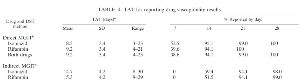
the MOP on all five occasions (Tables 1 and 2). Table 4
de-scribes the TATs with the three methods. The direct MGIT
system provided DST results 2 to 13 (mean, 6.1) days sooner
than the indirect MGIT method (
P
Ⰶ
0.001), which in turn
produced results 9 to 91 (mean, 44.3) days earlier than the
indirect MOP (
P
Ⰶ
0.001).
[image:2.612.52.293.92.206.2]The manufacturer instructs that indirect MGIT DSTs are
invalid and should be repeated if the GC tube does not
fluo-resce by day 12. No such invalid indirect MGIT tests occurred
in this study. A similar interval for invalidating direct MGIT
DSTs was not applied in this pilot evaluation. A review of the
data found that the GC tube became positive more than 12
days after inoculation for two (22.2%) of the nine specimens
producing discordant isoniazid or rifampin results by the direct
TABLE 1. Isoniazid and rifampin susceptibility results by direct
MGIT, indirect MGIT, and the proportion methods
Drug and MOP classificationa
MGIT results (no. of isolates)b Indirect R Indirect S
Direct R Direct S Direct R Direct S
Isoniazid
Resistant
75
Susceptible
2
1
23
Rifampin
Resistant
49
4
2
Susceptible
46
aGold standard results obtained by MOP in the Mariinsk laboratory with any discordant results confirmed in the Antwerp reference laboratory.
bMGIT results obtained by direct and indirect methods. R, resistant; S, sus-ceptible.
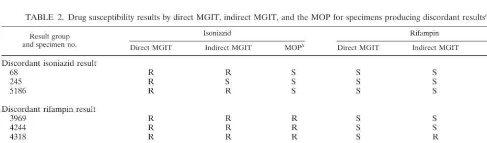
TABLE 2. Drug susceptibility results by direct MGIT, indirect MGIT, and the MOP for specimens producing discordant results
aResult group and specimen no.
Isoniazid Rifampin
Direct MGIT Indirect MGIT MOPb Direct MGIT Indirect MGIT MOPb
Discordant isoniazid result
68
R
R
S
S
S
S
245
R
S
S
S
S
S
5186
R
R
S
S
S
S
Discordant rifampin result
3969
R
R
R
S
S
R
4244
R
R
R
S
S
R
4318
R
R
R
S
R
R
4883
R
R
R
S
R
R
5114
R
R
R
S
R
R
5391
R
R
R
S
R
R
aDrug susceptibility result: R, resistant; S, susceptible.
bGold standard results obtained by MOP in the Mariinsk laboratory with any discordant results confirmed in the Antwerp reference laboratory.
on May 15, 2020 by guest
http://jcm.asm.org/
[image:2.612.54.550.565.711.2]MGIT method compared with only 7 (7.6%) of 92 concordant
specimens (
P
⫽
0.18).
DISCUSSION
This is the first published evaluation of direct DST using the
MGIT system. Similar trials of direct DST by radiometric
BACTEC were performed when that system was introduced in
the 1980s (14). Both systems share the advantages of being
rapid and of testing the actual mycobacterial population
caus-ing the patient’s disease instead of a selected subset that is
(preferentially) cultivated in vitro during primary isolation.
Fortunately, the direct MGIT DST system does not appear to
have some of the disadvantages that have limited the
wide-spread use of direct radiometric BACTEC DST (8). For
ex-ample, unlike the direct MGIT DST system which used a
different “critical proportion” to define resistance than the
indirect radiometric BACTEC DST, the criteria for defining
resistance in the indirect MGIT DST also appears appropriate
for the direct MGIT DST. The manufacturer stipulates that
indirect MGIT DST results are only valid if the GC tube
becomes positive within 12 days of inoculation. The present
study found that discordant results tended to occur more
fre-quently with the direct MGIT method among specimens
incu-bated beyond this time. However, this association did not reach
statistical significance, with only nine discordant results.
Fur-ther experience with the direct MGIT DST method is required
to define an upper limit for the incubation time that optimizes
test performance.
Contamination did not prove to be a problem in the direct
MGIT DST despite the enriched Middlebrook medium that is
used in the tubes. As in the previous direct radiometric
BACTEC DST evaluations, PANTA antibiotic solution was
added to limit contamination. The high concordance (i.e., 96 to
99.0%) between the direct and indirect MGIT methods
sug-gests that the addition of PANTA has had little effect on the
direct DST results. Unnecessary performance of DSTs on
non-tuberculous mycobacteria was not a problem in this Siberian
prison population with a high prevalence of TB but, as with the
direct radiometric BACTEC DST, would presumably be a
problem in low-prevalence populations. Finally, this evaluation
found that the direct MGIT system produced DST results for
both isoniazid and rifampin 2 to 13 (mean, 6.1) days earlier
than when using MGIT for primary isolation and then an
indirect DST. Though statistically significant, the actual clinical
benefit of this 6-day time-saving remains to be defined.
[image:3.612.53.550.84.175.2]The present study does have some limitations. First, this
initial evaluation of direct MGIT DST used only strongly
smear-positive specimens to ensure that a significant quantity
of acid-fast bacilli was present in each DST. Second, we only
evaluated the direct MGIT system for obtaining isoniazid and
rifampin susceptibility results. This approach was adopted
be-cause these two drugs are the key elements in short-course
TABLE 3. Accuracy and reliability of direct and indirect MGIT compared with the MOP
aDrug and
MGIT method % Sensitivity(95% CI) % Specificity(95% CI) % PVR (95% CI) PVS (95% CI) Accuracy (95%CI)
Isoniazid
Direct
100 (95.2–100)
88.5 (69.8–97.6)
96.2 (89.2–99.2)
100 (85.2–100)
97.0 (91.6–99.4)
Indirect
100 (95.2–100)
92.3 (74.9–99.1)
97.4 (90.9–99.7)
100 (85.8–100)
98.0 (93.0–99.8)
Rifampin
Direct
89.1 (77.8–95.9)
100 (92.3–100)
100 (92.7–100)
88.5 (76.6–95.6)
94.1 (87.5–97.8)
Indirect
96.4 (87.5–99.6)
100 (92.3–100)
100 (93.3–100)
95.8 (85.7–99.5)
98.0 (93.0–99.8)
aGold standard results were obtained by MOP in the Mariinsk laboratory with any discordant results confirmed in the Antwerp reference laboratory. 95% CI, 95% confidence interval calculated using the exact binomial method.
TABLE 4. TAT for reporting drug susceptibility results
Drug and DST method
TAT (days)a % Reported by day:
Mean SD Range 7 14 21 28 42
Direct MGIT
bIsoniazid
8.5
3.4
3–23
52.5
95.1
99.0
100
Rifampin
9.2
3.4
4–21
39.6
94.1
100
Both drugs
9.2
3.4
4–23
38.6
94.1
99.0
100
Indirect MGIT
cIsoniazid
14.7
4.2
8–30
0
59.4
94.1
98.0
100
Rifampin
15.3
4.2
9–29
0
51.5
94.1
99.0
100
Both drugs
15.3
4.2
9–30
0
51.4
94.1
98.0
100
MOP
b,c,d59.6
21.2
26–101
0
0
0
1.0
17.8
aTAT calculated in days from the date of specimen processing to the date of the DST report (including the time required for primary isolation by MGIT for the indirect MGIT DST and on solid media for the MOP).
bSignificant difference in TAT for obtaining isoniazid, rifampin, and both drug susceptibility results by direct MGIT compared with the MOP results (PⰆ0.001 for all three comparisons).
cSignificant difference in TAT for obtaining isoniazid, rifampin, and both drug susceptibility results by indirect MGIT compared with the MOP results (PⰆ0.001 for all three comparisons).
dThe cumulative percentages of DST results available by the proportion method after 56, 70, 84, and 112 days were 59.4, 68.3, 80.2, and 100%, respectively.
on May 15, 2020 by guest
http://jcm.asm.org/
[image:3.612.51.548.534.669.2]chemotherapy and provide the most robust DST results (13).
Third, the study cohort contained only 26 isoniazid-susceptible
specimens, so the estimated performance of the direct MGIT
system for isoniazid susceptibility testing is inexact, with wide
95% confidence intervals (e.g., specificities of 69.8 to 97.6%;
Table 3). Further studies will therefore be required to assess
the performance and TAT of direct MGIT DST for weakly
smear-positive specimens and for performing streptomycin and
ethambutol susceptibility tests and to evaluate in more detail
isoniazid susceptibility testing by the direct MGIT method.
Finally, the present study did not compare direct MGIT
DST with direct DST on a solid medium because the Mariinsk
laboratory did not routinely perform such tests. Direct agar
dilution susceptibility testing is a recognized inexpensive
alter-native that can provide DST results within 3 to 4 weeks (8, 11,
14). However, direct agar DST can be confounded by bacterial
contamination, under- or overgrowth in controls that
invali-date about 15% of tests, and potential inactivation of the test
drug during prolonged incubation. For example, Libonati et al.
(14) found that direct agar DST provided reportable results in
only 41% of smear-positive cases and 62% of culture-positive
cases. Other low-technology techniques, such as the
colorimet-ric Alamar Blue assay, microscopic observation of broth
cul-tures, and direct DST on novel agar media (5, 6, 8, 20), have
also been developed for rapid DST in low-income countries,
but these “in-house” alternatives may not be as robust as
MGIT and do require considerable laboratory expertise.
In contrast, the MGIT system was quickly and easily
imple-mented in this low-resource prison TB laboratory. Only one
modification to the laboratory’s standard practices was
re-quired. In the training period before this study commenced,
some growth failures in the MGIT system were attributed to
pH variations in inocula processed by the Petroff method (the
usual decontamination method used in the prison laboratory);
use of the NALC-NaOH method as recommended by the
man-ufacturer quickly resolved this problem, and no growth failures
occurred during the study.
In summary, this study has demonstrated that the
nonauto-mated MGIT system is a dependable, rapid method for
per-forming direct DST. This evaluation has also confirmed that
the excellent performance and rapid TAT reported for indirect
MGIT DST in other studies (2, 3, 18, 19, 21, 22, 24, 27) can be
reproduced in a low-resource setting. The MGIT system
there-fore represents appropriate technology for laboratories in
these countries. Cost is the only prohibitive factor. The MGIT
system and similar nonradiometric techniques are becoming
the accepted gold standard methods for mycobacterial
cultiva-tion in high-income countries with low prevalences of TB (1, 9,
15, 16, 25). These techniques are even more necessary in areas
with a high prevalence of MDRTB. International
organiza-tions, biomedical companies, and governments must develop
arrangements that give low-income countries access to these
new technologies if TB care is to be seen as globally equitable
and the (MDR)TB epidemic controlled.
ACKNOWLEDGMENTS
This study was funded by BD Biosciences–Europe. I.B. was
sup-ported by a Neil Hamilton Fairley Fellowship (987069) awarded by the
National Health and Medical Research Council of Australia.
We thank Natalya Vezhnina, the prison medical personnel, and the
MSF staff in Mariinsk for their help, the laboratory personnel at
Colony 33 and at the Institute of Tropical Medicine in Antwerp for
their technical assistance, and Ulrike Kunert for her support.
REFERENCES
1.Alcaide, F., M. A. Benı´tez, J. M. Escriba`, and R. Martı´n.2000. Evaluation of the BACTEC MGIT 960 and the MB/BacT systems for recovery of myco-bacteria from clinical specimens and species identification by DNA Accu-Probe. J. Clin. Microbiol.38:398–401.
2.Bergmann, J. S., and G. L. Woods.1997. Mycobacterial growth indicator tube for susceptibility testing ofMycobacterium tuberculosisto isoniazid and rifampin. Diagn. Microbiol. Infect. Dis.28:153–156.
3.Bergmann, J. S., G. Fish, and G. L. Woods.2000. Evaluation of the BBL MGIT (mycobacterial growth indicator tube) AST SIRE system for antimy-cobacterial susceptibility testing ofMycobacterium tuberculosisto 4 primary antituberculous drugs. Arch. Pathol. Lab. Med.124:82–86.
4.Canetti, G., W. Fox, A. Khomenko, H. T. Mahler, M. K. Menon, D. A. Mitchison, N. Rist, and N. A. Sˇmelov.1969. Advances in techniques of testing mycobacterial drug sensitivity, and the use of sensitivity tests in tuberculosis control programmes. Bull. W. H. O.41:21–43.
5.Caviedes, L., T.-S. Lee, R. H. Gilman, P. Sheen, E. Spellman, E. H. Lee, D. E. Berg, S. Montenegro-James, and the Tuberculosis Working Group in Peru.
2000. Rapid, efficient detection and drug susceptibility testing of Mycobac-terium tuberculosisin sputum by microscopic observation of broth cultures. J. Clin. Microbiol.38:1203–1208.
6.Franzblau, S. G., R. S. Witzig, J. C. McLaughlin, P. Torres, G. Madico, A. Hernandez, M. T. Degnan, M. B. Cook, V. K. Quenzer, R. M. Ferguson, and R. H. Gilman.1998. Rapid, low-technology MIC determination with clinical Mycobacterium tuberculosisisolates by using the microplate Alamar Blue assay. J. Clin. Microbiol.36:362–366.
7.Frieden, T. R., L. F. Sherman, K. L. Maw, P. I. Fujiwara, J. T. Crawford, B. Nivin, V. Sharp, D. Hewlett, Jr., K. Brudney, D. Alland, and B. N. Kreis-worth.1996. A multi-institutional outbreak of highly drug-resistant tubercu-losis: epidemiology and clinical outcomes. JAMA276:1229–1235. 8.Heifets, L.2000. Conventional methods for antimicrobial susceptibility
test-ing ofMycobacterium tuberculosis, p. 133–143.InI. Bastian and F. Portaels (ed.), Multidrug-resistant tuberculosis. Kluwer Academic Publications, Am-sterdam, The Netherlands.
9.Heifets, L., T. Linder, T. Sanchez, D. Spencer, and J. Brennan.2000. Two liquid medium systems, Mycobacteria Growth Indicator Tube and MB Re-dox tube, forMycobacterium tuberculosisisolation from sputum specimens. J. Clin. Microbiol.38:1227–1230.
10. Huebner, R. E., R. C. Good, and J. I. Tokars.1993. Current practices in mycobacteriology: results of a survey of state public health laboratories. J. Clin. Microbiol.31:771–775.
11. Kent, P. T., and G. P. Kubica (ed.).1985. Public health mycobacteriology: a guide for the level III laboratory. U.S. Department of Health and Human Services, Atlanta, Ga.
12. Kimerling, M. E., H. Kluge, N. Vezhnina, T. Iacovazzi, T. Demeulenaere, F. Portaels, and F. Matthys.1999. Inadequacy of the current WHO re-treat-ment regimen in a central Siberian prison: treatre-treat-ment failure and MDR-TB. Int. J. Tuberc. Lung Dis.3:451–453.
13. Laszlo, A., M. Rahman, M. Raviglione, F. Bustreo, and The WHO/IUATLD Network of Supranational Reference Laboratories.1997. Quality assurance programme for drug susceptibility testing ofMycobacterium tuberculosisin the WHO/IUATLD supranational laboratory network: first round of profi-ciency testing. Int. J. Tuberc. Lung Dis.1:231–238.
14. Libonati, J. P., C. E. Stager, J. R. Davis, and S. H. Siddiqi.1988. Direct antimicrobial drug susceptibility testing ofMycobacterium tuberculosisby the radiometric method. Diagn. Microbiol. Infect. Dis.10:41–48.
15. Lumb, R., and I. Bastian.1998. Recent developments in the epidemiology and laboratory diagnosis of tuberculosis. Rec. Adv. Microbiol.6:122–187. 16. Metchock, B., F. S. Nolte, and R. J. Wallace, Jr.1999.Mycobacterium, p.
399–437.InP. R. Murray, E. J. Baron, M. A. Pfaller, F. C. Tenover, and R. H. Yolken (ed.), Manual of clinical microbiology, 7th ed. ASM Press, Washington, D.C.
17. Pablos-Me´ndez, A., M. C. Raviglione, A. Laszlo, N. Binkin, H. I. Rieder, F. Bustreo, D. L. Cohn, C. S. B. Lambregts-van Weezenbeek, S. J. Kim, P. Chaulet, and P. Nunn for the World Health Organization-International Union against Tuberculosis and Lung Disease Working Group on Antitu-berculosis Drug Resistance Surveillance.1998. Global surveillance for an-tituberculosis-drug resistance, 1994–1997. N. Engl. J. Med.338:1641–1649. 18. Palaci, M., S. Y. M. Ueki, D. N. Sato, M. A. da Silva Telles, M. Curcio, and
E. A. M. Silva.1996. Evaluation of mycobacteria growth indicator tube for recovery and drug susceptibility testing ofMycobacterium tuberculosis iso-lates from respiratory specimens. J. Clin. Microbiol.34:762–764. 19. Palomino, J. C., H. Traore, K. Fissette, and F. Portaels.1999. Evaluation of
Mycobacteria Growth Indicator Tube (MGIT) for drug susceptibility testing ofMycobacterium tuberculosis. Int. J. Tuberc. Lung Dis.3:344–348. 20. Palomino, J. C., and F. Portaels.1999. Simple procedure for drug
suscepti-bility testing ofMycobacterium tuberculosisusing a commercial colorimetric
on May 15, 2020 by guest
http://jcm.asm.org/
assay. Eur. J. Clin. Microbiol. Infect. Dis.18:380–383.
21.Reisner, B. S., A. M. Gatson, and G. L. Woods.1995. Evaluation of myco-bacteria growth indicator tubes for susceptibility testing ofMycobacterium tuberculosisto isoniazid and rifampin. Diagn. Microbiol. Infect. Dis.22:325– 329.
22.Ru¨sch-Gerdes, S., C. Domehl, G. Nardi, M. R. Gismondo, H.-M. Welscher, and G. E. Pfyffer.1999. Multicenter evaluation of the mycobacterial growth indicator tube for testing susceptibility ofMycobacterium tuberculosis to first-line drugs. J. Clin. Microbiol.37:45–48.
23. Tenover, F. C., J. T. Crawford, R. E. Huebner, L. J. Geiter, C. R. Horsburgh, Jr., and R. C. Good.1993. The resurgence of tuberculosis: is your laboratory ready? J. Clin. Microbiol.31:767–770.
24. Walters, S. B., and B. A. Hanna.1996. Testing of susceptibility of Mycobac-terium tuberculosisto isoniazid and rifampin by mycobacterium growth indi-cator tube method. J. Clin. Microbiol.34:1565–1567.
25. Woods, G. L., G. Fish, M. Plaunt, and T. Murphy.1997. Clinical evaluation of Difco ESP Culture System II for growth and detection of mycobacteria. J. Clin. Microbiol.35:121–124.
26. World Health Organization.1998. Laboratory services in tuberculosis con-trol. WHO/TB/98.258. World Health Organization, Geneva, Switzerland. 27. Zapata, P., M. Arbeloa, and J. Aznar.1999. Evaluation of mycobacteria
growth indicator tube (MGIT) for drug susceptibility testing of Mycobacte-rium tuberculosisisolates from clinical specimens. Clin. Microbiol. Infect.
5:227–230.