R E S E A R C H A R T I C L E
Open Access
Regulatory effects of glycyrrhizae radix
extract on DSS-induced ulcerative colitis
Yong-Deok Jeon
1, Keuk-Soo Bang
1, Min-Kyoung Shin
1, Jong-Hyun Lee
2, Young-Nam Chang
1and Jong-Sik Jin
1*Abstract
Background:Glycyrrhizae Radix (GR) is a Korean traditional herb medicine that is widely-used in clinical health care. The clinical functions of GR include relief of toxicity, anti-cancer, regulating blood cholesterol and anti-inflammation. This study investigated the role of GR on ulcerative colitis in a dextran sulfate sodium (DSS)-induced mouse model of colitis.
Method:Western blot analysis and enzyme-linked immunosorbent assay (ELISA) analyses were done on male BALB/c mice administered 5 % DSS during the experimental period. Ethanol extracts of GR were orally administered at same time daily to control mice. The severity of colitis was measured by body weight change and colon length. Result:DSS-treated mice displayed weight loss and shortened colon length compared with control mice. Mice were administered GR showed less weight loss and longer colon length than the DSS-treated group. Inflammatory cytokines were decreased by GR treatment. Treatment also reduced DSS-induced microscopic damage to colon tissue. GR regulated the phosphorylation of transcription factors such as NF-κB p65 and IκBα.
Conclusions:GR has beneficial effects in a colitis model. GR might be a useful herb medicine in the treatment of ulcerative colitis.
Keywords:Glycyrrhizae radix, Ulcerative colitis, Colon, Dextran sulfate sodium, COX-2, PGE2
Background
Ulcerative colitis (UC) is a chronic and relapsing inflammatory disease characterized by dysregulation of the immune function response and imbalanced release of cytokines and unresolved inflammatory progress associated with intestinal mucosa [1, 2]. The inflamma-tion reacinflamma-tion may be initiated by proinflammatory cyto-kines [3]. Patients with bowel disease have increased interleukin-(IL) 6 in the intestinal mucosa and tumor necrosis factor-alpha (TNF-α) in the blood and colon tissue. Immune cells (T cells, intestinal epithelial cells, macrophages) secrete various cytokines including TNF-α, IL-1, IL-6, IL-8 and interferon-gamma (IFN-γ), which regulate the inflammatory response in UC. Elevated levels of cytokines have also been implicated in the pathogenesis of bowel disease [4, 5].
Blocking the generation of inflammatory cytokines is an area of robust research in ulcerative colitis therapy [6]. In chronic colitis, inflammation extends into the colon muscle layer with acute manifestations, followed by an asymptomatic period and a relapse that occurs months or even years later [7]. UC affects the rectum and extends in a retrograde manner with extensive areas of superficial lesions [8]. The pathogenic factors of UC are unclear, but heredity, immune system dysfunction, and dietary habits could be influential [9].
Dextran sulfate sodium (DSS)-induced colitis is a suitable mice model characterized by the morphologic-ally and biochemicmorphologic-ally. DSS induces bloody stools, ulcerations, epithelial injury and infiltration of inflam-matory cells [10]. Histologically, DSS influences crypt abscesses and epithelioglandular hyperplasia in mice. DSS induced crypt architecture, disappearance of crypts and inflammatory infiltration. During DSS colitis, inter-cellular distance of mucosal cells in crypt and vascular endothelial cell was increased [11]. These colon condi-tions are similar to acute and chronic UC in humans.
* Correspondence:jongsik.jin@jbnu.ac.kr
1Department of Oriental Medicine Resources, Chonbuk National University, 79 Gobongro, Iksan, Jeollabuk-do 570-752, Republic of Korea
Full list of author information is available at the end of the article
DSS is toxic to gut epithelial cells of the basal crypts and affects the integrity of the mucosal barrier [12]. Thus, the DSS-induced colitis model is especially suit-able to study mechanism of inflammatory colitis.
Cyclooxygenase is a constitutive enzyme that catalyzed the production of prostaglandins (PGs), which protect the stomach from damage. COX-2 is induced by inflam-matory stimuli, such as cytokines, and also catalyzed PG production that contributes to inflammation-related swelling [13]. COX-2 and PGE2 levels are raised in the inflamed mucosa of patients with UC [14]. So, to eluci-date reduction of COX-2 and PGE2 shows another pathway to ameliorate inflammation.
Glycyrrhiza Uralensis is a medical plant grown in Europe and Asia. It has been used as a herbal medicine respiratory system and gastrointestinal tract medicine [15]. Glycyrrhizae Radix (GR) has been traditionally used for treatment of cough, anti-allergy and bowel dis-ease [16]. GR possesses various bioactive constitutents that include saponins, flavonoids and coumarins [17]. Constituents of GR have anti-inflammatory effects in lipopolysaccharide-stimulated macrophage. Glycyrrhi-zin suppressed nitric oxide, TNF-α and PGE2levels in LPS-stimulated macrophage. Glycyrrhetic acid and glycyrol have anti-inflammatory effect by regulation of NF-κB pathway in LPS-treated RAW264.7 macro-phage through inhibition of inflammatory cytokines such as IL-6, IL-1β, and TNF-α. Also glycyrol regu-lates COX-2 and iNOS expression in LPS-treatment [18]. These results supposed that GR has anti-inflammatory effects in vitro. Therapeutic activities attributed to GR include anti-cancer, anti-diabetes, antioxidant and anti-inflammation [19]. However, it is unknown whether GR or its constitutents can regulate intestinal inflammation.
To explore the potential of GR as a useful therapeutic in UC, we examined its effects on DSS-induced colitis in a mouse model. The goals were to evaluate the effect of GR on clinical signs including weight loss, colon shortening, diarrhea and gross bleeding, and to investi-gate the role of GR on inflammatory mediators in DSS-treated mice.
Methods
Preparation of GR
GR purchased from Humanherb (kyuong San, Korea). GR extract was prepared by decocting with 70 % ethanol for 3 h (100 g/L). The solvent ethanol was filtered and allowed to evaporate using a rotary evaporator at a temperature of 40–45 °C. The yield of dried extract was 3.022 %. The extract was diluted in 0.9 % saline and filtered through a 0.22μm syringe filter (HYUNDAI Micro, Seoul, Korea).
Mice
Male BALB/c, 6-week-old mice obtained from SAMTACO (Osan, Kyungki-Do, Korea) were acclimatized in a specific pathogen-free environment under controlled conditions (22 ± 2 °C with a 12 h light/dark cycle) for a week. The mice were housed in five colony cages with six mice per cage.
Experimental design
All experimental protocols (CBNU2015-089) were ap-proved by the Committee on the Care of Laboratory Animal Resources, Chonbuk National University and were conducted in accordance with the Guide for the Care and Use of Laboratory Animals. Acute colitis was induced by administering drinking water containing 5 % (w/v) DSS (MP Bio, Santa Ana, CA, USA) to mice for 10 days. Thirty mice were weighted before the experi-ment, and were divided into five groups with six mice per group. Group 1 comprised untreated mice. Mice in group 2 received DSS. In group 3 and group 4, mice with induced colitis were treated with GR 100 mg/kg or 50 mg/kg, respectively. In group 5, mice with induced colitis were treated with 50 mg/kg5-aminosalicylic acid (5-ASA; Sigma-Aldrich, St. Louis, MO, USA) as the ref-erence drug. Mice were checked daily for body weight, stool consistency and the presence of gross bleeding. GR was diluted with purified water and orally administered once a day during the 10 days of DSS treatment, after which the mice were sacrificed.
Disease Activity Index (DAI)
[image:2.595.305.541.648.731.2]Intestinal disease activity was assessed based on the weight loss, presence of diarrhea accompanied by blood and mucus, and colonic shortening [20]. DAIs were cal-culated by scoring weight loss, diarrhea and rectal bleed-ing based on the scorbleed-ing system shown in Table 1 that was previously described by Murthy et al. [21]. Weight loss was defined as the difference between initial and final weights, and diarrhea as the absence of fecal pellet formation and the presence of continuous fluid fecal material in the colon. Rectal bleeding was assessed based on the presence of diarrhea containing visible blood and on the presence of gross rectal bleeding. DAI values were calculated as {(weight loss score) + (diar-rhea score) + (rectal bleeding score)}/4. The DAI was
Table 1Criteria for disease activity index
Score Weight loss (%) Stool Consistency Bloodstain or gross Bleeding
0 None Normal Negative
1 1-5 Loose stool Negative
2 5-10 Loose stool Positive
3 10-15 Diarrhea Positive
determined by three investigators blinded to the proto-col. The clinical parameters used in the present study were chosen to represent the subjective clinical symp-toms observed in human UC.
Cytokine assays
Levels of IL-6 and TNF-α in the serum and tissue were measured using an enzyme-linked immunosorbent assay (ELISA), as previously described [22]. Briefly, 96-well
[image:3.595.55.291.100.192.2]plates (SPL Life Science, Seoul, Korea) were coated with 100 μL of anti-mouse monoclonal antibody (1.0 mg/mL at pH 7.4 in phosphate buffered saline [PBS]) and incu-bated overnight at 4∘C. After additional washes, 50μL of sample, or IL-6 and TNF-α (BD Bioscience, San Diego, CA, USA) standard was added and incubated at room temperature for 2 h. Plates were washed and 0.2μg/mL of biotinylated anti-mouse antibody was added and in-cubated at room temperature for 2 h. After washing, avidin-peroxidase (Sigma-Aldrich, St. Louis, MO, USA) was added and plates were incubated for 30 min at 37∘C. The plates were then washed again and (2,2′-azino-bis [3-ethylbenzothiazoline-6-sulfonic acid]-diammonium salt) substrate (BD Bioscience, San Diego, CA, USA) was added. Color development was measured at 405 nm using an automated microplate ELISA reader (Molecular Devices, Sunnyvale, CA, USA). Standard curves were pre-pared using serial dilutions of recombinant antibody (BD Bioscience, San Diego, CA, USA). Protein concentrations
Table 2Criteria for assessment of microscopic rectal damage
Score Remarks
0 Normal colonic mucosa
1 Loss of one-third of the crypts
2 Loss of two-third of the crypts
3 Lamina propria covered with single layer of epithelial cells with mild inflammatory cell infiltration
4 Erosions and marked inflammatory cell infiltration
[image:3.595.57.539.327.676.2]were measured using bicinchoninic acid (BCA) protein assay reagent (Sigma-Aldrich, St. Louis, MO, USA).
Western blot
The end of experiment, Mice were sacrificed and the section of colons (5–8 mm) were dissected and flushed with PBS. Distal colons were homogenized in lysis buffer (iNtRON Biotech, Republic of Korea) and centrifuged at 16,582 g for 5 min. The supernatants were transferred to fresh tubes and protein concentrations were determined using BCA protein assay reagent (Sigma-Aldrich, St. Louis, MO, USA). Lysates (50μg protein) were separated by 10 % SDS-PAGE and transferred to membranes (Amersham Pharmacia Biotech, Piscataway, NJ, USA), which were blocked with 5 % skim milk in 0.05 % PBS-Tween-20 (PBST) for 1 h at room temperature. Mem-branes were incubated overnight with primary antibodies against phospho-extracellular signal regulated kinase (pERK), phospho-3-Jun, N-terminal kinase (p-JNK),
p38 (Cell Signaling technology, Danvers, MA, USA) and glyceraldehyde-phosphate dehydrogenase (GAPDH) (Thermo scientific, Waltham, MA, USA) and washed three times with PBST. Blots were incubated with secondary antibody for 1 h at room temperature. Antibody-specific proteins were visualized using an enhanced chemilumines-cence detection system (Amersham, Newark, NJ, USA). Protein densities were quantified by densitometry.
Histological processing
Mice were sacrificed at the end of the experiment. The en-tire colon was dissected and flushed with ice-cold PBS. Sections of rectums were taken and fixed in 10 % neutral-buffered formalin (Sigma-Aldrich) for 24 h at room temperature and embedded in paraffin to provide sections for histological evaluation. Severity of colitis was evaluated in sections stained with Hematoxylin and Eosin (H&E; Muto Pere Chemicals, Tokyo, Japan and Sigma-Aldrich) by two independent observers blinded to the experimental Fig. 2Effect of GR on DSS-induced colon shortening. Ulcerative colitis was induced and GR and 5-ASA administered as described in the legend to Fig. 1. Colon length steadily shortened in mice receiving DSS.aColon was harvested on day 10, and colon lengths were measured.bColon lengths in the five groups. Values represent mean ± S.E.M (n= 6). Data were analyzed by Tukey post hoc test (#P< 0.05 versus control and *
[image:4.595.60.537.87.442.2]conditions according to the modified criteria of Hama-moto et al. [23] summarized in Table 2. Mucosal damage was scored as 0–4 based on the loss of crypt (mucosa) and infiltration of inflammatory cells (maximum score = 4).
PGE2assay
Distal colons were homogenized in lysis buffer (iNtRON Biotech, South Korea), and centrifuged at 16,582 g for 5 min. PGE2levels were quantified using immunoassay kits according to the manufacturer’s instructions (Enzo Life Science, East Farmingdale, NY, USA).
Identification of constituents of GR extract
GR extract lycyrrhizae was dissolved in methanol (1 mg/ml). Glycyrrhizin and glycyrrhetic acid (Tokyo Chemical Industry, Tokyo, Japan) were dissolved in methanol for high-performance liquid chromatography (HPLC) analysis. HPLC was performed on a Waters 2695 separation module (Waters, Milford, MA, USA) coupled with a model 2487 dual λ absorbance detector
(Waters) under the following conditions: column, TSK-gel ODS-80Ts (4.6 mm × 150 mm; Tosoh Co., Tokyo, Japan); mobile phase, 0.1 % formic acid (solvent system A) and acetonitrile (solvent system B) in a gradient mode (B from 30 to 60 % in 15 min, B from 60 to 100 % from 15 to 30 min, B 100 % from 30 to 40 min); flow rate, 0.5 ml/min; temperature, 30 °C and ultraviolet wavelength of 254 nm.
Statistical analysis
All results are presented as the mean ± S.E.M. Results were analyzed using Graph Pad Prism version 5.0 pro-gram (Graph Pad Software, Inc, La Jolla, CA, USA). One-way analysis of variance with Tukey post hoc test was used to determine statistically significant differences.
P<0.05 was considered significant.
Result
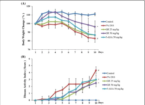
Effects of GR on clinical signs in DSS-induced colitis
[image:5.595.58.541.87.434.2]respectively, compared to the control group. Both GR and 5-ASA alleviated the DSS effects on body weight loss and colon shortening (Figs. 1a, 2a and b). GR also attenuated the DSS-mediated increase in DAI scores (Fig. 1b).
Effect of GR on IL-6 and TNF-αlevels in DSS-induced UC
Serum IL-6 level was significantly higher in the DSS treatment group (85.403 ± 10.719 pg/mL) than in the control group (9.44 ± 0.942 pg/mL). IL-6 levels were lower in the GR groups (25 mg/kg; 63.620 ± 7.942 pg/mL, 50 mg/kg; 36.143 ± 6.652 pg/mL) (Fig. 3a). The serum TNF-α level was also increased in the DSS treatment group (828.623 ± 110.387 pg/mL) compared to control group (95.786 ± 11.485 pg/mL), but was lower in the GR groups (50 mg/kg; 391.052 ± 63.914 pg/mL) (Fig. 3b). In addition, tissue IL-6 and TNF-α levels were significantly higher in the DSS treatment group (524.809 ± 28.643, 5648.749 ± 395.143 pg/mL) than in the control group (61.640 ± 9.275, 1316.768 ± 156.371 pg/mL), but were significantly decreased by GR (25 mg/kg; 241.031 ±
31.794, 3258.119 ± 502.022 pg/mL, 50 mg/kg; 127.148 ± 16.579, 2303.215 ± 90.442 pg/mL) (Fig. 3c and d).
Effect of GR on transcription factors in DSS-induced colitis
Nuclear factor-kappa B (NF-κB) is a important transcrip-tion factor of inflammatranscrip-tion reactranscrip-tions [24]. The effect of GR on the activation of NF-κB was investigated. Phos-phorylation of IκBα and NF-κB in the colon tissue was inhibited by GR treatment (Fig. 4).
Effect of GR on COX-2 and PGE2abundance in
DSS-induced colitis
[image:6.595.58.541.86.414.2]affects PGE2levels. PGE2levels were enhanced by DSS, and this increase was significantly inhibited by GR admin-istration (Fig. 5c).
Effects of GR on epithelial injury in DSS-induced colitis
Mucosal thickness is regarded as a parameter of normal mucosal condition. DSS caused epithelial injury and in-filtration of inflammatory cells, including mast cells [25]. We observed histopathological changes of colon tissue between DSS-treatment and control groups. And then examined the effect of GR treatment. GR treatment at-tenuated these effects induced by DSS treatment (Fig. 6a). GR treatment also reduced the DSS-mediated microscopic damage to the colonic tissue (Fig. 6b).
Identification of GR constituents
When standards compounds were analyzed by HPLC, the retention times of glycyrrhizin and glycyrrhetic acid were
19.5 and 29.8 unit, respectively. The two compounds were detected in GR extracts (Fig. 7). Quantitative analysis of glycyrrhetic acid indicated a concentration of 0.027 mg/g in the extract.
Discussion
[image:7.595.63.541.87.440.2]inflammatory chronic disease [32]. Approximately 10 indi-viduals per 100,000 people are diagnosed annually with UC [33].
There are over the 20 animal models of colitis. DSS-induced colitis is one of the most suitable experimental models [34]. Using this model, we presently demonstrate
that GR alleviates clinical signs of UC (weight loss, colon shortening, diarrhea and occult/gross bleeding) (Figs. 1 and 2).
IL-6, TNF-α and IFN-γ are pathogenic mediators of murine colitis [35, 36]. In IBD, immune cells including T lymphocytes and macrophages secrete inflammatory Fig. 6Effect of GR on epithelial injury in DSS-induced colitis. Ulcerative colitis was induced and GR and 5-ASA administered as described in the legend to Fig. 1.aParaffin sections of colonic tissue were stained with hematoxylin and eosin observed by microscope (100× and 400×).
bMicroscopic scores. Values represent mean ± S.E.M. (n= 6). Data were analyzed by Tukey post hoc test (#P< 0.05 versus control and*P< 0.05 versus DSS alone)
[image:8.595.58.541.86.422.2] [image:8.595.57.539.573.720.2]cytokines in the area of inflammation. These activated cells modulate the balance between pro- and anti-inflammatory cytokines. IL-6 and TNF-α expression is elevated in the rectal mucosa of UC patients [37]. Presently, GR suppressed the DSS-induced increase in IL-6 and TNF-αin mouse serum and tissues (Fig. 3).
In the inflammation reaction, COX-1 protein activity rarely changes, but COX-2 production dramatically in-creases, leading to the increased production of PGs [38]. Among the PGs, PGE2is increased in the intestines of IBD patients [39]. 5-ASA is used to treat IBD by inhibiting COX-2 activation [40]. COX-2 and PGE2play key role as mediators in UC. Presently, GR inhibited the DSS-induced activation of COX-2 and PGE2(Fig. 5). The results suggest that the anti-inflammatory effect of GR is attributable to the regulation of COX-2 in DSS-induces colitis. We also found that GR reduced epithelial injury and inflammatory cell infiltration into colon tissue (Fig. 6).
Pro-inflammatory mediators, such as inflammatory cytokines and chemokines are controlled by the activity of transcriptional factors. The activation of NF-κB is important in the pathogenesis of IBD [41–43]. DSS-induced IκBα phosphorylation and phosphorylation of NF-κB p65 in colonic tissues were suppressed by GR treatment. These results suggested that GR can inhibit the activation of transcription factors in UC.
Along with IL-6, TNF-α, COX-2 and NF-κB p65 phosphorylation, inflammation related factors including caspases and mitogen-activated protein kinases affect clinical signs [44]. Further investigation of GR effects on colitis is needed to clarify the discrepancy.
GR is used as a traditional herbal medicine in many Asian countries. Glycyrrhizin is the principal component of GR, and has been used for treatment of liver disease, allergic reaction and gastric ulcers [45]. Glycyrrhetic acid is another component, which has an anti-inflammatory effect on lipopolysaccharide-induced liver injury and inhibition of hepatic lipotoxicity [46]. These compounds might be useful for the prevention of inflammatory disease.
In summary, the anti-inflammatory activities of GR were as effective as those of 5-ASA. GR may inhibit in-flammation by regulating the inflammatory mediators COX-2 and NF-κB. GR might be a useful therapeutic agent for the treatment of inflammatory diseases.
Conclusions
GR extract reduces the clinical signs and levels of in-flammatory mediators in DSS-induced colitis in mice. GR extract suppresses PGE2production. This study pro-vides experimental evidence to show that GR might be a useful therapy in the treatment of UC.
Acknowledgement
This research was supported by Basic Science Research Program through the National Research Foundation of Korea (NRF) funded by the Ministry of
Science, ICT & Future Planning (2015R1C1A1A09054675) and the Industrial Technology Research Infrastructure Program (N0000004) funded by the Ministry of trade, Industry and Energy.
Availability of data and materials
The datasets supporting the conclusions of this article are included within the article.
Authors’contributions
Y-DJ and J-SJ performed the in vivo mouse model experiment and analyzed the data. J-HL, J-SJ and M-KS provided technical and material support. Y-DJ and M-KS collected the data, undertook the statistical analyses, and wrote the manuscript. Y-NC and J-SJ designed and supervised the study, including editing of the manuscript. All authors contributed to and have approved the final manuscript.
Competing interests
The authors declare that they have no competing interests.
Consent for publication
Not relevant in this study.
Ethics approval and consent to participate
All experimental protocols (CBNU2015-089) were approved by the Committee on the Care of Laboratory Animal Resources, Chonbuk National University and were conducted in accordance with the Guide for the Care and Use of Laboratory Animals.
Author details
1Department of Oriental Medicine Resources, Chonbuk National University, 79 Gobongro, Iksan, Jeollabuk-do 570-752, Republic of Korea.2Department of Pharmacy, College of Pharmacy, Dongduk Woman’s University, 23-1 Wolgok-Dong, SungBuk-Gu, Seoul, Republic of Korea.
Received: 23 November 2015 Accepted: 13 October 2016
References
1. Arita M, Yoshida M, Hong S, Tjonahen E, Glickman JN, Petasis NA, et al. Anendogenous lipid mediator derived from omega-3 eicopentanoic acid, protects against 2,4,6-trini-trobenzene sulfonic acid-induced colitis. Proc Natl Acad Sci U S A. 2005;102:7671–6.
2. Libby P. Inflammation in atherosclerosis. Arterioscler Thromb Vasc Biol. 2012; 32:2045–51.
3. Li Y, de Haar C, Chen M, Deuring J, Gerrits MM, Smits R, et al. Disease-related expression of the IL6/STAT3/SOCS3 signalling pathway in ulcerative colitis and ulcerative colitis-related carcinogenesis. Gut. 2010;59:227–35. 4. Oqata H, Hibi T. Cytokine and anti-cytokine therapies for inflammatory
bowel disease. Curr Pharm Des. 2003;9:1107–13.
5. Papadakis KA, Targan SR. Role of cytokines in the pathogenesis of inflammatory bowel disease. Annu Rev Med. 2000;51:589–98. 6. Mueller C. Tumour necrosis factor in mouse models of chronic intestinal
inflammation. Immunology. 2002;105:1–8.
7. Braus NA, Elliott DE. Advances in the pathogenesis and treatment of IBD. Clin Immunol. 2009;132:1.
8. Lascano CA, Soto F, Carrodeguas L, Szomstein S, Rosenthal RJ, Wexner SD. Management of ulcerative colitis in the morbidly obese epatient : is bariatric surgery indicated ? Obes Surg. 2006;16:783–6.
9. Baumgart DC, Carding SR. Inflammatory bowel disease: cause and immunobiology. Lancet. 2007;369:1627–40.
10. Cooper HS, Murthy SN, Shah RS, Sedergran DJ. Clinicopathologic study of dextran sulfate sodium experimental murine colitis. Lab Invest. 1993;69:238–49. 11. Toiyama Y, Mizoguchi A, Yoshinaga O, Koike Y, Morimoto Y, Kusunoki M,
et al. Intravital imaging of DSS-induced cecal damage in GFP-transgenic mice using two-photon microscopy. J Gastoenterol. 2010;45:544–53. 12. Okayasu I, Hatakeyama S, Yamada M, Ohkusa T, Inagaki Y, Nakaya R. A novel
method in the induction of reliable experimental acute and chronic ulcerative colitis in mice. Gastroenterology. 1990;98:694–702.
14. MacDonald TT, Murch SH. Aetiology and pathogenesis of chronic inflammatory bowel disease. Baillieres Clin Gastroenterol. 1994;8:1–34. 15. Asl MN, Hosseinzadeh H. Review of pharmacological effects of Glycyrrhiza
sp. and its bioactive compounds. Phytother Res. 2008;22:709–24. 16. Wang W, Luo M, Fu Y, Wang S, Efferth T, Zu Y. Glycyrrhizic acid
nanoparticles inhibit LPS-induced inflammatory mediators in 264.7 mouse macrophages compared with unprocessed glycyrrhizic acid. Int J Nanomedicine. 2013;8:1377–83.
17. Chang YL, Chen CL, Kuo CL, Chen BC, You JS. Glycyrrhetinic acid inhibits ICAM-1 expression via blocking JNK and NF-κB pathways in TNF-α-activated endothelial cells. Acta Pharmacol Sin. 2010;31:546–53.
18. Shin EM, Zhou HY, Guo LY, Kim JA, Lee SH, Merfort I, et al. Anti-inflammatory effects of glycyrol isolated from Glycyrrhiza uralensis in LPS-stimulated RAW264.7 macrophages. Int Immunopharmacol. 2008;8:1524–32. 19. Fiore C, Eisenhunt M, Krausse R, Ragazzi E, Pellati D, Armanini D, et al.
Antiviral effects of Glycyrrhiza species. Phytother Res. 2008;22:141–8. 20. Hendrickson BA, Gokhale R, Cho JH. Clinical aspects and pathophysiology of
inflammatory bowel disease. Clin Microbiol Rev. 2002;15:79–94. 21. Murthy SN, Cooper HS, Shim H, Shah RS, Ibrahim SA, Sedergran DJ.
Treatment of dextran sulfate sodium induced murine colitis by intracolonic cyclosporine. Dig Dis Sci. 1993;38:1722–34.
22. Kim SJ, Kim MC, Um JY, Hong SH. The beneficial effect of vanillic acid on ulcerative colitis. Molecules. 2010;15:7208–17.
23. Hamamoto N, Maemura K, Hirata I, Murano M, Sasaki S, Katsu K. Inhibition of dextran sulphate sodium (DSS)-induced colitis in mice by intracolonically administered antibodies against adhesion molecules (endothelial leucocyte adhesion molecule-1 (ELAM-1) or intercellular adhesion molecule-1 (icam-1)). Clin Exp Immunol. 1999;117:462–8.
24. Barnes PJ, Karin M. Nuclear factor-kappaB: a pivotal transcription factor in chronic inflammatory diseases. N Engl J Med. 1997;336:1066–71.
25. Kim DS, Ko JH, Jeon YD, Han Y, Kim HJ, Hong SH, et al. Ixeris dentate NAKAI reduces clinical score and HIF-1 expression in experimental colitis in mice. Evid Based Complement Altemat Med. 2013;2013:671281.
26. Dharmani P, Chadee K. Biologic therapies against inflammatory bowel disease: a dysregulated immune system and the cross talk with gastrointestinal mucosa hold the key. Curr Mol Pharmacol. 2008;1:195–212. 27. Strober W, Fuss I, Mannon P. The fundamental basis of inflammatory bowel
disease. J Clin Invest. 2007;117:514–21.
28. Sartor RB. Microbial influences in inflammatory bowel diseases. Gastroenterology. 2008;134:577–94.
29. Podolsky DK. Inflammatory bowel disease. N Engl J Med. 2002;347:417–29. 30. Rufo PA, Bousvaros A. Current therapy of inflammatory bowel disease in
children. Pediatric Drugs. 2006;8:279–302.
31. Ishiguro Y, Ohkawara T, Sakuraba H, Yamagata K, Hiraga H, Yamaguchi S, et al. Macrophage migration inhibitory factor has a proinflammatory activity via the p38 pathway in glucocorticoid-resistant ulcerative colitis. Clin Immunol. 2006;120:335–41.
32. Sandborn WJ, Targan SR. Biologic therapy of inflammatory bowel disease. Gastroenterology. 2002;122:1592–608.
33. Russel MG, Stockbrügger RW. Epidemiology of inflammatory bowel disease: an update. Scand J Gastroenterol. 1996;31:417–27.
34. Wirtz S, Neufert C, Weigmann B, Neurath MF. Chemically induced mouse models of intestinal inflammation. Nat Protoc. 2007;2:541–6.
35. Myers KJ, Murthy S, Flanigan A, Witchell DR, Butler M, Murray S, et al. Antisense oligonucleotide blockade of tumor necrosis factorαin two murine models of colitis. J Pharmacol Exp Ther. 2003;304:411–24. 36. Naito Y, Takagi T, Uchiyama K, Kuroda M, Kokura S, Ichikawa H, et al.
Reduced intestinal inflammation induced by dextran sodium sulfate in interleukin- 6-deficient mice. Int J Mol Med. 2004;14:191–6.
37. Peng JC, Shen J, Ran ZH. Novel agents in the future: Therapy beyond anti-TNF agents in inflammatory bowel disease. J Dig Dis. 2014;15:585–590. 38. Morita I. Distinct functions of COX-1 and COX-2. Prostaglandins Other Lipid
Mediat. 2002;68–69:165–75.
39. Wiercińska-Drapało A, Flisiak R, Prokopowicz D. Effects of ulcerative colitis activity on plasma and mucosal prostaglandin E2concentration. Prostaglandins Other Lipid Mediat. 1999;58:159–65.
40. Lauritsen K, Laursen LS, Kjeldsen J, Bukhave K, Hansen TK, Rask-Madsen J. Effects of mesalazine on the formation of lipoxygenase and cyclooxygenase products. Adv Exp Med Biol. 1995;371B:1301–6.
41. Perkins ND, Gilmore TD. Good cop, bad cop: the different faces of NF-κB. Cell Death Differ. 2006;13:759–72.
42. Hayden MS, Ghosh S. Signaling to NF-κB. Genes Dev. 2004;18:2195–224. 43. Lawrence T, Bebien M, Liu GY, Nizet V, Karin M. IKKαlimits macrophage
NF-κB activation and contributes to the resolution of inflammation. Nature. 2005;434:1138–43.
44. Karhausen J, Furuta GT, Tomaszewski JE, Johnson RS, Colgan SP, Haase VH. Epithelial hypoxia-inducible factor-1 is protective in murine experimental colitis. J Clin Invest. 2004;114:1098–106.
45. Fiore C, Eisenhut M, Ragazzi E, Zanchin G, Armanini D. A history of the therapeutic use of liquorice in Europe. J Ethnopharmacol. 2005;99:317–24. 46. Yoshida T, Abe K, Ikeda T, Matsushita T, Wake K, Sato T, et al. Inhibitory
effect of glycyrrhizin on lipopolysaccharide and D-galactosamine-induced mouse liver injury. Eur J Pharmacol. 2007;576:136–42.
• We accept pre-submission inquiries
• Our selector tool helps you to find the most relevant journal • We provide round the clock customer support
• Convenient online submission • Thorough peer review
• Inclusion in PubMed and all major indexing services • Maximum visibility for your research
Submit your manuscript at www.biomedcentral.com/submit