R E S E A R C H A R T I C L E
Open Access
Acute and repeated toxicological study of
Myelophil, an ethanol extract of a mixture
of
Astragali Radix
and
Salviae Miltiorrhizae
Radix, in beagle dogs
Jin-Yong Joung
1, Jin-Seok Lee
1, Jung-Hyo Cho
1, Dong-Soo Lee
2and Chang-Gue Son
1*Abstract
Background:To evaluate the pharmaceutical safety of Myelophil, an ethanol extract of a mixture ofAstragali Radix andSalviae Miltiorrhizae Radix, using both acute and repeated toxicological studies.
Methods:A total of 40 beagle dogs (20 each male and female) were fed doses up to 5,000 mg/kg for the acute study and up to 1,250 mg/kg for the 13-week repeated dose toxicological study. Adverse effects were examined intensively by comparing the differences between normal and drug-administered groups using clinical signs, autopsies, histopathological findings, hematology, urinalysis, and biochemical analysis.
Results:No mortality or drug-related clinical signs were observed in the Myelophil-treated groups, except for vomiting due to an excessive dose (5,000 mg/kg). Likewise, in the repeated toxicity test, compound-colored stools in the Myelophil-treated groups and soft stools in all groups, including the control, were observed. No drug-related abnormalities were found in the histopathology, hematology, urinalysis, and biochemical analyses for any doses of Myelophil.
Conclusion:These results support the safety of Myelophil with a no observed adverse effect level (NOAEL) of 1250 mg/kg in beagle dogs, which corresponds to a human equivalent dose (HED) of 694 g/kg.
Keywords:Myelophil, Astragali Radix, Salviae Miltiorrhizae Radix, Safety, Toxicological study, NOAEL, HED
Background
The use of complementary and alternative medicine is increasing worldwide, and many medicinal plants are be-ing used for disease treatment and health improvement purposes [1, 2]. Since medicinal plants are derived from nature and have been used for a long time, they are gen-erally regarded as safe [3]. However, recent studies have warned about the safety of medicinal plants [4, 5]. In particular, the potential hepatotoxicity and renal toxicity of medicinal plants have been reported [6,7].
Myelophil is a 1:1 mixture of the 30% ethanol extracts ofAstragali radixandSalvia radixand is used clinically to treat patients with chemotherapy/radiation
therapy-induced myelosuppression or chronic fatigue-related disorders [8, 9]. In particular, in addition to the results in an animal model, Myelophil showed anti-fatigue effectiveness in a clinical trial on idiopathic chronic fatigue [9, 10]. In addition, a previous preclinical study showed partial evidence for the safety of Myelophil from a sub-chronic toxicological study using Sprague Dawley (SD) rats [11]. Regarding the wide spectrum of Myelo-phil applications with respect to age, period and sub-jects, there is a strong demand for further evidence on the safety of Myelophil. In particular, comparisons with non-rodent-derived studies are needed due to the limita-tions of rodent-based toxicity studies [12].
Individually, both Astragali radix and Salvia radix,
which compose Myelophil, have been reported to be safe in several toxicological studies [13, 14]. These medicinal plants are known as two representative herbs used to treatQi- andBlood- disorders, respectively, and they are
© The Author(s). 2019Open AccessThis article is distributed under the terms of the Creative Commons Attribution 4.0 International License (http://creativecommons.org/licenses/by/4.0/), which permits unrestricted use, distribution, and reproduction in any medium, provided you give appropriate credit to the original author(s) and the source, provide a link to the Creative Commons license, and indicate if changes were made. The Creative Commons Public Domain Dedication waiver (http://creativecommons.org/publicdomain/zero/1.0/) applies to the data made available in this article, unless otherwise stated. * Correspondence:ckson@dju.ac.kr
1Liver and Immunology Research Center, Oriental Medical Collage of Daejeon University, 75, Daedeok-daero 176beon-gil, Seo-gu, Daejeon 35235, Republic of Korea
frequently prescribed together in a formula in clinical practice [9]. However, to date, no non-rodent animal studies have been conducted to evaluate the safety of the combination.
Myelophil is an anti-fatigue therapeutics candidate, which its effects will be evaluated via clinical study in the future. In order to provide the safety evidence according to Korea Food and Drug Administration (KFDA), this study aimed to evaluate the tolerance range in a single acute study and to estimate the no observed adverse effect level (NOAEL) of Myelophil using a 13-week repeated toxicological test on beagle dogs.
Materials and methods
Preparation and fingerprinting of Myelophil
Myelophil was prepared in powder form by Kyung-Bang Pharmacy (Incheon, Korea) as follows according to the approved good manufacturing practice (GMP) guidelines of the KFDA [15]. Myelophil is the 1:1 mixture of
Astragali Radix(Astragalus membranaceus Bunge, culti-vated in Jecheon, South Korea Ser. No. 20101106-JC-HG) andSalviae Miltiorrhizae Radix(Salvia miltiorrhiza Bunge, cultivated in Hebei, China; Ser. NO. 20110302-CHN-DS). These two herbal materials were purchased from Daeyeon Pharmacy (Supplier of standardized herbs, Incheon, Korea), and they were confirmed by expert for herbology who is an herbal pharmacist. Mye-lophil was extracted using 30% ethanol for 20 h at 80 °C and the final product obtained with a yield of 20.52% (w/w) was stored for future use. To confirm the
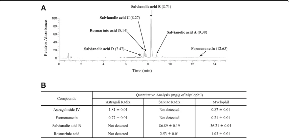
reproducibility of Myelophil’s components, ultra-high-performance liquid chromatography tandem mass spec-trometry (UHPLC-MS/MS, Thermo Scientific, San Jose, CA, USA) method was re-conducted as described previ-ously [16] (Fig. 1a). Liquid chromatography-mass spec-trometry (LC/MS, LTQ ORbitrap XL linear ion-trap MS system, Thermo Scientific, San Jose, CA, USA) was also performed on the Myelophil as well as 4 reference com-pounds (astragaloside IV and formononetin for Astragali Radix and salvianolic acid B and rosmarinic acid for Salviae Miltiorrhizae Radix, respectively) for quantitative analysis as previously described [17] (Fig.1b).
Animals
A total of 40 beagle dogs (20 males and 20 females) were purchased from Woojung BSG (Gyeonggi-do Suwon, Korea) and used for the study. Each dog was acclimated to conditions in a stainless-steel mesh box (700 mm W × 750 mm L × 750 mm H) for 3 weeks and was subjected to routine examination daily. The environment was maintained at 21.9 ± 0.8 °C with a 12-h light/dark cycle, and the air was exchanged between 10 and 15 times/h. Each dog received 300 g/day of a standard dry diet (Biopia; Gyeoggi-do Gunpo, Korea) and had free access to automatically filtered tap water that had undergone a purification process. At the time of drug administration, the dogs averaged 6–7 months of age and ranged from 5.52 to 6.72 kg for males and 5.04 to 6.42 kg for females. All animals were checked for health before the drug administration. This study was conducted in Korea
A
B
[image:2.595.60.538.466.695.2]Conformity Laboratories (KCL, Incheon, Korea), an au-thorized institute for toxicological research, and adhered to the Testing Guidelines of KFDA [18]. Experts including histopathologist, animal specialist, and drug manager con-firmed that this toxicity study was performed correctly.
Acute toxicity
After an overnight fast, one male and one female in each of the four groups received 5,000 mg/kg, 2,500 mg/kg, 1, 250 mg/kg, and 0 mg/kg doses of Myelophil using an oral capsule (single dose). Clinical signs were observed for 6 h after drug administration; thereafter, daily mortality and symptoms were observed for 2 weeks. Body weights were measured 1, 3, 7, and 14 days after administration, and necropsy was performed on day 14.
Repeated toxicity
For the repeated toxicity test, 32 beagles (16 male and 16 female) were dived into 4 groups (Control: 10 dogs at 0 mg/kg, low dose: 6 dogs at 312.5 mg/kg, middle dose: 6 dogs at 625 mg/kg and high dose: 10 dogs at 1,250 mg/kg). Over 13 weeks, each dog was administered Myelophil by using an oral capsule once a day. In the two groups with 5 males and 5 females (1,250 mg/kg and 0 mg/kg), the extra 2 males and 2 females of each group were set up as recov-ery groups for 4 weeks.
Clinical signs of toxicity were checked once a day, and body weights and feed intake were measured once a week. Before the drug administration and within 1 week prior to the necropsy, ophthalmological examination, electrocardiography, urinalysis, hematological, and vari-ous biochemical parameters were performed. After nec-ropsy, the external findings were recorded, and all organs, such as abdominal organs, thoracic organs and brain, were weighed. Histopathological examinations were performed for the following organs: brain, pituitary, heart, lungs, liver, gallbladder, kidney, bladder, mesen-teric lymph nodes, thymus, spleen, pancreas, salivary glands, submandibular lymph nodes, thyroid, adrenal gland, esophagus, aorta, Spinal cord, sciatic nerve, skeletal muscles, skin, mammary gland, eye ball, stom-ach, pancreas, thymus, thyroid gland, parathyroid gland, duodenum, jejunum, ileum, appendix, colon, rectum, femur, sternum, trachea, tongue, prostate gland, testis, epididymis, ovary, bladder, uterus, and vagina.
Statistical analysis
The analysis of the continuous data (organ weight, food intake, and hematological and biochemical parameters) was performed using one-way ANOVA. Statistical differ-ences between the groups were analyzed using Dunnett’s multiple comparison test [19]. Dunnett’s t-test was performed when the dispersion was not homogeneous. The analysis of discontinuous data used the chi-square
test after re-input of data after scale conversion. All analyses were performed using SPSS 12.0 K program, which is a widely used statistical package.
Results Acute toxicity
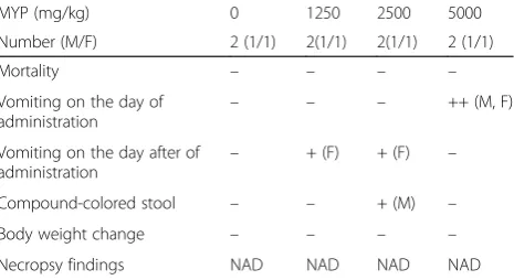
No animals died during the test. In female animals, vomiting was observed on the day of administration in the 5,000 mg/kg group and the day after administration in the 2,500 mg/kg and 1,250 mg/kg groups. Compound-colored stools were observed on the 2nd day after administration in the 2,500 mg/kg group. These symp-toms were also observed in the male animals; vomiting on the day of administration in the 5,000 mg/kg group and compound-colored stools in the 2,500 mg/kg group until day 4 after administration. There were no changes in body weight during the study period, and no abnor-mal lesions were observed at necropsy (Table1).
Repeated toxicity
Clinical signs and mortality
No deaths were observed in any group during the study period. Drug compound-colored stools were observed in the 625 and 1,250 mg/kg groups, which appeared to be dose-related in both males and females. Soft stools were observed in all the dose groups (including the control) in a dose-related pattern in both males and females. Anorexia was sporadically observed all groups but without a dose-correlation (Table2).
Weight change and feed intake
[image:3.595.306.539.553.680.2]Body weight increased gradually during the study period in all groups. However, there were no significant differ-ences in the body weights or food intake in groups administered with Myelophil compared to the controls during the study period (Fig.2).
Table 1Summary of the acute toxicity test in beagle dogs
MYP (mg/kg) 0 1250 2500 5000
Number (M/F) 2 (1/1) 2(1/1) 2(1/1) 2 (1/1)
Mortality – – – –
Vomiting on the day of administration
– – – ++ (M, F)
Vomiting on the day after of
administration –
+ (F) + (F) –
Compound-colored stool – – + (M) –
Body weight change – – – –
Necropsy findings NAD NAD NAD NAD
Beagle dogs in each group were fed Myelophil (MYP). Clinical signs were observed for 6 h after drug administration; thereafter, daily mortality and symptoms were observed for 2 weeks. At the end of the experiment, necropsy was performed
Hematologic tests
At the 13th week of administration, no abnormal param-eters were observed in the complete blood counts (CBC), including the blood-clotting time tests, in male or female animals of any group (Table3).
Biochemical tests
At the 13th week of administration, the serum concen-trations of sodium and chloride were significantly de-creased in the 312.5 and 1,250 mg/kg groups of females compared to the control group (P< 0.05). Those findings were not observed in the male groups. Other abnormal-ities in the biochemical parameters were not observed in the Myelophil-administered groups (Table4).
Urinalysis
At the 13th week of administration, occult blood was in-creased in a statistically significant manner in 312.5 mg/kg males compared with the control group, and a statistically significant decrease was observed in the 312.5, 625, and 1, 250 mg/kg females compared to the control group. These changes had no dose-relatedness or male-to-female corre-lations. No other abnormal parameters were observed in
the urinalysis in the male or female animals of all groups (data not shown).
Gross autopsy findings
Adhesion between the left lobe and the medial lobe of liver was observed in 1 animal in the 625 mg/kg group, but there was no dose correlation or histopathological abnormality. No other abnormalities associated with the administration of Myelophil were observed. No changes associated with test drug administration were observed in organ weight. No abnormalities in either eye examin-ation or electrocardiography were observed in any group before or after the test (data not shown).
Histopathologic examinations
In all of the Myelophil-treated groups and control group, infiltrations of inflammatory cells in several organs (liver, lung, thyroid, testicle), white pulp atrophy of the spleen, vacuolization in interstitium of the kidney, degranulation in the thymus, and mineral deposition in the kidney were observed sporadically. All of these abnormalities were infrequent and were not related to the dose (Additional file 1: Table S1). The normal histopatho-logical findings in the main organ (liver, lung and kidney) were shown in Fig.3.
Discussion
[image:4.595.56.292.100.237.2]Our previous studies partially proved the safety of Mye-lophil in rats [11]. Rodent-derived studies are generally thought to provide only limited information on the adverse effects of drugs in humans [20]. In preclinical studies of drugs, the U.S. Food and Drug Administration (FDA) recommended that acute and repeated toxicity testing should be conducted in at least two mammalian species, including a non-rodent species [21]. In this study, we therefore evaluated the toxicity of Myelophil using beagle dog-based acute and repeated toxicological tests. Regarding the duration for repeated dose toxicity studies, the FDA generally recommends the same duration as is used for clinical purposes but a longer
Table 2Summary of the repeated toxicity test in beagle dogs
MYP (mg/kg per day) 0 312.5 625 1250
Number (M/F) 10 (5/5) 6(3/3) 6(3/3) 10 (5/5)
Mortality – – – –
Anorexia −+ −+ −+ −+
Soft stool + + ++ +++
Compound-colored stool – – ++ +++
Food intake NAD NAD NAD NAD
Ophthalmological findings NAD NAD NAD NAD
Relative organ weight NS NS NS NS
Necropsy findings NAD NAD NAD NAD
Beagle dogs in each group were fed MYP, and the clinical symptoms, including mortality, were monitored for 13 weeks. At the end of the experiment, ophthalmological, necropsy-based examinations were performed
NADNo abnormality detected,NSNot significant;−: Absent; +: Slight; ++: Moderate; +++: Severe;−+: Sporadically detected
[image:4.595.57.539.585.705.2]period than that required for the authorization of marketing [22]. For drugs used for more than 2 weeks and less than 1 month, FDA recommends a 3-month toxicity test in non-rodent. The common prescription-period of Myelophil is 4-week; therefore we designed a 13-week repeated toxicological test in our study.
Myelophil is a mixture of Astragali radix and Salvia radix extracts that has been prescribed to a wide spectrum of patients complaining of chronic fatigue and bone marrow dysfunctions in Daejeon University Hospital since 2002. This formula (the combination of
Astragali radix and Salviae Radixextracts) was derived based on TCM theory to maintain the balance between
Qi and Blood, and this combination is supported by experimental data that showed that it improved bone marrow function [8]. Astragali radixis a medicinal herb that TCM indicates enhances Qi[23] and is reported to have immunomodulatory, anti-aging and antitumor effects [24–26]. Salviae Radix is a representative herb that is used in TCM to treat blood-related disorders [27] and has been studied for antiplatelet aggregation, anti-oxidant, and anti-inflammatory effects [28–30].
[image:5.595.57.550.100.407.2]In general, the clinical dose of Myelophil is 2,000–4, 000 mg/day for a 60 kg adult. For evaluation of this clin-ical dose, we decided the maximum dose (1250 mg/kg) in repeated toxicological test based on NOAEL and safety factor (1/10) [31]. For the acute toxicity test, we de-termined the much larger maximum dose (5,000 mg/kg) to predict approximate lethal dose (ALD). In the present re-sults, no beagle dog died following the single administration of 75 times the clinical dose of Myelophil (5,000 mg/kg), even in those dogs in which vomiting or drug compound-colored stool was observed. These symptoms were pre-sumed to be due to the inability to absorb excess drug, and no abnormalities related to these side effects were found at the time of necropsy. Accordingly, the ALD was estimated to be greater than 2,500 mg/kg. The main purpose of a pre-clinical toxicological study is to evaluate the NOAEL value, which provides the safety range of a test drug in clinical practice [32]. The NOAEL values can be determined using a repeated toxicological test, and the resulting animal-derived value can be converted to a human equivalent dose (HED) [33]. These results provide key information on the range of clinical doses [34]. The calculation of the NOAEL
Table 3Hematological analysis after administration of Myelophil for 13 weeks
MYP (mg/kg) Male Female
0 312.5 625 1250 0 312.5 625 1250
Number 5 3 3 5 5 3 3 5
WBC (103/μL) 9.4 ± 0.8 10.6 ± 2.2 10.0 ± 2.1 8.4 ± 1.3 7.6 ± 0.7 8.1 ± 1.6 8.7 ± 1.6 9.0 ± 1.7
WBC Differential Counts (%)
NE 52.0 ± 3.5 54.3 ± 4.6 49.3 ± 8.7 62.1 ± 3.0 50.3 ± 8.1 55.5 ± 5.2 50.5 ± 4.0 50.1 ± 10.2
LY 36.8 ± 6.2 37.0 ± 4.2 41.5 ± 11.4 38.8 ± 2.4 39.2 ± 7.7 35.3 ± 6.7 41.9 ± 3.9 42.1 ± 10.5
MO 4.9 ± 1.2 4.0 ± 0.7 4.4 ± 1.9 4.5 ± 1.3 4.5 ± 0.8 5.0 ± 1.4 4.5 ± 0.9 4.0 ± 1.0
EO 5.8 ± 3.3 4.2 ± 1.1 4.3 ± 1.2 4.1 ± 0.9 5.3 ± 2.9 3.6 ± 1.2 2.3 ± 0.7 3.2 ± 1.3
BA 0.3 ± 0.2 0.4 ± 0.1 0.3 ± 0.2 0.4 ± 0.1 0.6 ± 0.2 0.4 ± 0.3 0.6 ± 0.2 0.5 ± 0.2
RBC (106/μL) 7.3 ± 0.6 7.8 ± 0.3 7.5 ± 0.6 8.0 ± 0.3 8.3 ± 0.6 8.2 ± 0.8 8.4 ± 0.7 8.3 ± 0.7
Hemoglobin (g/dL) 16.3 ± 0.9 16.4 ± 0.6 16.0 ± 1.1 17.1 ± 0.9 18.5 ± 1.1 17.6 ± 1.2 18.9 ± 1.6 18.3 ± 1.2
Hematocrit (%) 49.6 ± 2.6 50.7 ± 1.4 49.7 ± 3.1 52.8 ± 2.4 57.4 ± 3.7 55.4 ± 3.8 57.6 ± 4.5 56.4 ± 3.9
MCV (fL) 68.2 ± 2.5 64.8 ± 2.1 66.7 ± 2.2 66.1 ± 3.2 69.0 ± 1.4 67.7 ± 1.8 68.4 ± 1.1 68.4 ± 1.7
MCH (pg) 22.5 ± 0.7 20.9 ± 0.8 21.4 ± 1.1 21.4 ± 1.3 22.3 ± 0.5 21.5 ± 0.7 22.4 ± 0.0 22.2 ± 0.7
MCHC (g/dL) 32.9 ± 0.2 32.3 ± 0.3 32.1 ± 0.5 32.4 ± 0.5 32.3 ± 0.4 31.7 ± 0.5 32.8 ± 0.5 32.4 ± 0.3
RDW (%) 14.1 ± 0.6 14.4 ± 0.2 14.7 ± 1.5 14.6 ± 0.9 13.4 ± 0.9 13.6 ± 0.4 13.8 ± 0.6 14.2 ± 1.1
Platelet (103/μL) 327.0 ± 44.2 326.3 ± 68.3 380.7 ± 35.6 349.6 ± 73.9 301.8 ± 54.0 352.7 ± 87.8 329.0 ± 62.4 326.4 ± 52.9
MPV (fL) 16.3 ± 1.4 17.8 ± 1.1 17.0 ± 0.3 16.6 ± 1.3 16.5 ± 1.1 16.4 ± 2.4 17.2 ± 0.8 16.6 ± 0.7
Reticulocytes (%) 1.1 ± 0.3 1.1 ± 0.2 1.1 ± 0.6 1.0 ± 0.3 0.8 ± 0.2 1.0 ± 0.3 1.1 ± 0.3 0.9 ± 0.2
PT (sec) 6.0 ± 0.2 6.00 ± 0.1 6.0 ± 0.2 6.0 ± 0.2 6.0 ± 0.2 6.1 ± 0.1 6.3 ± 0.3 6.1 ± 0.2
aPTT (sec) 12.3 ± 0.7 12.1 ± 0.3 12.2 ± 1.0 12.2 ± 0.8 12.8 ± 1.3 12.9 ± 0.8 11.5 ± 1.5 12.5 ± 0.6
After administration of MYP for 13 weeks, hematology was analyzed to compare the MYP-treated groups to the control group
WBCWhite blood cell,NENeutrophil,LYLymphocyte,MOMonocyte,EOEosinophil,BABasophil,RBCRed blood cell,MCVMean corpuscular volume,MCHMean corpuscular hemoglobin,MCHCMean corpuscular hemoglobin concentration,RDWRed cell distribution width,MPVMean platelet volume,PTProthrombin time,
Table 4Serum chemistry analysis after administration of Myelophil for 13 weeks
MYP (mg/kg, Number) Male Female
0 (5) 312.5 (3) 625 (3) 1250 (5) 0 (5) 312.5 (3) 625 (3) 1250 (5)
AST (IU/L) 38.4 ± 9.6 48.0 ± 7.2 42.3 ± 12.1 47.8 ± 6.8 40.6 ± 5.1 44.0 ± 2.0 47.3 ± 16.4 44.0 ± 6.0
ALT (IU/L) 40.0 ± 10.2 37.7 ± 9.1 31.3 ± 6.0 47.8 ± 19.8 36.8 ± 5.7 37.0 ± 5.6 40.0 ± 14.8 40.4 ± 10.4
ALP (IU/L) 334.8 ± 97.7 494.0 ± 103.5 330.3 ± 116.6 345.2 ± 60.2 336.8 ± 120.0 219.3 ± 50.5 295.0 ± 10.4 289.4 ± 79.7
BUN (mg/dL) 16.6 ± 2.4 17.4 ± 2.6 15.0 ± 2.3 14.3 ± 1.6 17.4 ± 4.8 17.0 ± 1.7 14.4 ± 2.7 14.4 ± 2.4
Creatinine (mg/dL) 0.72 ± 0.03 0.66 ± 0.11 0.77 ± 0.06 0.82 ± 0.10 0.72 ± 0.13 0.63 ± 0.03 0.75 ± 0.05 0.72 ± 0.08
Glucose (mg/dL) 89.8 ± 7.3 79.3 ± 3.8 84.3 ± 7.8 84.4 ± 9.5 86.6 ± 4.0 84.7 ± 3.1 92.0 ± 3.0 86.4 ± 9.6
Total cholesterol (mg/dL) 184.0 ± 35.2 169.7 ± 27.2 151.7 ± 19.7 170.6 ± 37.3 160.0 ± 28.9 165.7 ± 34.6 150.3 ± 3.2 162.6 ± 32.2
Total protein (g/dL) 5.5 ± 0.4 5.6 ± 0.2 5.6 ± 0.4 5.9 ± 0.3 5.8 ± 0.3 5.7 ± 0.4 5.8 ± 0.2 5.8 ± 0.4
CPK (U/L) 242.6 ± 45.3 372.0 ± 152.2 307.0 ± 77.2 369.2 ± 122.9 256.0 ± 56.2 319.0 ± 79.7 362.3 ± 249.4 256.0 ± 75.6
Albumin (g/dL) 2.6 ± 0.2 2.7 ± 0.1 2.6 ± 0.1 2.8 ± 0.1 2.7 ± 0.3 2.7 ± 0.3 2.9 ± 0.2 2.9 ± 0.2
Total bilirubin (mg/dL) 0.04 ± 0.01 0.06 ± 0.02 0.04 ± 0.02 0.06 ± 0.03 0.07 ± 0.02 0.05 ± 0.02 0.06 ± 0.03 0.07 ± 0.02
A/G ratio 0.89 ± 0.19 0.94 ± 0.02 0.87 ± 0.14 0.88 ± 0.07 0.88 ± 0.07 0.93 ± 0.09 0.98 ± 0.05 0.99 ± 0.04
Triglyceride (mg/dL) 24.4 ± 3.6 44.0 ± 17.8 23.0 ± 4.0 30.4 ± 9.8 28.0 ± 2.9 28.0 ± 4.4 31.3 ± 6.1 30.2 ± 9.3
Calcium (mg/dL) 10.3 ± 0.4 10.4 ± 0.4 10.2 ± 0.5 10.6 ± 0.5 10.3 ± 0.4 10.2 ± 0.5 10.3 ± 0.4 10.2 ± 0.6
Inorganic phosphorus (mg/dL) 6.0 ± 0.7 6.0 ± 0.1 5.7 ± 0.7 5.8 ± 0.6 5.2 ± 0.4 4.9 ± 0.3 5.1 ± 1.3 4.8 ± 0.3
Chloride (mmol/L) 112.4 ± 1.5 112.3 ± 0.6 110.7 ± 1.5 111.0 ± 2.0 109.8 ± 10.3 107.0 ± 1.7* 109.7 ± 1.2 108.6 ± 0.9*
Sodium (mmol/L) 149.2 ± 1.3 148.0 ± 0.0 148.0 ± 1.0 148.0 ± 2.4 146.4 ± 1.5 144.0 ± 1.0* 148.0 ± 1.7 145.8 ± 0.8*
Potassium (mmol/L) 5.3 ± 0.3 5.4 ± 0.2 5.4 ± 0.2 5.7 ± 0.2 5.0 ± 0.4 5.3 ± 0.2 5.3 ± 0.2 5.1 ± 0.2
After the administration of MYP for 13 weeks, the serum chemistry was analyzed and compared to the control group The data were expressed as the means ± sd
ASTAspartate aminotransferase,ALTAlanine aminotransferase,ALPAlkaline phosphatase,BUNBlood urea nitrogen,CPKCreatine phosphokinase,A/G ratio
Albumin globulin ratio
*A statistical significance was considered ifP< 0.05 compared with the vehicle control
[image:6.595.60.539.447.702.2]is derived from the test material and metabolic rate in animals, and then the values can be converted into HED according to the comparison of body surface index and body weight between animals and human [35].
In our study, the 13-week repeated toxicity studies founded that the NOAEL of Myelophil in beagle dogs was greater than 1,250 mg/kg for both males and females. The soft compound-colored stools appeared to be a dose-related side effect in the testing groups; how-ever, no weight changes or any histopathological findings were observed. These stool changes completely disap-peared during the recovery period. Therefore, these effects were not considered to be toxicological changes but were presumed to be due to the excessive dose of testing materials. In the biochemical tests and urinalysis, electrolyte changes (sodium and chloride) and occult blood were observed in the test group. However, the range of changes was within the normal range and had no dose relatedness and no male-female correlation. These changes are likely to occur in normal beagle dogs and considered not to be related to Myelophil. Sporadic and rare histopathological findings, including the infiltra-tion of inflammatory cells in the liver and lung, were observed in all groups including the control group, which would indicate accidental or spontaneous lesions inde-pendent of test substance administration. Abnormal stool formation and sporadic histopathological legions are gen-erally common findings in repeated toxicological studies, even for edible materials [36–38]. The results described above may indicate that Myeloplil is very tolerable and safe for the tested animals, namely, beagle dogs.
In fact, several toxicological studies have been conducted on the individual herbs, Astragali radix or
Salviae Radix. A subchronic toxicity study reported on the safety of Astragali radix extracted with an organic solvent in Sprague Dawley (SD) rats and beagle dogs up to 39.9 g/kg and 19.95 g/kg [39]. Another toxicological study (acute and subchronic) showed that the Salvia radix aqueous extract had a NOAEL of 5.76 g/kg in SD rats [14]. In these studies, the administration routes were intraperitoneal or intravenous injection, and their extrac-tion condiextrac-tions (organic solvent or water) were also dif-ferent from our study (30% ethanol extract). Our toxicity study examined the combination of two common herbs and thus considered the possibility of drug-drug interac-tions in the toxicity, even though each drug is safe [40].
Conclusion
In summary, the NOAEL of Myelophil was over 1250 mg/kg in beagle dogs, which corresponded to an HED of 694 mg/kg. This result provides evidence for the safety of Myelophil at a clinical dose, which is an oral adminis-tration of 2,000–4,000 mg/day for a 60 kg adult. The present study provided the ALD and NOAEL values of
Myelophil and toxicological information for the combin-ation ofAstragali radixandSalviae Radix.
Additional file
Additional file 1:Table S1.Summary of histopathological findings. (DOCX 18 kb)
Abbreviations
ALD:Approximate lethal dose; CBC: Complete blood counts; FDA: U.S. Food and Drug Administration; HED: Human equivalent dose; KFDA: Korea Food and Drug Administration; MYP: Myelophil; NOAEL: No observed adverse effect level; SD rat: Sprague Dawley rat; TCM: Traditional Chinese medicine; UHPLC: Ultra-high-performance liquid chromatography
Acknowledgements
None.
Authors’contributions
JYJ and JSL both participated mainly in the design of the experiments and the manuscript preparation. JHC and DSL conducted the assays and analyses. CGS supervised the overall processes of the experiments and manuscript preparation. All authors read and approved the final manuscript.
Funding
This research was supported by Traditional Korean Medicine R&D Project, Ministry of Health & Welfare, South Korea (HI15C0112) and the Bio-medical Technology Development Project (2017M3A9E4065193).
Availability of data and materials
The datasets used and/or analyzed during the current study are available from the corresponding author on reasonable request.
Ethics approval and consent to participate
The beagle dogs were supplied by the Beijing Marshall Biotechnology (Beijing, China) and the study protocols were approved by the Institutional Animal Care and Use Committee of Korea Conformity Laboratories (Approval Number: GT11– 00021). Animals were handled according to the guidelines by the National Institutes of Health and all efforts were made to alleviate animal suffering.
Consent for publication
Not applicable.
Competing interests
The authors declare that they have no competing interests.
Author details
1
Liver and Immunology Research Center, Oriental Medical Collage of Daejeon University, 75, Daedeok-daero 176beon-gil, Seo-gu, Daejeon 35235, Republic of Korea.2Department of Internal Medicine, Daejeon St. Mary’s Hospital of Catholic University, Daejeon, Republic of Korea.
Received: 26 July 2018 Accepted: 1 July 2019
References
1. Eardley S, Bishop FL, Prescott P, Cardini F, Brinkhaus B, Santos-Rey K, et al. A systematic literature review of complementary and alternative medicine prevalence in EU. CMR. 2012;19(Suppl. 2):18–28.
2. Posadzki P, Watson LK, Alotaibi A, Ernst E. Prevalence of use of complementary and alternative medicine (CAM) by patients/consumers in the UK: systematic review of surveys. Clin Med. 2013;13:126–31.
3. Karimi A, Majlesi M, Rafieian-Kopaei M. Herbal versus synthetic drugs; beliefs and facts. J Nephropharmacol. 2015;4:27–30.
4. Ekor M. The growing use of herbal medicines: issues relating to adverse reactions and challenges in monitoring safety. Front Pharmacol. 2014;4.
https://doi.org/10.3389/fphar.2013.00177.
6. Teschke R, Wolff A, Frenzel C, Schulze J. Review article: herbal hepatotoxicity –an update on traditional Chinese medicine preparations. Aliment Pharmacol Ther. 2014;40:32–50.
7. Asif M. A brief study of toxic effects of some medicinal herbs on kidney. Adv Biomed Res. 2012;1.https://doi.org/10.4103/2277-9175.100144. 8. Shin JW, Lee MM, Son JY, Lee NH, Cho CK, Chung WK, et al. Myelophil, a
mixture of Astragali Radix and Salviae Radix extract, moderates toxic side effects of fluorouracil in mice. World J Gastroenterol. 2008;14:2323–8. 9. Cho JH, Cho CK, Shin JW, Son JY, Kang W, Son CG. Myelophil, an extract
mix of Astragali Radix and Salviae Radix, ameliorates chronic fatigue: a randomised, double-blind, controlled pilot study. Complement Ther Med. 2009;17:141–6.
10. Lee J-S, Kim H-G, Han J-M, Kim Y-A, Son C-G. Anti-fatigue effect of Myelophil in a chronic forced exercise mouse model. Eur J Pharmacol. 2015; 764:100–8.
11. Jung J-M, Shin J-W, Son J-Y, Seong N-W, Seo D-S, Cho J-H, et al. Four-week repeated dose toxicity test for Myelophil in SD rats. J Korean Med. 2009; 30(3):79-85.http://www.dbpia.co.kr/journal/articleDetail?nodeId=NODE021 02497. Accessed 5 July 2019.
12. Box RJ, Spielmann H. Use of the dog as non-rodent test species in the safety testing schedule associated with the registration of crop and plant protection products (pesticides): present status. Arch Toxicol. 2005;79:615–26. 13. Liu Y, Zhang Y, Sun Y, Zhang J. Long term toxicity experimental study of
traditional Chinese herb Huangqi. Modern J Integr Tradit Chin West Med. 2009;18(29):3545–7.
14. Wang M, Liu J, Zhou B, Xu R, Tao L, Ji M, et al. Acute and sub-chronic toxicity studies of Danshen injection in Sprague-Dawley rats. J Ethnopharmacol. 2012;141:96–103.
15. KFDA. Guidelines for Good Manufacturing Practice (GMP). Notification no. 2015–35. Seoul: Korea Food and Drug Administration; 2015.http://www.law. go.kr/LSW/admRulInfoP.do?admRulSeq=2100000021585&lsId=48642 &chrClsCd=010202. Accessed 16 Sept 2018.
16. Lee J-S, Kim H-G, Han J-M, Son S-W, Ahn Y-cC, Son C-G. Myelophil ameliorates brain oxidative stress in mice subjected to restraint stress. Prog Neuro-Psychopharmacol Biol Psychiatry. 2012;3(39):339–47.
17. Lee J-S, Kim H-G, Han J-M, Kim D-W, Yi M-H, Son S-W, et al. Ethanol extract of Astragali Radix and Salviae Miltiorrhizae Radix, Myelophil, exerts anti-amnesic effect in a mouse model of scopolamine-induced memory deficits. J Ethnopharmacol. 2014;153:782–92.
18. KFDA. Guidelines for toxicity studies of drugs. Notification no. 2017–71. Seoul: Korea Food and Drug Administration; 2017.http://www.law.go.kr/ admRulLsInfoP.do?admRulSeq=2100000097873.
19. Mukerjee H, Robertsonm T, Wright FT. A multiple comparison procedure for comparing several treatments with a control on JSTOR. J Am Stat Assoc. 1987;82:902–10.
20. Morgan SJ, Elangbam CS, Berens S, Janovitz E, Vitsky A, Zabka T, et al. Use of animal models of human disease for nonclinical safety assessment of novel pharmaceuticals. Toxicol Pathol. 2013;41:508–18.
21. FDA. Guidance for industry M3(R2) nonclinical safety studies for the conduct of human clinical trials and marketing authorization for pharmaceuticals. 2010.https://www.fda.gov/downloads/drugs/
guidancecomplianceregulatoryinformation/guidances/ucm073246.pdf. Accessed 18 July 2018.
22. Denny KH, Stewart CW. Acute, subacute, subchronic, and chronic general toxicity testing for preclinical drug development. In: Faqi AS, editor. A comprehensive guide to toxicology in nonclinical drug development. 2nd ed. Michigan: Academic; 2016. p.96-7.
23. Liu J, Zhao Z, Chen H. Review of Astragali Radix. Chinese Herbal MEdicines. 2011;3:90–105.
24. Ryu M, Kim EH, Chun M, Kang S, Shim B, Yu Y-B, et al. Astragali Radix elicits anti-inflammation via activation of MKP-1, concomitant with attenuation of p38 and Erk. J Ethnopharmacol. 2008;115:184–93.
25. Wang P, Zhang Z, Ma X, Huang Y, Liu X, Tu P, et al. HDTIC-1 and HDTIC-2, two compounds extracted from Astragali Radix, delay replicative senescence of human diploid fibroblasts. Mech Ageing Dev. 2003;124:1025–34. 26. Lin YW, Chiang BH. Anti-tumor activity of the fermentation broth of
Cordyceps militaris cultured in the medium of Radix astragali. Process Biochem. 2008;43(3):244–50.
27. Wu B, Liu M, Zhang S. Dan Shen agents for acute ischaemic stroke. Cochrane Database Syst Rev. 2004;(4):CD004295.
28. Tang MK, Ren DC, Zhang JT, Du GH. Effect of salvianolic acids from Radix Salviae miltiorrhizae on regional cerebral blood flow and platelet aggregation in rats. Phytomedicine. 2002;9:405–9.
29. Hung Y-C, Wang P-W, Pan T-L, Bazylak G, Leu Y-L. Proteomic screening of antioxidant effects exhibited by Radix Salvia miltiorrhiza aqueous extract in cultured rat aortic smooth muscle cells under homocysteine treatment. J Ethnopharmacol. 2009;124:463–74.
30. Ma S, Zhang D, Lou H, Sun L, Ji J. Evaluation of the anti-inflammatory activities of tanshinones isolated from Salvia miltiorrhiza var. alba roots in THP-1 macrophages. J Ethnopharmacol. 2016;188:193–9.
31. Clegg DJ, Gemert M. Determination of the reference dose for chlorpyrifos: proceedings of an expert panel. J Toxicol Environ Health B Crit Rev. 1999; 2(3):211–55.
32. Dorato MA, Engelhardt JA. The no-observed-adverse-effect-level in drug safety evaluations: use, issues, and definition(s). Regul Toxicol Pharmacol. 2005;42:265–74.
33. Nair AB, Jacob S. A simple practice guide for dose conversion between animals and human. J Basic Clin Pharm. 2016;7:27–31.
34. Bae J-W, Kim D-H, Lee W-W, Kim H-Y, Son C-G. Characterizing the human equivalent dose of herbal medicines in animal toxicity studies. J Ethnopharmacol. 2015;162:1–6.
35. Blanchard OL, Smoliga JM. Translating dosages from animal models to human clinical trials--revisiting body surface area scaling. FASEB J. 2015;29: 1629–34.
36. Park H, Hwang Y-H, Ma JY. Single, repeated dose toxicity and genotoxicity assessment of herb formula KIOM2012H. Integr Med Res. 2017;6:361–71. 37. Cha E, Lee J, Lee S, Park M, Song I, Son I, et al. A 4-week repeated dose oral
toxicity study of Mecasin in Sprague-Dawley rats to determine the appropriate doses for a 13-week, repeated toxicity test. Aust J Pharm. 2015; 18:45–50.
38. Lee MJ, Kim MJ, Park Y-C, Choi JJ, Jin M, Jung IC. A thirteen-week oral toxicity study of so-ochim-tang-gami-bang, a traditional Korean medicine, in Sprague-Dawley rats. J Ethnopharmacol. 2018;213:26–30.
39. Yu S-Y, OuYang H-T, Yang J-Y, Huang X-L, Yang T, Duan J-P, et al. Subchronic toxicity studies of Radix Astragali extract in rats and dogs. J Ethnopharmacol. 2007;110:352–5.
40. Tannenbaum C, Sheehan NL. Understanding and preventing drug–drug and drug–gene interactions. Expert Rev Clin Pharmacol. 2014;7:533–44.
Publisher’s Note


