Abstract— Conventionally, radiographic images have low contrast due to less illumination of the light. X-Ray imaging is one of the least expensive, easy to read method used by most of the doctors and radiologists. A method based on dual tree complex wavelet transform (DTCWT), contrast limited adaptive histogram equalization (CLAHE) and Wiener filter is proposed for enhancing the visual quality of the X-Ray images. Quantitative analysis of proposed algorithm is done by evaluating MSE, PSNR, SNR and Contrast Ratio (CR).
Index Terms— Dual Tree Complex Wavelet Transform (DTCWT); resolution enhancement; contrast enhancement.
I. INTRODUCTION
Medical images require enhancement, for improving the visual quality. Radiographic images such as X-Ray need to be enhanced for providing better and effective medication to the patient. The procedure of conducting the X-Ray is simple, less time taking and as far as cost is concerned from patient point of view, it is cheap as compared to MRI, CT scan.
While doing the X-Ray, noise can be included in the X-Ray film due to patient‟s motion or due to low illumination of the light. This will make doctors and radiologists to read the X-Ray film in a difficult mode. So, this requires improvement of the visual quality of the film.
With time, various methods are available for enhancement of medical images. These methods are applied depending on the radiographic image considered for diagnosing the patient. These methods are divided depending on whether pixels are considered or the image is transformed. Spatial domain method considers pixels for enhancement whereas Frequency domain based method perform enhancement on orthogonal transformation of the image instead of original image itself.
Some of the common methods for enhancing the contrast of the image are Histogram Equalization (HE) [6] [7], Adaptive Histogram Equalization (AHE) [6] [11] [12], Contrast Limited Adaptive Histogram Equalization (CLAHE) [12] and these methods are Spatial domain based method. Frequency domain methods are fourier transform, wavelet transform, curvelet transform and contourlet transform. For enhancing the X-Ray image, combination of frequency domain and spatial domain based method is proposed here.
Manuscript received June , 2015.
Renu Sharma, Department of Electronics and Communication Engineering, Ajay Kumar Garg Engineering College, Ghaziabad, U.P., India
Prof. (Dr.) P.K. Chopra, Head of Department, E.C.E., Ajay Kumar Garg Engineering College, Ghaziabad, U.P., India
Resolution enhancement based on Dual tree complex wavelet transform (DTCWT) [2] [4] is proposed, which has the property of nearly shift invariance. DTCWT uses two trees, one for the rows of the image and other for the columns of the image. If two filters used in both trees are different, then one will provide real coefficients and other will provide imaginary coefficients. Apart from DTCWT, there are various transforms such as stationary wavelet transform (SWT) [3], which uses un-decimated decomposition for obtaining high frequency wavelet coefficients whereas discrete wavelet transform (DWT) produce artifacts due to shift variance property and requires proper filtering of the coefficients to overcome this problem.
II. PRELIMINARIES
This paper is divided in two sections, resolution enhancement and contrast enhancement of radiographic image. Resolution enhancement is done in frequency domain, which requires transformation of the input image in frequency/transform domain. To fulfill this requirement, wavelet transform is used.
A. Wavelet Transform
A wavelet is a small wave of short duration of a signal, which is considered stationary during that duration. In speech processing, as signal varies continuously with time, small duration of signal is considered stationary for analyzing the speech. These are classified as
1) Continuous Wavelet Transform : Signal having finite energy needs to be projected on frequency bands of continuous family. Some of commonly used continuous wavelet transform are Shannon wavelet, Morlet wavelet, Poisson wavelet, Meyer wavelet etc.
2) Discrete Wavelet Transform (DWT): If the wavelet
series expansion is expanded in sequence of numbers, than resulting wavelet transform is discrete wavelet transform.
3) Stationary Wavelet Transform (SWT) : This type of
wavelet transform is designed to overcome the flaws of discrete wavelet transform. DWT does not have translation-invariance property. This property is achieved in SWT by removing downsamplers and upsamplers from DWT and upsampling the coefficients of filter by 2(j-1) in jth level of algorithm. SWT is digitally implemented using following flow diagram in Fig. 1
RENU SHARMA, PROF. (DR.) P.K. CHOPRA
ENHANCING X-RAY IMAGES BASED ON DUAL
TREE COMPLEX WAVELET TRANSFORM
Fig. 1 Implementation of SWT
In Fig. 1, filters used in each stage are upsampled version of the filter used in previous stage.
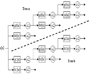
1) Dual Tree Complex Wavelet Transform (DTCWT): It has the property of nearly shift-invariance. This property overcomes the problem related with DWT, that it produces artifacts. Fig. 2 shows, two trees for performing the wavelet transform. If filters used in both trees are different, than upper filter provide real coefficients and lower tree provide imaginary coefficients.
Fig. 2 Implementation of DTCWT
For enhancing the contrast of the input image, various techniques are used based on histogram
B. Histogram Processing
To enhance the brightness or contrast of the image, histogram is the process of adjusting the intensity values. Histogram is used to define the intensity distribution of the image.
1) Histogram Equalization: Intensities of the image are
distributed well in histogram. This help to improve the intensity distribution of the image in local areas. Histogram equalization deals with the probability of occurrence of the gray level values to be equal.
2) Adaptive Histogram Equalization (AHE): In this case, each image is divided into several subsections. For each subsection, histogram is designed. Then, these histograms are used to improve the contrast of the image locally.
3) Contrast Limited Adaptive Histogram Equalization
(CLAHE): Due to AHE, noise gets over amplified. To prevent
this situation, CLAHE is designed. This is used to improve the contrast globally.
I. PROPOSED WORK
To enhance the resolution and contrast of the radiographic image such as X-Ray image, a combination of spatial domain and frequency domain based technique is proposed. Block diagram describing the proposed work is shown in Fig. 3.
Input image is decomposed using dual tree complex wavelet transform (DTCWT). By decomposing the image we get low subband image through low pass filtering and high subband image through high pass filtering. Due to low pass filtering, high frequency components are lost, which contain information of edges, fine line details etc. So, interpolation is performed on high frequency components.
For interpolating the coefficients, lanczos interpolation is used. This type of interpolation is not linear, but it is sinc function type interpolation. Advantage of using the sinc function type interpolation that it varies with the change in the signal information so better reconstruction can be achieved.
For improving the contrast of the image, adaptive histogram equalization is proposed. While using the adaptive histogram equalization (AHE), certain input parameters are used such as clip limit and distribution.
Further the AHE image is applied to singular value decomposition (SVD) block. SVD [2] [9] is a decomposition tool, which decompose the input image into three (S, U and V) equal size image. U and V image are orthogonal to each other whereas S image is a diagonal image, on its diagonal there are intensity values of respective pixels. By considering the SVD, a weighting function is evaluated.
A. Image Decomposition
Wavelet transform is a frequency domain based technique, used for decomposition of input image. There are various wavelet transform available for such operation. Stationary wavelet transform (SWT) [3] based method is used for fingerprint enhancement. In this the parameters used are , mean, standard deviation, smoothness, uniformity and entropy.
Combination of discrete wavelet transform (DWT) and SWT [13] is proposed. Subbands are interpolated using lanczos interpolation which provides better results. But artifacts are present while using the DWT, so filtering is required.
B. Coefficient Filtering
After decomposing the image by wavelet transform, wavelet coefficients need to be interpolated. As DWT produce artifacts, dual tree complex wavelet transform (DTCWT) [2], can be used for evaluating the wavelet coefficients.
To reduce the artifacts, high frequency subband images need to be interpolated and then filtered. Wiener filter [7] , Non Local Mean (NLM) [2] filter, Gaussian filter , Median filter can be used for this purpose.
These filters have their own pros and cons and have to be used depending on the images (medical image, satellite image etc.) considered, their dimension, class etc.
Fig. 3 Block Diagram of Proposed Work
C. Image Reconstruction
To enhance the input image, image reconstruction is required. This is done using inverse of the wavelet transform used for image decomposition. The output enhanced image, has higher pixel count which increases by the factor used for interpolation.
III. RESULTS AND DISCUSSION
To simulate the proposed algorithm, grayscale images of 8 patients is collected from hospital. These images are reduced to size 512x512.
A. Parameter Calculation
Analysis of
proposed algorithm is done qualitatively and quantitatively. For quantitative analysis, certain parameters are calculated
based on the following formulas. To calculate the Peak signal-to-noise ratio, Eq. 1 is used,
(1)
where, MAXI is the maximum value of intensity present
in the image, by default it is 255 in case of 8 bits per pixel. This is evaluated in terms of decibels (dB). MSE in Eq. 1 is mean square error, which is the difference between input image and reconstructed image as shown in Eq. 2.
2 1 1 0 01
( , )
( , )
m n i jMSE
I i j
K i j
mn
(2) CLAHE SVD SVD S U Vmax( ) max( 1)
2* max( 1)
S
S
w
S
1*( * 1) * 1
LLnew U
w S
V
Input Image (AxB) Image Decomposition (DTCWT) Low Subband Image High Subband Image Wiener Filter Interpolation by factor α/2 Interpolation by factor α Super Resolution Image (αAxαB) Image Reconstruction (IDTCWT) S1 U1 V1 210 * log(
MAX
I)
PSNR
MSE
Here I(i,j) is the input image and K(i,j) is reconstructed image.
Contrast ratio (CR) is defined as the ratio of difference between the maximum and minimum intensity value of reconstructed image K(i,j) to the sum of maximum and minimum intensity value of K(i,j) image as shown in Eq. 3.
max[ ( , )] min[ ( , )]
max[ ( , )] min[ ( , )]
K i j
K i j
CR
K i j
K i j
(3) B. DiscussionFor enhancing the medical image,
DTCWT-CLAHE-Wiener filter based algorithm is proposed. To evaluate the results, chest PA view X-Ray image of 8 patients is collected from hospital.
To improve the contrast of the image, contrast limited adaptive histogram equalization (CLAHE) and SVD based technique is proposed here. To enhance the resolution of the image DTCWT and Wiener filter is used.
Due to lanczos interpolation by factor α, size of the enhanced output image is 4 times that of the input image. As DTCWT is nearly shift invariant, it provides better result as compared to DWT and SWT. While using DTCWT, artifacts appear, to remove these artifacts Wiener filter is used. Non Local Mean (NLM) filter can also be used for filtering the coefficients and to remove the artifacts, but it increases the complexity of the algorithm by more time consumption. So, Wiener filter is proposed to filter the coefficients.
C. Simulated Results
Chest PA view X-Ray image is enhanced using proposed method. The result is shown in Fig. 4. Proposed algorithm result is compared with the other methods to show the comparison among proposed method and those methods.
Histogram equalization (HE) , can be used for improving the contrast of the image, but this provides less resolution as well as parameters such as PSNR, CR, MSE and SNR is also less.
Adaptive histogram equalization (AHE) along with wavelet transform can be used for improving the contrast of the image, but it produces artifacts.
Fig. 4(a), shows the input image of size 512x512. Fig. 4(b), shows the histogram equalized image. Fig. 4(c) shows the adaptive histogram equalized image (AHE). Fig. 4(d) shows DTCWT-HE-NLM based algorithm result and it has low resolution and contrast, also it takes 10630.4 sec. of computational time which is larger than other methods. Fig. 4(e) shows the DTCWT-AHE-NLM method for enhancing the visual quality of the image. Fig. 4(f) shows result of DTCWT-CLAHE-Wiener filter based proposed algorithm, which outperforms other methods. These results are simulated on MATLAB R2014b, i5 processor.
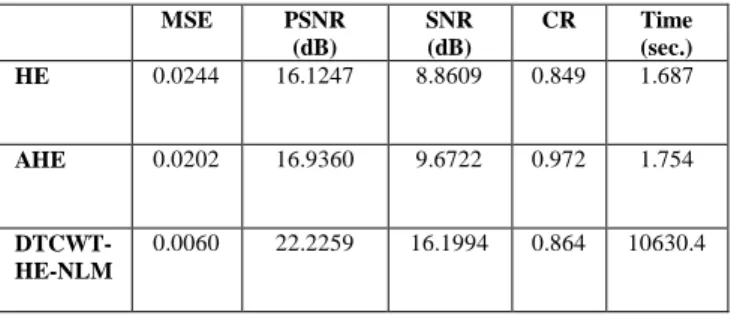
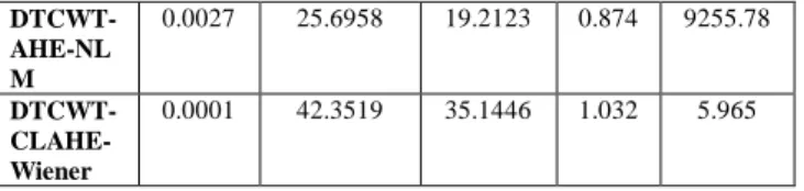
For quantitative analysis, Table I shows comparative results of various methods used. Five different methods for improving the contrast and resolution are compared based on different parameters such as peak signal-to-noise ratio (dB), signal-to-noise ratio (SNR) (dB), mean square error (MSE), contrast ratio (CR) and total time required for computation of respective algorithms.
Computational time of HE and AHE is less as compared to proposed method, but resolution and contrast of the proposed method is improved. DTCWT-HE-NLM and DTCWT-AHE-NLM takes large time but their results are better than HE and AHE.
Proposed algorithm DTCWT-CLAHE-Wiener
produces better result as compared to other methods. PSNR is 42.35 dB, SNR is 35.144 dB, MSE is 0.0001, contrast ratio is 1.0322 but computational time is 5.965 sec. which is quiet high than HE and AHE method.
IV. CONCLUSION
X-Ray images need to be enhanced for better visual interpretation. Resolution could be enhanced using DWT, SWT and DTCWT. Contrast can be improved using histogram equalization (HE), adaptive histogram equalization (AHE), contrast limited adaptive histogram equalization (CLAHE).
Proposed algorithm DTCWT-CLAHE-Wiener shows that it outperforms other conventional method for improving visual quality of the X-Ray image. Wiener filter takes less time as compared to NLM filter, which is the advantage in emergency situations.
V. FUTURE SCOPE
The enhancement method is proposed here for the purpose of medical image enhancement has considered some transform techniques, also some medical images such as X-Ray, Ultrasound and MRI. But with the ever increasing technology, new methods will be introduced in the market and that may provide better results than this proposed work. So, with the increasing time, new transforms and images will be tested for providing more benefit to various areas of interests.
In the proposed work, for enhancing the contrast, contrast limited adaptive Histogram Equalization (CLAHE) is used and it showed better results. Other techniques such as Local Histogram Equalization, Generalized Histogram Equalization can be used for enhancing the results. Resolution is improved using dual tree complex wavelet. etc.
TABLE I. Comparison of proposed method with other methods MSE PSNR (dB) SNR (dB) CR Time (sec.) HE 0.0244 16.1247 8.8609 0.849 1.687 AHE 0.0202 16.9360 9.6722 0.972 1.754 DTCWT- HE-NLM 0.0060 22.2259 16.1994 0.864 10630.4
DTCWT-AHE-NL M 0.0027 25.6958 19.2123 0.874 9255.78 DTCWT- CLAHE-Wiener 0.0001 42.3519 35.1446 1.032 5.965 ACKNOWLEDGMENT
I sincerely thanks ECE department of Ajay Kumar Garg Engineering College, Ghaziabad for providing the opportunity and guidance for research work.
(a) Input image (b) HE Image
(c) AHE Image (d) DTCWT-HE-NLM
(e) DTCWT-AHE-NLM (f) DTCWT-CLAHE-Wiener Fig. 4 (a) Input Image, (b) Histogram Equalized Image, (c) Adaptive histogram equalized Image, (d) DTCWT-HE-NLM Image, (e) DTCWT-AHE-NLM, (f) DTCWT-CLAHE-Wiener.
REFERENCES
[1] R.C. Gonzalez and R. E. Woods, Digital Image Processing, 3rd Edition, Prentice Hall, NJ, 2008.
[2] Muhammad Zafar Iqbal, Abdul Ghafoor, Adil Masood Siddiqui, Muhammad Mohsin Riaz and Umar Khalid, “Dual-tree complex wavelet transform and SVD based medical image resolution enhancement,” Elsevier, Signal processing, 2014, vol. 10, pp 430-437.
[3] K. V. Kale, R. R. Manza, S. S. Gornale, P. D. Deshmukh and Vikas Humbe, “ SWT based Composite Method for fingerprint image enhancement,” IEEE International symposium in signal processing and information technology, 2006, pp 162-167.
[4] Hasan Demriel and Gholamreza Anbarjafari, “Satellite image Resolution enhancement using complex wavelet transform,” IEEE Geoscience and remote sensing letters, 2010, vol. 7, no. 1, pp 123-126.
[5] Dah-Chung Chang and Wen-Rong Wu, “ Image contrast enhancement based on a histogram transformation of local standard deviation,” IEEE Transactions on Medical Imaging, 1998, vol. 17,no. 4, pp 518-531.
[6] J.C. Fu, H.C. Lien and S.T.C. Wong, “Wavelet-based histogram equalization enhancement of gastric sonogram,” Elsevier, Computerized Medical Imaging and Graphics, 2000, vol. 25, no. 2, pp 59-68.
[7] Janaki Sivakumar, Dr. K. Thangavel and P. Saravanan, “ Computed radiography skull image enhancement using weiner filter,” IEEE International Conference on Pattern recognition, informatics and medical engineering(PRIME), 2012, pp 307-311.
[8] Sudipto Dolui, Alan Kuurstra, Ivan C. Salgado Patarroyo and Oleg V. Michailovich, “ A new similarity measure for non-local means filtering of MRI images,” Elsevier, J. Vis. Commun. Image R., 2013, vol. 24, no.7, pp 1040-1054.
[9] Randa Atta, Rabab Farouk Abdel-Kader, “ Brightness preserving based on singular value decomposition for image contrast enhancement,” Elsevier, Optik, 2015, vol. 126, pp 799-803.
[10] Tzong-Huei Lin and Tsair Kao, “ Adaptive local contrast enhancement method for medical images displayed on a video monitor,” Elsevier, Medical Engineering & Physics, 2000, vol. 22, no. 2, pp 79-87. [11] Garima Vyas, Anita Thakur and Anupama Bhan, “Analysis of
histogram based contrast enhancement with noise reduction method for endodontic therapy,” IEEE International Conference on Reliability, infocom technologies and optimization (ICRITO), 2014, pp 1-5.
[12] Li Yang, Yanmei Liang and Hailun Fan, “Study on the methods of image enhancement for liver CT images,” Elsevier, Optik, 2010, vol. 121, pp 1752-1755.
[13] M. Priyadarshini, Ms. R. Sasikala and Dr. R. Meenakumari, “Novel approach for satellite resolution and contrast enhancement using wavelet transform and brightness preserving dynamic histogram equalization,” IEEE International Conference on green computing communication and electrical engineering, 2014, pp 1-4.
Renu Sharma received the B.Tech
degree in Electronics and
Communication Engineering from ABES Institute of technology, GBTU, in 2011. She is pursuing M.Tech in Electronics and Communication Engineering from Ajay Kumar Garg Engineering College, UPTU, from 2013. She completed Engineering with Honors and her area of interest are Image processing.
She qualified GATE exam in 2011. She has presented a paper in a National Conference held at Ajay Kumar Garg Engineering College, Ghaziabad during 6-7 February 2015. Currently she is working on project, enhancement of medical image for improving the quality of the image visually.
Gp. Capt. Pradeep Kumar Chopra (R), VSM entered the field of education in the year 2004 after 24 years of exemplary service in the technical branch of the Indian Air Force. He earned his Bachelors degree in Engineering (Electronics) from Delhi College of Engineering in the year 1979 and Masters in Technology from IIT Delhi in the year 1985. He also graduated from the prestigious Defence Services Staff College, Wellington and earned Masters degree in Defence Studies in the year 1993 from Madras University. While he was in the Indian Air Force he was part of, and headed, a number of important technical projects. For his exemplary services he was awarded “Vishist Seva Medal” by the President of India for engineering skills of highest order in the year 1993. He took voluntary retirement from the IAF in the year 2004 and entered the field of education. He has done his Ph.D in the field of „Electronics & Communication Engineering‟ from the prestigious G.G.S.I.P University, New Delhi. Presently he is the Dean and Head of Department of Electronics & Communication Engineering and Electronics & Instrumentation Engineering. His areas of specialisation are Radar and Satellite Communication. He is Fellow of Institution of Electronics & Telecommunication Engineers, New Delhi.