Original Article
Increased expression of miR-24 is associated
with acute myeloid leukemia with t(8;21)
Jia-Yu Yin
*, Qin Tang
*, Wei Qian, Jun Qian, Jiang Lin, Xiang-Mei Wen, Jing-Dong Zhou, Ying-Ying Zhang,
Xiao-Wen Zhu, Zhao-Qun Deng
Affiliated People’s Hospital of Jiangsu University, Zhenjiang, Jiangsu, China. *Equal contributors.
Received September 1, 2014; Accepted October 18, 2014; Epub October 15, 2014; Published November 1, 2014
Abstract: This study was designed to learn the expression status of miR-24 and its clinical relevance in patients with acute myeloid leukemia (AML). We detected the miR-24 expression levels using real-time quantitative PCR in 84 AML patients and investigated the clinical significance of miR-24 expression in AML. There was no difference in clinical parameters between cases with miR-24 high expression and with miR-24 low expression. The frequency of miR-24 high expression was higher in patients with t(8;21) than in others (82% (9/11) versus 44% (32/72),
P=0.026). The levels of miR-24 expression had no correlation with the mutations of nine genes (FLT3-ITD, NPM1, C-KIT, IDH1/IDH2, DNMT3A, N/K-RAS and C/EBPA). Meanwhile, among the group who obtained CR, the cases with miR-24 high expression had no difference in overall survival (OS) and relapse-free survival (RFS) than those with miR-24 low expression (P=0.612 and 0.665, respectively). These findings implicated that miR-24 high regulation is a common event in AML with t(8;21), and it might serve as a novel and selective therapeutic target for the treatment of AML with t(8;21).
Keywords: miR-24, acute myeloid leukemia, microRNA
Introduction
Acute myeloid leukemia (AML) is a
heteroge-neous malignant disease, which is
character-ized by arrested differentiation and abnormal
proliferation, leading to bleeding, fatal
infec-tion, or organ infiltration [1]. Recurring
chromo-somal aberrations and gene mutations
contrib-ute to the pathogenesis of AML [2]. Recently,
many researches have indicated that miRNAs
play important roles in myeloid
leukemogene-sis [3-5].
MicroRNAs (miRNAs) are known as small
non-coding single-stranded RNAs of 20-22
nucleo-tides that regulate the expression of over 60%
of all human genes and are involved in pivotal
biological processes, including development,
differentiation, proliferation, as well as
apopto-sis [6-10]. A large body of evidences implicate
that dysregulation of miRNA expression may
take part in oncogenesis of human malignant
cancers, such as non-small cell lung cancer
(NSCLC), gastrointestinal carcinoma,
hepato-cellular carcinoma, pancreatic cancer, breast
cancer, cervical cancer and the like [11-17].
Particular microRNA signatures have been
dis-covered in myeloid and lymphoid leukemia, and
have relationships with the pathogenesis,
diag-nosis and progdiag-nosis of myeloid and lymphoid
leukemia, such as 124-1, let-7a-3,
miR-181, miR-29B [4, 18-23].
Many researches have shown miR-24 takes
control of cell cycle distribution and apoptosis
[24-26]. Overexpression of miR-24 has been
found in oral carcinoma and non-small cell lung
cancer (NSCLC) [11, 27]. MiR-24 was reported
to enhance invasion and metastasis in cancer
cell [28]. However, the role of miR-24 in AML
should be explored more. Here we addressed
the question whether miR-24 expression is
related to AML.
Materials and methods
Patients and samples
Hospital of Jiangsu University. The diagnosis
and classification of AML patients were based
on French-America-British (FAB) and World
Health Organization (WHO) criteria [23, 29].
[image:2.612.91.518.96.651.2]Treatment protocol was described as reported
previously [21]. Written informed consent was
obtained from all patients. The study was
approved by the Institutional Review Board of
Table 1.
Clinical characteristics at diagnosis of AML patients divided according to miR-24 expression
status
miR-24 high expression
+ - P
Sex (male/female) 21/21 27/15 0.270
Median age at diagnosis, years (range) 53 (10-86) 57 (15-87) 0.164
Median WBC at diagnosis, ×109 L-1 (range) 8.4 (0.3-185.4) 11.7 (1.1-528.0) 0.697
Median hemoglobin at diagnosis, g/L (range) 75.0 (34.0-131.0) 77.0 (40.0-138.0) 0.333 Median platelets at diagnosis, ×109 L-1 (range) 33.0 (3.0-140.0) 38.0 (4.0-264.0) 0.707
FAB 0.053
M1 2 4
M2 24 14
M3 6 11
M4 4 11
M5 5 2
WHO 0.080
AML with t(8;21) 9 2
AML with t(15;17) 6 11
AML without maturation 2 4
AML with maturation 15 12
Acute myelomonocytic leukemia 5 11
Acute monoblastic and monocytic leukemia 4 2
Karyotype classification 0.919
Favorable 15 13
Intermediate 23 23
Poor 3 4
No date 1 2
Karyotyping 0.250
Normal 20 20
t(8;21) 9 2
t(15;17) 6 11
Complicated 3 3
Others 3 4
No date 1 2
Gene mutation
NPM1 (+/-) 5/37 4/37 1.000
FLT3 ITD (+/-) 6/36 6/35 1.000
C-KIT (+/-) 1/41 0/41 1.000
IDH1/IDH2 (+/-) 3/35 1/38 0.358
DNMT3A (+/-) 3/35 3/36 1.000
NRAS/KRAS (+/-) 4/34 3/36 0.711
C/EBPA (+/-) 6/36 3/38 0.483
CR (+/-) 23/18 20/19 0.823
the Affiliated People’ Hospital of Jiangsu Uni-
versity. The main clinical and laboratory featu-
res of the patient cohort were collected in
Table
1
. 16 healthy donors were collected as cont-
rols.
RNA extraction and reverse transcription
Using the mirVana miRNA isolation kit (Ambion,
Austin, TX, USA), we extracted the total RNA.
According to the manufacturer’s protocol using
miScript Reverse Transcription Kit (Qiagen, Du-
esseldorf, Germany), total RNA was reverse
transcribed to cDNA.
Real-time quantitative PCR
Real-time quantitative PCR (RQ-PCR) was
car-ried out according to the Manufacturer’s in-
structions using miScript SYBR green PCR kit
(Qiagen, catalog no. 218073) with the
manu-facturer-provided miScript Universal primer and
miRNA-specific forward primer: TGGCTCAGTTC-
AGCAGGAACA (miR-24).
RQ-PCR was performed on a 7300 Thermo cy-
cler (Applied Biosystems, Foster City, CA, USA),
using 50 ng of cDNA in a 20 μl reaction volume
with 1× QuantiTect SYBR Green PCR Master
Mix, 1× miScript Universal Primer, and 1.0 μM
of the specific forward primer. PCR program
conditions were 95°C for 15 min, followed by
40 cycles at 94°C for 15 s, 55°C for 30 s and
72°C for 34 s. At the end of the PCR cycles,
melting program (95°C for 15 s, 60°C for 60 s,
95°C for 15 s, and 60°C for 15 s) was
per-formed to validate the specificity of the
expect-ed PCR product. PCR amplicons were also
con-firmed by direct DNA sequencing in three
ran-domly selected patients. The relative
expres-sion level of miR-24 was calculated by the
com-parative 2
-△△Ctmethod using U6 small nuclear
RNA level for normalization.
Gene mutation detection
NPM1 and C-KIT mutations were detected by
high-resolution melting analysis (HRMA) as
reported previously [3]. Briefly, genomic DNA
samples were amplified using gene-specific
primers. Mutation scanning was performed for
PCR products using HRMA with the Light-
ScannerTM platform (Idaho Technology Inc.,
Salt Lake City, Utah). All positive samples were
directly DNA sequenced to confirm the resul-
ts of HRMA. FLT3 internal tandem duplication
(ITD) and C/EBPA mutations were detected
using direct DNA sequencing [30, 31].
Statistical analyses
All statistical analyses were implemented using
spss17.0. Chi-square analysis or Fisher exact
test was executed to compare the distinction of
categorical variables. The comparison of miRNA
expression status between patients and
con-trols was executed using Mann-Whitney test.
Survival was analyzed according to the
Kaplan-Meier method. All values showed two-sided
with a
P
-value < 0.05 considering statistically
significant.
Results
MiR-24 expression in AML
In our 84 samples, miR-24 expression levels
represented a continuum ranging from 0.000-
011 to 130.192 (median 0.123342). To
evalu-ate the impact of miR-24 expression levels on
clinical outcome without seeking an optimal
cut-point, patients were divided into low and
high expressers according to the median
exp-ression level of miR-24.
Association of miR-24 expression with clinical
and laboratory characteristics in AML
There was no significant difference in sex, age,
WBC, hemoglobin and platelets between pati-
ents with and without miR-24 high expression
(
Table 1
). MiR-24 high expression could be
[image:3.612.90.287.73.219.2]observed in each AML subtype analyzed (
Table
1
). The level of miR-24 was higher in AML with
t(8;21) than in t(15;17) (
P
=0.0127), in normal
(
P
=0.0214), in others (
P
=0.1237), and in
com-plex (
P
=0.8802) (
Figure 1
). Meanwhile, the
fre-quency of miR-24 high expression was higher in
patients with t(8;21) than in others (82% (9/11)
versus 44% (32/72),
P
=0.026). There was no
significant difference in the mutations of nine
genes between low expressers and high
expressers (
Table 1
).
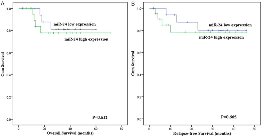
Impact of miR-24 expression on outcome of
AML patients
To investigate the prognostic impact of miR-24
high expression in AML, survival analysis was
performed in 80 cases with follow-up data.
There was no definite difference in the rates of
CR between patients with and without miR-24
high expression (56% versus 51%,
P
=0.823).
The patients with and without miR-24 high
expression were similar in the overall survival
(OS) (
P
=0.929). Among the group who obtained
CR, although the OS of AML patients with
miR-24 high expression (median 20.5 months, 95%
confidence interval 17-32 months) was shorter
than those with miR-24 low expression (median
30 months, 95% confidence interval 22-38
months), the difference was not statistically
significant (
P
=0.612) (
Figure 2A
). In addition,
the cases with miR-24 high expression had no
difference in relapse-free survival (RFS) than
those with miR-24 low expression (
P
=0.665)
(
Figure 2B
). The size of cases with t(8;21) was
small and most cases were still alive, so the OS
and RFS could not be analyzed. In cases with
cytogenetically normal AML, there was no
sig-nificant difference in the OS and RFS between
patients with and without miR-24 high
expres-sion (
P
=0.532 and 0.772, respectively).
Discussion
[image:4.612.90.522.71.297.2]High endogenous expression levels of miR-24
were more abundant in myeloid cells compared
with lymphoid cells [32]. Overexpression of
miR-24 has been found in oral carcinoma,
non-small cell lung cancer (NSCLC) and breast
can-cer [11, 27, 33]. The function of miRNAs was
highly tissue-dependent, which means that in
different types of tissues one specific miRNA
might get involved in different functions [26]. In
our study, we didn’t see obvious difference
between AML patients and controls. Meanwhile,
we didn’t find miR-24 expression had any
influ-ences on outcome of AML patients. Though the
OS of AML patients with miR-24 high
expres-sion (median 20.5 months, 95% confidence
interval 17-32 months) was shorter than those
with miR-24 low expression (median 30 months,
95% confidence interval 22-38 months), the
difference was not statistically significant
(
P
=0.612) due to the small size of samples.
Furthermore, more cases should be analyzed
to further confirm its clinical significance in
AML.
In our previous studies, we found some
microR-NAs had close relationships with AML involving
t(8;21) as well [3, 21]. However, lacking of
defi-nite mechanisms, regarding microRNA and
AML with t(8;21), it seemed that there were no
clinically meaningful findings. Recently, few
studies have reported the expression status of
miR-24 in AML. Zaidi et al. have reported that
miR-24 may act as a novel and selective
thera-peutic target for the treatment of AML. The
t(8;21)-encoded AML1-ETO hold the
miR-24-23-27 locus and control miR-24 transcription.
Disruption of Runx1/AML1 subnuclear
localiza-tion, by a chromosomal translocation t(8;21), is
connected to the etiology of acute myeloid
leu-kemia. Modified Runx1 subnuclear targeting by
leukemia-related translocation t(8;21)
tran-scriptionally deregulates the miR-24. By
acti-vating a miR-24/MKP-7/MAP kinase network,
modified Runx1 subnuclear targeting may
enhance proliferation and block granulocytic
differentiation [34]. In the present study, we
investigated the expression status of miR-24 in
patients with AML. Interestingly, high
expres-sion of miR-24 showed more frequently in AML
patients with translocation t(8;21) than in
oth-ers (82% (9/11) voth-ersus 44% (32/72),
P
=0.026).
Zaidi et al. provided exact evidences for us to
prove that miR-24 played an important role in
AML involving t(8;21) translocation. It was a
coincidence that the experiment we conducted
corroborated association between miR-24 and
AML with t(8;21) in clinic. Nowadays, a growing
body of evidences showed microRNAs really
had close relationships with this chromosomal
translocation [5, 35-37]. According to those
researches, they focused on the AML1/ETO,
which is a fusion protein having functions of
inhibiting differentiation and apoptosis, and
triggering signals for cell proliferation [38, 39].
Next, we should take attentions to this fusion
protein then we study the networks between
microRNA and AML with t(8;21) translocation.
Taken together with existing evidence from
microRNA and AML with t(8;21) studies, the
current results support a relationship between
microRNA and AML with t(8;21) translocation,
which merits further study.
In summary, we suggest that high expression of
miR-24 in AML patients might imply miR-24 can
serve as a valuable source for biomarker
dis-covery and validation in AML patients with
t(8;21), meanwhile, miR-24 might serve as a
novel and selective therapeutic target for the
treatment of AML patients with t(8;21).
Acknowledgements
This study was supported by National Natural
Science Foundation of China (81172592, 812-
70630).
Disclosure of conflict of interest
None.
Address correspondence to: Dr. Zhao-Qun Deng, Af- filiated People’s Hospital of Jiangsu University, 8 Dianli Road, Jiangsu 212002, Zhenjiang, People’s Republic of China. Tel: +86 511 88915586; Fax: +86 511 85234387; E-mail: dengqian2012@gmail. com
References
[1] Estey E, Döhner H. Acute myeloid leukaemia. Lancet 2006; 368: 1894-1907.
[2] Chen J, Odenike O, Rowley JD. Leukaemoge- nesis: more than mutant genes. Nat Rev Cancer 2010; 10: 23-36.
[3] Qian J, Lin J, Qian W, Ma JC, Qian SX, Li Y, Yang J, Li JY, Wang CZ, Chai HY, Chen XX, Deng ZQ. Overexpression of miR-378 is frequent and may affect treatment outcomes in patients with acute myeloid leukemia. Leuk Res 2013; 37: 765-768.
[4] Emmrich S, Katsman-Kuipers JE, Henke K, Khatib ME, Jammal R, Engeland F, Dasci F, Zwaan CM, den Boer ML, Verboon L, Stary J, Baruchel A, de Haas V, Danen-van Oorschot AA, Fornerod M, Pieters R, Reinhardt D, Klu- smann JH, van den Heuvel-Eibrink MM. miR-9 is a tumor suppressor in pediatric AML with t(8;21). Leukemia 2014; 28: 1022-1032. [5] Hager M, Pedersen CC, Larsen MT, Andersen
MK, Hother C, Grønbæk K, Jarmer H, Borre- gaard N, Cowland JB. MicroRNA-130a-media- ted down-regulation of Smad4 contributes to reduced sensitivity to TGF-beta1 stimulation in granulocytic precursors. Blood 2011; 118: 6649-6659.
[6] Calin GA, Croce CM. MicroRNA signatures in human cancers. Nat Rev Cancer 2006; 6: 857-866.
[7] Visone R, Croce CM. MiRNAs and cancer. Am J Pathol 2009; 174: 1131-1138.
[9] Deng Z, Yang X, Fang L, Rutnam ZJ, Yang BB. Misprocessing and functional arrest of microR-NAs by Pirate: roles of 378 and miR-17. Biochem J 2013; 450: 375-386.
[10] Deng Z, Du WW, Fang L, Shan SW, Qian J, Lin J, Qian W, Ma J, Rutnam ZJ, Yang BB. The inter-mediate filament vimentin inter-mediates microRNA miR-378 function in cellular self-renewal by regulating the expression of the Sox2 tran-scription factor. J Biol Chem 2013; 288: 319-331.
[11] Franchina T, Amodeo V, Bronte G, Savio G, Ric- ciardi GR, Picciotto M, Russo A, Giordano A, Adamo V. Circulating 22, 24 and miR-34a as novel predictive biomarkers to peme-trexed-based chemotherapy in advanced non-small cell lung cancer. J Cell Physiol 2014; 229: 97-99.
[12] Jiao F, Jin Z, Wang L, Wang L. Research and clinical applications of molecular biomarkers in gastrointestinal carcinoma (Review). Biomed Rep 2013; 1: 819-827.
[13] Salvi A, Abeni E, Portolani N, Barlati S, De Petro G. Human hepatocellular carcinoma cell-spe-cific miRNAs reveal the differential expression of miR-24 and miR-27a in cirrhotic/non-cir-rhotic HCC. Int J Oncol 2013; 42: 391-402. [14] Song S, Zhou J, He S, Zhu D, Zhang Z, Zhao H,
Wang Y, Li D. Expression levels of microRNA- 375 in pancreatic cancer. Biomed Rep 2013; 1: 393-398.
[15] Deng ZQ, Qian J, Liu FQ, Lin J, Shao R, Yin JY, Tang Q, Zhang M, He L. Expression level of miR-93 in formalin-fixed paraffin-embedded tissues of breast cancer patients. Genet Test Mol Biomarkers 2014; 18: 366-70.
[16] Deng ZQ, Yin JY, Tang Q, Liu FQ, Qian J, Lin J, Shao R, Zhang M, He L. Over-expression of miR-98 in FFPE tissues might serve as a valu-able source for biomarker discovery in breast cancer patients. Int J Clin Exp Pathol 2014; 7: 1166-1171.
[17] Wang X, Tang S, Le SY, Lu R, Rader JS, Meyers C, Zheng ZM. Aberrant expression of oncogen-ic and tumor-suppressive moncogen-icroRNAs in cervi-cal cancer is required for cancer cell growth. PLoS One 2008; 3: e2557.
[18] Landgraf P, Rusu M, Sheridan R, Sewer A, Iovino N, Aravin A, Pfeffer S, Rice A, Kamphorst AO, Landthaler M, Lin C, Socci ND, Hermida L, Fulci V, Chiaretti S, Foà R, Schliwka J, Fuchs U, Novosel A, Müller RU, Schermer B, Bissels U, Inman J, Phan Q, Chien M, Weir DB, Choksi R, De Vita G, Frezzetti D, Trompeter HI, Hornung V, Teng G, Hartmann G, Palkovits M, Di Lauro R, Wernet P, Macino G, Rogler CE, Nagle JW, Ju J, Papavasiliou FN, Benzing T, Lichter P, Tam W, Brownstein MJ, Bosio A, Borkhardt A, Russo JJ, Sander C, Zavolan M, Tuschl T. A mammalian
microRNA expression atlas based on small RNA library sequencing. Cell 2007; 129: 1401-1414.
[19] Volinia S, Calin GA, Liu CG, Ambs S, Cimmino A, Petrocca F, Visone R, Iorio M, Roldo C, Ferracin M, Prueitt RL, Yanaihara N, Lanza G, Scarpa A, Vecchione A, Negrini M, Harris CC, Croce CM. A microRNA expression signature of human solid tumors defines cancer gene targets. Proc Natl Acad Sci U S A 2006; 103: 2257-2261. [20] Chen XX, Lin J, Qian J, Qian W, Yang J, Ma JC,
Deng ZQ, Xie D, An C, Tang CY, Qian Z. Dysre- gulation of miR-124-1 predicts favorable prog-nosis in acute myeloid leukemia. Clin Biochem 2014; 47: 63-66.
[21] Li Y, Lin J, Yang J, Qian J, Qian W, Yao DM, Deng ZQ, Liu Q, Chen XX, Xie D, An C, Tang CY. Overe- xpressed let-7a-3 is associated with poor out-come in acute myeloid leukemia. Leuk Res 2013; 37: 1642-1647.
[22] Li Z, Huang H, Li Y, Jiang X, Chen P, Arnovitz S, Radmacher MD, Maharry K, Elkahloun A, Yang X, He C, He M, Zhang Z, Dohner K, Neilly MB, Price C, Lussier YA, Zhang Y, Larson RA, Le Beau MM, Caligiuri MA, Bullinger L, Valk PJ, Delwel R, Lowenberg B, Liu PP, Marcucci G, Bloomfield CD, Rowley JD, Chen J. Up-regu- lation of a HOXA-PBX3 homeobox-gene signa-ture following down-regulation of miR-181 is associated with adverse prognosis in patients with cytogenetically abnormal AML. blood 2012; 119: 2314-2324.
[23] Xiong Y, Li Z, Ji M, Tan AC, Bemis J, Tse JV, Hu- ang G, Park J, Ji C, Chen J, Bemis LT, Bunting KD, Tse W. MIR29B regulates expression of MLLT11 (AF1Q), an MLL fusion partner, and low MIR29B expression associates with ad-verse cytogenetics and poor overall survival in AML. Br J Haematol 2011; 153: 753-757. [24] Lal A, Navarro F, Maher CA, Maliszewski LE,
Yan N, O'Day E, Chowdhury D, Dykxhoorn DM, Tsai P, Hofmann O, Becker KG, Gorospe M, Hide W, Lieberman J. miR-24 Inhibits cell pro- liferation by targeting E2F2, MYC, and other cell-cycle genes via binding to “seedless” 3’UTR microRNA recognition elements. Mol Cell 2009; 35: 610-625.
[25] Chen L, Zhang A, Li Y, Zhang K, Han L, Du W, Yan W, Li R, Wang Y, Wang K, Pu P, Jiang T, Jiang C, Kang C. MiR-24 regulates the prolifer-ation and invasion of glioma by ST7L via beta-catenin/Tcf-4 signaling. Cancer Lett 2013; 329: 174-180.
[26] Akbari Moqadam F, Boer JM, Lange-Turenhout EA, Pieters R, den Boer ML. Altered expression of miR-24, miR-126 and miR-365 does not af-fect viability of childhood TCF3-rearranged leu-kemia cells. Leuleu-kemia 2014; 28: 1008-14. [27] Lin SC, Liu CJ, Lin JA, Chiang WF, Hung PS,
carci-noma: positive association from clinical and in vitro analysis. Oral Oncol 2010; 46: 204-208. [28] Du WW, Fang L, Li M, Yang X, Liang Y, Peng C,
Qian W, O’Malley YQ, Askeland RW, Sugg SL, Qian J, Lin J, Jiang Z, Yee AJ, Sefton M, Deng Z, Shan SW, Wang CH, Yang BB. MicroRNA miR-24 enhances tumor invasion and metastasis by targeting PTPN9 and PTPRF to promote EGF signaling. J Cell Sci 2013; 126: 1440-1453. [29] Bennett JM, Catovsky D, Daniel MT, Flandrin G,
Galton DA, Gralnick HR, Sultan C. Proposed re-vised criteria for the classification of acute my-eloid leukemia. A report of the French-Ame- rican-British Cooperative Group. Ann Intern Med 1985; 103: 620-625.
[30] Kottaridis PD, Gale RE, Frew ME, Harrison G, Langabeer SE, Belton AA, Walker H, Wheatley K, Bowen DT, Burnett AK, Goldstone AH, Linch DC. The presence of a FLT3 internal tandem duplication in patients with acute myeloid leu-kemia (AML) adds important prognostic infor-mation to cytogenetic risk group and response to the first cycle of chemotherapy: analysis of 854 patients from the United Kingdom Medical Research Council AML 10 and 12 trials. Blood 2001; 98: 1752-1759.
[31] Lin LI, Chen CY, Lin DT, Tsay W, Tang JL, Yeh YC, Shen HL, Su FH, Yao M, Huang SY, Tien HF. Characterization of CEBPA mutations in acute myeloid leukemia: most patients with CEBPA mutations have biallelic mutations and show a distinct immunophenotype of the leukemic cells. Clin Cancer Res 2005; 11: 1372-1379. [32] Kong KY, Owens KS, Rogers JH, Mullenix J,
Velu CS, Grimes HL, Dahl R. MIR-23A microR-NA cluster inhibits B-cell development. Exp Hematol 2010; 38: 629-640. e621.
[33] Yin JY, Deng ZQ, Liu FQ, Qian J, Lin J, Tang Q, Wen XM, Zhou JD, Zhang YY, Zhu XW. Asso- ciation between mir-24 and mir-378 in forma-lin-fixed paraffin-embedded tissues of breast cancer. Int J Clin Exp Pathol 2014; 7: 4261-7.
[34] Zaidi SK, Dowdy CR, van Wijnen AJ, Lian JB, Raza A, Stein JL, Croce CM, Stein GS. Altered Runx1 subnuclear targeting enhances myeloid cell proliferation and blocks differentiation by activating a miR-24/MKP-7/MAPK network. Cancer Res 2009; 69: 8249-8255.
[35] Li Y, Gao L, Luo X, Wang L, Gao X, Wang W, Sun J, Dou L, Li J, Xu C, Wang L, Zhou M, Jiang M, Zhou J, Caligiuri MA, Nervi C, Bloomfield CD, Marcucci G, Yu L. Epigenetic silencing of mi-croRNA-193a contributes to leukemogenesis in t(8;21) acute myeloid leukemia by activating the PTEN/PI3K signal pathway. Blood 2013; 121: 499-509.
[36] Mrozek K, Marcucci G, Paschka P, Bloomfield CD. Advances in molecular genetics and treat-ment of core-binding factor acute myeloid leu-kemia. Curr Opin Oncol 2008; 20: 711-718. [37] Brioschi M, Fischer J, Cairoli R, Rossetti S,
Pezzetti L, Nichelatti M, Turrini M, Corlazzoli F, Scarpati B, Morra E, Sacchi N, Beghini A. Down-regulation of microRNAs 222/221 in acute myelogenous leukemia with deranged core-binding factor subunits. Neoplasia 2010; 12: 866-876.
[38] Gardini A, Cesaroni M, Luzi L, Okumura AJ, Biggs JR, Minardi SP, Venturini E, Zhang DE, Pelicci PG, Alcalay M. AML1/ETO oncoprotein is directed to AML1 binding regions and co-lo-calizes with AML1 and HEB on its targets. PLoS Genet 2008; 4: e1000275.