Original Article
Upregulation of
URI/RMP
gene expression in cervical
cancer by high-throughput tissue microarray analysis
Junxia Gu1*, Xiaoyun Li1*, Yuting Liang1, Longwei Qiao1, Deyuan Ran1, Yaojuan Lu1,2, Xingang Li3, Wenxiang
Wei4, Qiping Zheng1,2
1Department of Hematology and Hematological Laboratory Science, School of Medical Science and Laboratory
Medicine, Jiangsu University, Zhenjiang 212013, China; 2Department of Anatomy and Cell Biology, Rush University
Medical Center, Chicago, IL 60612, USA; 3Department of Hematology, Anyang District Hospital, Anyang 455000,
China; 4Department of Cell Biology, School of Medicine, Soochow University, Suzhou 215123, China. *These
authors contribute equal to the paper.
Received February 17, 2013; Accepted March 7, 2013; Epub March 15, 2013; Published April 1, 2013
Abstract: URI, or RMP, is a RNA polymerase II subunit RPB5-associated protein known to play essential roles in ubiquitination and transcription. Recently, we and others have shown that URI/RMP is also important for progres-sion of hepatocellular carcinoma, ovarian, and prostate cancers. To identify the mechanistic basis of URI/RMP during multiple cellular processes, we investigated URI/RMP expression in a tissue microarray (TMA) containing multiple normal human tissues. The results showed that URI/RMP is ubiquitously but differentially expressed in these human tissues which partially explains its multiple cellular functions. To elucidate the role of URI/RMP during oncogenesis of multiple malignancies, especially the tumors of reproductive system, we analyzed URI/RMP expres-sion in a TMA containing multiple reproductive system tumors. We did not observe significant difference of URI/ RMP expression between cancerous and adjacent tissues of the prostate, breast, ovarian, and endometrial cancers. However, increased URI/RMP expression was observed in two of the three cases of cervical SCC (squamous cell car-cinoma) cells compared to their adjacent epithelial cells. Moreover, we detected significantly upregulated URI/RMP expression not only in cervical cancers but also in pre-cancerous CINs (cervical intra-epithelial neoplasias) in a TMA that covers the whole spectrum of normal cervix, CINs, and cervical cancers. No difference of URI/RMP expression was observed between CINs and cervical cancers. Given the high risk of CINs (especially CIN3) turning into cervical cancer if left untreated, the increased URI/RMP expression in CINs as well as in cervical cancers suggest a clinical relevance of URI/RMP upon cervical cancer tumorigenesis and worth further investigation.
Keywords: URI/RMP, tissue microarray, immunohistochemistry, cervical cancer
Introduction
URI (unconventional prefoldin RPB5 interactor) or RMP (RPB5-mediating protein), is a member of the prefoldin family of molecular chaper-ones. It has been demonstrated that URI/RMP functions as a scaffolding protein and plays critical roles in ubiquitination and transcription. This may be partially attributed to URI/RMP interaction with the RNA polymerase II subunit RPB5, as well as through formation of protein complexes with many other protein peptides [1, 2]. Previous studies have also shown that URI/ RMP participates in the TOR (Target of Rapamycin) signaling pathway, suggesting its potential involvement in cancer development
RMP gene expression in multiple normal human tissues as well as in tumors of the reproductive system by focusing on tissues from precancer-ous cervical intraepithelial neoplasia (CIN) and invasive cervical cancer.
Materials and methods
Tissue microarrays (TMAs)
Three tissue microarrays (TMAs) were used for this study. The multiple normal human tissues TMA (FDA808-1) was purchased from the Ailina Biotechnology Co., Ltd. (Xi’an, China). This TMA contains 24 different kinds of human tissues that are partially listed in Table 1. Each tissue was dotted three times which is from three dif-ferent individuals. The TMA from tumors of reproductive system (OD-CT-Rp03-002) was purchased from Shanghai Outdo Biotech Co., Ltd (Shanghai, China). This TMA was prepared from 31 cases of tumors that include prostate (10 cases), breast (7 cases), ovarian (7 cases), endometrial (4 cases) and cervical (3 cases) cancers. The TMA of cervical cancer and cervi-cal intraepithelial neoplasias (CINs) was a prod-uct of Pantomics, Inc. (CIN481, Richmond, CA USA.). This tissue array contains tissues from 48 cases that cover the whole spectrum of
nor-mal cervix (4 cases), CINs (CIN 1-3, 32 cases) and invasive cervical cancers (12 cases).
Immunohistochemistry (IHC)
[image:2.612.90.522.84.341.2]Immunohistochemical staining was performed on arrayed tissue samples using RMP antibody (K-17, sc-103869, Santa Cruz, CA USA). Briefly, after deparaffin and rehydration, the TMA slides were pretreated with 10mM sodium citrate buf-fer (pH 6.0) for 10 minutes in a microwave for antigen retrieval. The endogenous peroxidase was quenched by adding the hydrogen peroxide (3% H2O2 in 100% ethanol) at room tempera-ture for 15 minutes. After washing, the slides were blocked by rabbit serum for 1 hour. The blocking buffer was removed and the slides were then incubated overnight with primary RMP antibody (K-17) at 1:100 dilutions at 4°C. Slides were washed with the 1xTBST (Tris Buffered Saline with 0.1% Tween-20) solution and further incubated with poly-HRP anti-Goat IgG (PV-9003, Beijing Zhongshan Golden Bridge Biotechnology Co., Ltd.) for 15 minutes. Detection was using the 3, 3′-diaminoben-zidene as instructed (DAB kit, ZLI-9032, Beijing Zhongshan Golden Bridge Biotechnology Co., Ltd.). Slides were counterstained with hema-toxylin before microscopic analysis.
Table 1. URI/RMP expression in cytoplasms of major cell type in normal human tissues
Tissues Cell Components Score Tissues Cell Components Score Tonsil LymphocytesEpithelial cells (+)(++) Brain Granulose cells,Pyramidal cells (+) Lung Alveolar cellsMacrophages (++)(+++) Liver Liver cells (++)
Testicle Spermatogonia, Spermato-cytes, Sperm cells (++) Bone marrow Granulocytes, Erythroid cells (++)
Sperm (++) Lymphocytes (+)
Parathyroid gland Chief cellsOxyphil cells (++)(+++) Thyroid Gland Follicular cells,Parafollicular cells (++)
Colon Columnar cells,Goblet cells (++) Spleen LymphocytesPlasma cells (+)(++) Pituitary gland Acidophil cells,Basophil cells (+++) Breast gland Epithelial cells (++) Prostate gland Epithelial cells (++) Adrenal gland Cortical cells,Medullary cells (+++) Kidney Epithelial cells (+++) Salivary gland Acinar cells (++) Esophagus Mucosal epithelial cells (+++) Small Intestine Epithelial cells (++) Stomach Gastric parietal cellsPrincipal cells (++) Myocardia Myocardial cells (++) Cerebellum Granule cells (++) Pancreas Islet cellsAcinar cells (+++)(+) Thymus Lymphocytes (+)
Microscopic analysis of URI/RMP in normal TMA
The URI/RMP expression in TMA of normal human tissues was subjected to microscopic analysis. Briefly, after IHC staining, a cell was recorded as positive if it was stained from light yellow to brown either in the nucleus or cyto-plasm of that cell. The cell type and subcellular (nucleus and cytoplasm) localization of URI/ RMP expression in different tissues were deter-mined by an experienced histologist. Cells from five randomly selected visions under high mag-nification (400x) were counted for statistical analysis. The expression levels of URI/RMP were quantified using a modified scoring meth-od as previously described [6]. Specifically, if the percentage of positively stained cells over total number of cells is ≤5%, the score is “0”; 6-25%, scores “1”; 26-50%, scores “2”; 51-75%, scores “3”, and more than 75%, scores “4”. The intensity of the staining signal which represents relative URI/RMP expression level was also scored: “0” means no expression and “1-3” rep-resents weak, moderate, and strong expres-sion respectively. We also calculated the weight score of each specimen by multiplying percent-age of positive cells and the score of the signal intensity. Weight score “0” means negative staining or no expression, while scores “1-4”, “5-8”, and “9-12” represents weak, moderate, and strong positive staining respectively.
Image analysis of URI/RMP in tumor TMA
The URI/RMP expression in tumor TMAs were measured by analyzing the staining signal intensity using the Image-Pro Plus 6.0 image analysis software (Media Cybernetics, Inc. Silver Spring, MD USA). The mean densitometry of the digital image (× 400) is designated as representative URI/RMP staining intensity. For the TMA of reproductive system tumors, the areas from both cancer and its adjacent normal tissue were selected for analysis of the staining intensity of URI/RMP. For the cervical cancer-related TMA, the cervical epithelium of normal cervix and the cervical lesions in CINs and inva-sive cervical cancers were analyzed.
Statistical analysis
One-way ANOVA (SPSS16.0) was used to assess the significance of URI/RMP staining intensity across sample groups of cervical
can-cer and CIN TMA. Values (scores or signal inten-sities) are presented as means ± SEM (stan-dard error of mean). If there is a statistical significance, SNK-q test will be used for pair-wise comparison among the multiple groups. p<0.05 was considered statistically significant. Results
URI/RMP expression in normal human tissue
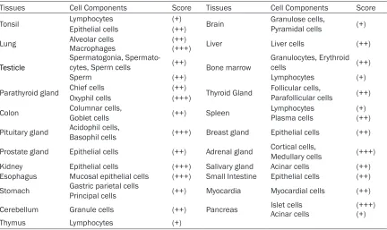
IHC analysis of the TMA containing multiple nor-mal human tissues (FDA808-1) indicates that, URI/RMP is ubiquitously expressed in all the human tissues examined, although more URI/ RMP in cytoplasms was detected than in nuclei. We analyzed URI/RMP expression by focusing on the major cell type of each tissue. As illus-trated in Table 1, URI/RMP were moderately expressed in cytoplasms of most of the major cell types examined, while weak or strong URI/ RMP expression can be occasionally detected in some of the major cell types of the corre-sponding tissues. URI/RMP may be differen-tially expressed in same cell type of a tissue. Take lymphocytes of the thymus as an example, no URI/RMP expression was appreciated in the nuclei. However, weak URI/RMP expression was observed in cytoplasm of most of the lym-phocytes, whereas there are lymphocytes that show either strong or negative URI/RMP expres-sion in the cytoplasm (Figure 1A). URI/RMP expression can be significantly different among different cell types of a tissue. For instance, URI/RMP is strongly expressed in islet cells of the pancreas, while URI/RMP is barely detect-able in the adjacent acinar cells (Figure 1B). URI/RMP is weakly expressed in nuclei of most of the tissues examined. We do not include data from ovarian tissue, as no follicular and luteal structure was observed except some connective tissue (Table 1).
URI/RMP expression in tumors of reproductive system
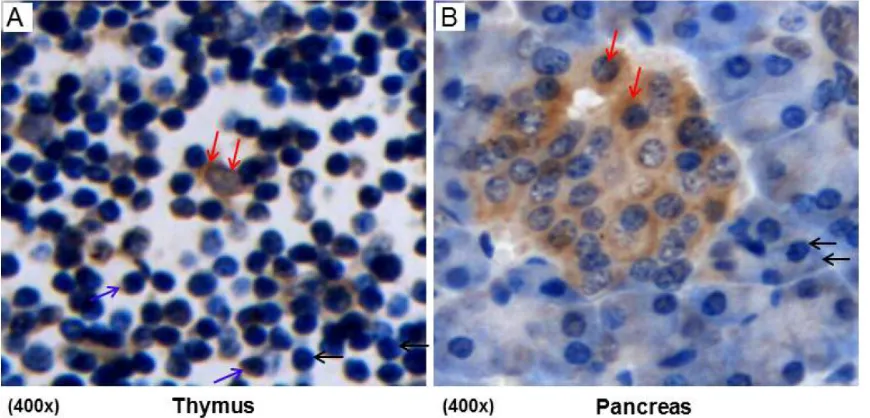
URI/RMP expression between cancerous and the adjacent tissues in prostate, breast, ovari-an, and the endometrial cancers. However, enhanced immunostaining indicating upregu-lated URI/RMP expression was observed in two of the three cervical SCC cells (Figure 2A, 2C) compared to their adjacent epithelial cells (Figure 2B, 2D).
URI/RMP expression in tissues of CINs and cervical cancers
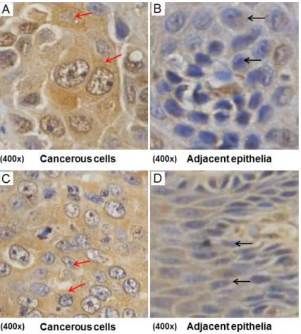
To attest above finding of upregulated URI/RMP expression in cervical SCC cells, we specifically performed URI/RMP immunostaining using a CIN and cervical cancer related TMA from Pantomics (Cat. No: CIN481, Set 1, California). This TMA covers the whole spectrum of tissues from normal cervix (4 cases), CIN 1 (12 cases), CIN 2 (11 cases), CIN3 (9 cases), and invasive cervical cancers (including 10 cases of cervical SCC, 1 case of mucinous adenocarcinoma, and 1 case of adenocarcinoma). As shown in Figure 3A, no or only weak URI/RMP expression was detected in the normal epithelial cells of the cervix, whereas high-level URI/RMP expression was shown in the nuclei and cytoplasms of the pre-cancerous cells of the CIN1, CIN2, and CIN3, as well as in cells from invasive cervical
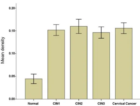
cancers (Figure 3B-F). We also quantified URI/ RMP expression in tissues of normal cervix, CINs and invasive cancers by analyzing their immunostaining signal intensity (densitometry). The results showed that URI/RMP expression in tissues of CINs and cervical SCC is significantly upregulated compared to its expression in tis-sues of normal cervix (Table 2 and Figure 4, p<0.05). There is no difference of the signal intensity between CINs and cervical SCC (Table 2 and Figure 4, p>0.05).
Discussion
According to the NCBI (National Center for
Biotechnology Information) database, URI/
[image:4.612.91.526.72.281.2]II by formation of a prefoldin-like complex [13]. To identify the potential mechanistic basis of URI/RMP and its correlation with multiple cel-lular functions, we investigated URI/RMP expression in a TMA containing multiple normal human tissues (FDA808-1). We observed ubiq-uitous URI/RMP expression in all the human tissues examined with more URI/RMP in cyto-plasms than in nuclei (Table 1). Notably, URI/
[image:5.612.95.522.72.545.2]pattern is not clear, it may partially explain the multiple cellular functions of URI/RMP. Meanwhile, the differential or specific expres-sion of URI/RMP in different cell types of a tis-sue provides a biological basis for human dis-eases. Here, our finding of high-level URI/RMP expression in islet (beta) cells, a cell population known to synthesize large amount of insulin [14], suggests a putative function of URI/RMP upon DM (Diabetes Mellitus) pathogenesis.
[image:6.612.94.521.73.348.2]While it is not clear whether URI/RMP is impor-tant for DM pathphysiology, there is growing evidence which suggests an essential role of URI/RMP upon carcinogenesis of multiple malignancies. We have previously shown that URI/RMP regulates the growth, apoptosis, and death of hepatocellular carcinoma cells (HCC) both in vitro and in vivo [4]. URI was also identi-fied as an oncogene that is ampliidenti-fied and over-expressed in ovarian cancer cell lines and human ovarian carcinomas. In addition, URI/ RMP has been shown to play important func-tion in meiotic cell cycle regulafunc-tion and partici-pate in AR (androgen receptor) signaling, a pathway critical for prostate cancer progres-sion [8, 15-17]. These findings suggest a role of URI/RMP in development and cancer formation that majorly involves the reproductive system. However, expression analysis of TMA (OD-CT-Rp03-002) containing multiple reproductive system tumors did not show difference between cancerous and adjacent tissues in prostate, breast, ovarian, and endometrial cancers. This Figure 3. URI/RMP expression in normal cervix, CINs, and cervical cancer. URI/RMP is barely detectable in nuclei and cytoplasms of the epithelial cells within the normal cervix (A, black arrows). URI/RMP is highly expressed both in nuclei and in cytoplasms of the epithelial cells at the CIN1 (B), CIN2 (C), and CIN3 (D) stages (red arrows). The epithelial cells of the CINs show nuclear atypia. URI/RMP is also highly expressed in nuclei and cytoplasms of the cervical SCC cells (E, red arrows). The cancer cells are enlarged and show irregular morphology and multiple nuclei. Illustrated are representative images from each of the normal cervix, CINs and cervical SCC groups.
Table 2. Densitometry calculation of URI/RMP in different groups
Groups Cases Mean Std Error Normal 4 0.044433 0.0104337 CIN1 12 0.151727 0.0119604 CIN2 11 0.159850 0.0155759 CIN3 9 0.146300 0.0125643 Cervical SCC 10 0.155833 0.0118321
[image:6.612.89.295.468.546.2]is possibly due to the limited number of speci-mens we analyzed and that contamination between cancerous and adjacent tissues may not be excluded. We did observe significant dif-ference of URI/RMP expression between can-cerous and adjacent tissues in two of the three cases of cervical SCC (Figure 3). Moreover, our further studies using TMA (CIN481) containing tissues from CINs and more cervical SCC cases demonstrated that URI/RMP is indeed signifi-cantly upregulated not only in cancerous cervi-cal tissues but also in pre-cancerous CINs com-pared to the normal epithelium of the cervix (Figures 3, 4).
The finding that CINs show upregulation of URI/ RMP suggests a potential clinical relevance of URI/RMP upon oncogenesis of cervical SCC. CIN, or cervical dysplasia, is the abnormal growth of epithelial cells on the cervix surface. CIN can be divided into LSIL (low-grade squa-mous intraepithelial lesions, or CIN1) and HSIL (high-grade squamous intraepithelial lesions, which subdivides into CIN2 and CIN3). The HSILs, especially cells at stage CIN3, are often referred to as cervical carcinoma in situ, given their high risk of turning into cervical cancer (usually cervical SCC) without proper treatment [18, 19]. We observed upregulated URI/RMP expression in cervical tissues from as early stage as in CIN1 to invasive cancers. Although,
there is no detectable difference of URI/RMP expression among CINs, or between CINs and cervical cancers, the increased URI/RMP expression in CINs compared to normal epithelial cells of the cervix suggests its involvement in cervical SCC initiation. We notice that cervical cancer has been largely attrib-uted to HPV (human papillomavirus) infection, while CINs, which may take years to turn into invasive cervical can-cers, are also associated with HPV infection. HPV oncoproteins E6 and E7 are the primary viral factors that play important roles in cervical cancer tumorigenesis, possibly through inacti-vation of pRB and P53, two famous tumor suppressor genes [20-23]. Correspondingly, reduced p53 and Rb expression was observed in cervical cancer [24]. More recently,mTOR inhibi-tors have been suggested as molecular targets for the treatment of HPV-associated malignancies including cer-Figure 4. Quantitaiton of URI/RMP expression in normal
cer-vix, CINs, and cancer cells. Densitometry of the immunostain-ing signal intensity of URI/RMP within normal cervix, CINs and cancers was calculated and subjected to statistical analysis. The mean signal intensity indicating URI/RMP expression in tissues of CINs and invasive cancers is significantly higher than that of normal cervix (p<0.05). There is no difference of the signal intensity between CINs and cervical cancers (p>0.05).
vical cancer [25]. However, whether URI1/RMP works through the canonical HPV-P53-RB or mTOR signaling pathway and plays a role in cer-vical cancer oncogenesis remains to be deter-mined and worth further investigation.
Acknowledgements
We are grateful to histologists Miao Chen, Yian Miao, and Lirong Duan for their technical help on IHC and histological analysis of the cell-types of the TMAs. We thank Dr. Hai Qian for his assistance in taking the photographic images. The tissue microarrays prepared from human normal or cancerous tissues were purchased from companies and the research protocol was approved by Jiangsu University. This work was partly supported by the Department of hema-tology and laboratory hemahema-tology, Jiangsu University and the NSFC grant (31271399) awarded to QZ and JG.
Declaration
All authors have no conflict of interest.
[image:7.612.91.320.68.241.2][email protected] or Dr. Qiping Zheng, Department of Anatomy and Cell Biology, Rush University Medical Center, 600 South Paulina Street, Chicago, IL 60612. Phone: 1-312-942-5514; Fax: 1-312-942-5744; E-mail: [email protected]
References
[1] Dorjsuren D, Lin Y, Wei W, Yamashita T, Nomu-ra T, Hayashi N, MuNomu-rakami S. RMP, a novel RNA polymerase II subunit 5-interacting protein, counteracts transactivation by hepatitis B vi-rus X protein. Mol Cell Biol 1998 Dec; 18: 7546-55.
[2] Gstaiger M, Luke B, Hess D, Oakeley EJ, Wirbe-lauer C, Blondel M, Vigneron M, Peter M, Krek W. Control of nutrient-sensitive transcription programs by the unconventional prefoldin URI. Science 2003 Nov 14; 302: 1208-12.
[3] Delgermaa L, Hayashi N, Dorjsuren D, Nomura T, Thuy le TT, Murakami S. Subcellular localiza-tion of RPB5-mediating protein and its putative functional partner. Mol Cell Biol 2004 Oct; 24: 8556-66.
[4] Yang H, Gu J, Zheng Q, Li M, Lian X, Miao J, Ji-ang J, Wei W. RPB5-mediating protein is re-quired for the proliferation of hepatocellular carcinoma cells. J Biol Chem 2011 Apr 1; 286: 11865-74.
[5] Theurillat JP, Metzler SC, Henzi N, Djouder N, Helbling M, Zimmermann AK, Jacob F, Solter-mann A, Caduff R, HeinzelSolter-mann-Schwarz V, Moch H, Krek W. URI is an oncogene amplified in ovarian cancer cells and is required for their survival. Cancer Cell 2011 Mar 8; 19: 317-32. [6] Sinicrope FA, Ruan SB, Cleary KR, Stephens
LC, Lee JJ, Levin B. bcl-2 and p53 oncoprotein expression during colorectal tumorigenesis. Cancer Res 1995 Jan 15; 55: 237-41. [7] Wei W, Gu JX, Zhu CQ, Sun FY, Dorjsuren D, Lin
Y, Murakami S. Interaction with general tran-scription factor IIF (TFIIF) is required for the suppression of activated transcription by RPB5-mediating protein (RMP). Cell Res 2003 Apr; 13: 111-20.
[8] Parusel CT, Kritikou EA, Hengartner MO, Krek W, Gotta M. URI-1 is required for DNA stability in C. elegans. Development 2006 Feb; 133: 621-9.
[9] Deplazes A, Möckli N, Luke B, Auerbach D, Pe-ter M. Yeast Uri1p promotes translation initia-tion and may provide a link to cotranslainitia-tional quality control. EMBO J 2009 May 20; 28: 1429-41.
[10] Djouder N, Metzler SC, Schmidt A, Wirbelauer C, Gstaiger M, Aebersold R, Hess D, Krek W. S6K1-mediated disassembly of mitochondrial URI/PP1gamma complexes activates a nega-tive feedback program that counters S6K1
survival signaling. Mol Cell 2007 Oct 12; 28: 28-40.
[11] Guicciardi ME, Gores GJ. Cell stress gives a red light to the mitochondrial cell death pathway. Sci Signal 2008 Feb 19; 1: pe9.
[12] Kirchner J, Vissi E, Gross S, Szoor B, Rudenko A, Alphey L, White-Cooper H. Drosophila Uri, a PP1alpha binding protein, is essential for via-bility, maintenance of DNA integrity and nor-mal transcriptional activity. BMC Mol Biol 2008 Apr 15; 9: 36.
[13] Boulon S, Pradet-Balade B, Verheggen C, Molle D, Boireau S, Georgieva M, Azzag K, Robert MC, Ahmad Y, Neel H, Lamond AI, Bertrand E. HSP90 and its R2TP/Prefoldin-like cochaper-one are involved in the cytoplasmic assembly of RNA polymerase II. Mol Cell 2010 Sep 24; 39: 912-24.
[14] Marchetti P, Bugliani M, Boggi U, Masini M, Marselli L. The pancreatic beta cells in human type 2 diabetes. Adv Exp Med Biol 2012; 771: 288-309.
[15] Feldman BJ, Feldman D. The development of androgen-independent prostate cancer. Nat Rev Cancer 2001Oct; 1: 34-45. Review. [16] Chen CD,Welsbie DS,Tran C,Baek SH, Chen
R, Vessella R, Rosenfeld MG, Sawyers CL. Mo-lecular determinants of resistance to antian-drogen therapy. Nat Med 2004 Jan; 10: 33-9. Epub 2003 Dec 21.
[17] Mita P,Savas JN,DjouderN, Yates JR 3rd, Ha S, Ruoff R, Schafler ED, Nwachukwu JC, Tanese N, Cowan NJ, Zavadil J, Garabedian MJ, Logan SK. Regulation of androgen receptor-mediated transcription by RPB5 binding protein URI/ RMP. Mol Cell Biol 2011 Sep; 31: 3639-52. [18] Park J,Sun D, Genest DR,Trivijitsilp P, Suh I,
Crum CP. Coexistence of low and high grade squamous intraepithelial lesions of the cervix: morphologic progression or multiple papillo-maviruses? Gynecol Oncol 1998 Sep; 70: 386-91.
[19] Agorastos T, Miliaras D, Lambropoulos AF, Chrisafi S, Kotsis A, Manthos A, Bontis J. Detec-tion and typing of human papillomavirus DNA in uterine cervices with coexistent grade I and grade III intraepithelial neoplasia: biologic pro-gression or independent lesions? Eur J Obstet Gynecol Reprod Biol 2005 Jul 1; 121: 99-103. [20] Duensing S, Münger K. Mechanisms of ge-nomic instability in human cancer: insights from studies with human papillomavirus onco-proteins. Int J Cancer 2004Mar 20; 109: 157-62. Review.
[22] ScottoL, Narayan G, Nandula SV, Arias-Pulido H, Subramaniyam S, Schneider A, Kaufmann AM, Wright JD, Pothuri B, Mansukhani M, Mur-ty VV. Identification of copy number gain and overexpressed genes on chromosome arm 20q by an integrative genomic approach in cervical cancer: potential role in progression. Genes Chromosomes Cancer 2008 Sep; 47: 755-65.
[23] McLaughlin-Drubin ME, Münger K. The human papillomavirus E7 oncoprotein. Virology 2009 Feb 20; 384: 335-44.
[24] Gouveris P,Weinberger PM,Psyrri A. Use of tis-sue microarray to facilitate oncology research. Methods Mol Biol 2010; 632: 239-50. [25] Molinolo AA,Marsh C,El Dinali M, Gangane N,