Original Article
Histone-modifying genes as biomarkers in
hepatocellular carcinoma
Shih-Ya Hung1,2, Hui-Hua Lin1, Kun-Tu Yeh3, Jan-Gowth Chang1,2,4,5
1Epigenome Research Center, China Medical University Hospital, Taichung 40447, Taiwan; 2Graduate Institute of
Integrated Medicine, College of Chinese Medicine, China Medical University, Taichung 40402, Taiwan; 3
Depart-mant of Pathology, Changhua Christian Hospital, Changhua 50006, Taiwan; 4Department of Laboratory
Medi-cine, China Medical University Hospital, Taichung 40447, Taiwan; 5School of Medicine, China Medical University,
Taichung 40447, Taiwan
Received March 9, 2014; Accepted April 5, 2014; Epub April 15, 2014; Published May 1, 2014
Abstract: Hepatocellular carcinoma (HCC) is the world’s fifth most common cancer and second leading cause of cancer-related death in Taiwan. Over 600,000 HCC patients die each year worldwide despite recent advances in surgical techniques and medical treatments. Epigenetic regulations including DNA methylation and histone modifi-cation control gene expressions and play important roles during tumorigenesis. This study evaluates association be-tween histone-modifying genes and prognosis of HCC to ferret out new diagnostic markers. We collected 50 paired HCC and adjacent non-cancerous tissues from Taiwanese patients for survey by RT-qPCR and tissue microarray-based immunohistochemistry (TMA-microarray-based IHC) staining. RT-qPCR data showed four of twenty-four genes over eight-fold up-regulated in tumor tissues: e.g., histone phosphorylation gene-ARK2, methylation genes-G9a, SUV39H2, and EZH2 (n = 50, all p < 0.0001). Results of TMA-based IHC staining showed proteins of ARK2, EZH2, G9a, and SUV39H2 also overexpressed in tumor tissues. Staining intensity of SUV39H2 correlated with HCV infection (p = 0.025). We further restricted the analysis only in tumor tissues, we found EZH2 staining intensity associated with tumor stage (p = 0.016) and survival (p = 0.007); SUV39H2 intensity associated with tumor stage (p = 0.044). Our findings indicate overexpression of histone-modifying genes EZH2 and SUV39H2 associated with prognosis of HCC cases. EZH2 expression can serve as a novel prognostic biomarker during HCC progression among Taiwanese.
Keywords: Hepatocellular carcinoma, histone acetylation, histone methylation, histone phosphorylation, histone modification
Introduction
Hepatocellular carcinoma (HCC; also called liver cancer) is a highly lethal cancer (third most
common cause of cancer mortality worldwide)
with rising incidence; approximately 750,000 new cases reported each year around the globe and overall 5-year survival rate is only 0-14% [1, 2]. In addition, the Cancer Registry Annual
Report in Taiwan of 2012 ranked HCC as sec -ond most common cancer-related death in Taiwanese. This illustrates the urgency to expose cellular mechanisms involved in this
aggressive cancer to develop more effective treatments and better prognosis. Chief risk fac
-tors include chronic viral hepatitis of B- or C-virus infection (HBV or HCV), alcohol abuse, nonalcoholic steatohepatitis, aflatoxin B1 expo
-sure, and liver cirrhosis [3, 4]. Currently, HCCs are diagnosed by the invasive (biopsy) and
non-invasive (images and tumor markers) methods [5]. The former include imaging diagnosis such
as computed tomography (CT), magnetic
reso-nance imaging (MRI), and tumor markers like α-fetaprotein and glypican-3 [5]. Yet detailed molecular mechanisms of hepatocarcinogene
-sis are not fully understood; molecular factors capable of predicting clinical outcome and act -ing as potential therapeutic targets remain limited.
Human genome is highly compacted by nucleo-protein complexes called chromatin, whose basic unit is a nucleosome; DNA is tightly
histones [6]. The term “epigenetics” means changes in gene expression and cellular
pheno-type without affecting DNA sequence [7].
Epigenetic mechanisms can be considered as
an interface between the genome and risk fac
-tors of lifestyle and the environment [8]. Histone modification is one of the epigenetic mecha
-nism belonging to post-translational modifica
-tion of N-terminal tails of histone proteins by
acetylation, methylation, phosphorylation, ubi- quitylation, sumoylation, ADP ribosylation,
bioti-nylation, and potentially other modifications; these appear to work together with other epi -genetic mechanisms in establishing and main-taining gene activity states, thus regulating a
gamut of cellular processes [9]. So that several families of enzymes catalyze post-translational modifications of histones, including acetyl
-transferases and deacetylases, methyltrans
-ferases and demethylases, etc. play key roles in histone modification [10]. Recent studies prove the importance of histone-modifying enzymes and chromatin remodeling complexes in regulating chromatin activities of transcrip -tion, replica-tion, and DNA repair. Histone
modi-fication plays essential roles in generating the dynamic state of chromatin [6]. Alternations in the function of histone-modifying molecules and protein complexes are found to disrupt the pattern and levels of histone marks and conse
-quently deregulate the control of chromatin-based processes leads to development of
many cancers [11].
Hepatocarcinogenesis is viewed as a complex and multi-step process entailing epigenetic and genetic events [12]. Recent reports show
his-tone-modifying genes associate with HCC prog
-nosis. Besides histone acetylation-related pro -teins, epigenetic aberrations which caused by
histone phosphorylation (like ARK2 and MSK1) and lysine or arginine methylation (like G9a, EZH2, SUV39H1, and SUV39H2) demonstrably associate with progression of many tumors
[13-15]. For example, histone phosphorylation
enzymes-Aurora kinases (subfamily of serine/ threonine mitotic kinases) are primary mole
-cules for maintaining accurate cell cycling and
genomic stability [16]. In HCC, ARK1 (Aurora-A
kinase; centrosome-associated serine/threo
-nine kinase) is overexpressed frequently in HCC
patients, and correlated with high grade and
high stage [17]. ARK2 (Aurora-B kinase, which
phosphorylates histone H3 at serine 10) is a chromosomal passenger protein and that is
essential for chromosome segregation and cytokinesis; mRNA level of ARK2 is an
indepen-dent molecular marker predicting tumor inva
-sion of HCCs [18]. Likewise, Aurora kinase inhibitors are potential therapeutic agents for HCC treatment [18, 19]. G9a, a histone methyl
-transferase for lysine 9 of histone 3 (H3K9), is
also a major player in gene silencing and
essen-tial for early embryogenesis to regulate devel
-opmental gene expression [20, 21]. EZH2 (a histone methyltransferase for lysine 27 of his -tone 3) belongs to the polycomb genes crucial
for transcriptional regulation via nucleosome modification, chromatin remodeling, and inter
-action with other transcription factors [22]. Positive EZH2 expression as a diagnostic bio
-marker detects aggressive phenotype or a phe
-notype with poor prognosis in patients of China population [23]. SUV39H1 was the first SET
domain-containing histone lysine
methyltrans-ferase (HKMT) discovered in 2000; SUV39H1 and SUV39H2 knockout mice have shown genomic instability [24]. The mRNA level of
SUV39H1 has been shown no different in 23
paired HCC and non-cancerous liver tissues
from Japanese population [25]. On the other
hand, SUV39H1 mRNA level shows
up-regulat-ed in 38 HCC patients from Hong Kong [26]. The present report examines expressions of histone-modifying genes in 50 HCC patients to
derive a relationship between gene expres-sions and clinicopathological parameters in these Taiwanese patients. Here, we assessed
mRNA expression of 24 histone-modifying genes further studied protein expressions in
HCC and corresponding non-cancerous liver tissues by RT-qPCR and immunohistochemistry staining. Once we better understand the
clini-cal relevance of those histone-modifying genes
in HCCs among the Taiwanese population,
clini-cians can offer effective cancer treatments, biomarkers for early detection, better progno
-sis, and novel therapeutic targets. Identifying molecular markers related to hepatocarcino
-genesis would benefit patients and serve as potential therapeutic target for novel HCC drug
treatments.
Materials and methods
Patients and clinical methods
This study was approved by the Institutional
Hospital (CMUH103-REC1-033). Fifty
Taiwan-ese patients diagnosed with HCC at Changhua Christian Hospital (Changhua, Taiwan) during 2002-2006 enrolled. All underwent surgery with or without radiotherapy and/or chemother-apy. Their 50 paired HCC tissues and corre-sponding adjacent non-cancerous liver tissues
were stored at -80°C until RNA extraction and
IHC staining, their medical records reviewed. All
tumors were classified according to the 7th edi
-tion of TNM Staging Manual of American Joint
Committee on Cancer [27].
RNA extraction, reverse-transcription, and real-time quantitative polymerase chain reaction (RT-qPCR)
Total RNA was extracted from tissues accord
-ing to instructions of the High Pure RNA
Isolation Kit (Roche Applied Science; Mann- heim, Germany). Single-strand cDNA was
syn-thesized by high-capacity cDNA reverse tran
-scription kit (Applied Biosystems, Foster City,
thorax homolog, drosophila), SET1 (SET domain containing 1A), SET7 (SET domain containing 7), G9a (euchromatic histone-lysine N-methy-
ltransferase 2), SETDB1 (SET domain, bifurcat -ed 1), SUV39H1 (suppressor of variegation 3-9
homolog 1, drosophila), SUV39H2 (suppressor
of variegation 3-9 homolog 2, drosophila),
EZH2 (enhancer of zeste homolog 2, drosophi
-la), DOT1 (disruptor of telomeric silencing-1),
and SET8 (SET domain containing lysine
meth-yltransferase 8); {3} histone arginine methyla -tion genes such as CARM1
(coactivator-associ-ated arginine methyltransferase 1) and PRMT1
(protein arginine methyltransferase 1); {4} his -tone serine/threonine phosphorylation genes such as ARK2 (aurora kinase B) and MSK1
(ribosomal protein S6 kinase, 90 kDa, polypep
-tide 5). We used GAPDH (glyceraldehyde-3-phosphate dehydrogenase) as the RT-qPCR internal control. Table 1 shows primer and
[image:3.612.91.426.85.430.2]probe sequences for each RT-qPCR reaction. The thermal cycling conditions: 95°C for 10 min, followed by 40 cycles of 95°C for 10 s,
Table 1. Primer and probe sequences of RT-qPCR assays
Gene Forward primer Reverse primer Probe
ADA ccttcgacaagcccaaagta gcccctctgctgtgttagc ctcctgcc
ARK2 attgctgacttcggctggt gtccagggtgccacacat cttcctcc
ATF2 cgaccgcagtcattacaaca gtggagttgtgtgagctgga tgtggctg
CARM1 cacaccgacttcaaggacaa aaaaacgacaggatcccaga tggctgtg
CBP/CREBBP gagatccagggcgagaatg aactgatcctttgaaattgtcgt ctggggct
DOT1 ccccactcacctcagacg ttgcaatttttctctcaattgc cccagcag
ESA1 aaggagctcatcgctacagtg caagaggacctgcttcacct gctggatc
EZH2 tgcacatcctgacttctgtga tcatctcccatataaggaatgttatg ggaggtgg
G9a gggaccttcatctgcgagta tctcacatcagcctcagcat gggagctg
GAPDH agccacatcgctcagacac gcccaatacgaccaaatcc cttcagcc
Gcn5 ccgaccagctctacacaacc aggcactggggtgagactt gggagctg
HAT1 tctaaagttgatgagaactttgactgt ttgtctaattttgccctcaaca ctgcctct
MLL/TRX gcgccaagctctttgcta catcatcatcataacatttgtcacag cttctgcc
MOF gaagttcctcatcgctttcag tcagacagcggcttctcc ctccagct
MSK1 tggtgctgacagattttggt caaaaggaatatgctctttcagtttc cctggcgc
P300 gcagcctgcaactccact gaggatttgatacctgtccttca cctggagc
PCAF cagagaccctgaccagcttt gcgctttgatggctcttc acctgctg
PRMT1 ggagaattttgtagccaccttg cctggccacaggacactt ctccagcc
SET1 cactgggtccatcacacaat ctgagcctgtctggtgctc ctggggcc
SETDB1 tgaaaaagagcccttgaagc ccaggaagggaagacatgc tcctctcc
SET7 atggatagcgacgacgaga taatccgtcatcgtccaggt tggaggag
SET8 ggatttctaccctgtccgaag ttcaatcaattcatctattcttttcc cctggagc
SRC1 tctcaaaacagaagcagatgga gacgtcagcaaacacctgaa acctgctg
SUV39H1 tcatggagtacgtgggagaga cctgacggtcgtagatctgg ctctgcct
SUV39H2 gcattgttttccacaagaacc tgggctgtggtcaatagaatc ctccagca
CA). The RT-qPCR
was performed to analyze mRNA
ex-pression in 4 groups:
{1} histone acetyla -tion genes- such as
ADA (adenosine dea- minase), ATF2 (acti-vating transcription
factor 2), CREBBP
(CREB binding pro -tein), ESA1 (histone
acetyltransferase
ES-A1), GCN5 (general
control of amino-acid synthesis 5-like 2,
yeast), HAT1 (histone
acetyltransferase 1),
MOF (lysine
acetyl-transferase 8), p30-
0/CBP (E1A binding
protein P300), PCAF
(TAF6-like RNA poly -merase II, P300/
CBP-associated fac -tor), and SRC1; {2}
histone lysine meth-ylation genes such as
MLL/TRX (myeloid/
lymphoid or
-60°C for 30 s, and 72°C for 10 s. Expression levels of mRNA were first normalized to mRNA of GAPDH
and then expressed as folds of nor -mal by 2-∆∆Ct method according to
Liu et al [28].
Tissue microarray-based immuno-histochemistry (TMA-based IHC) staining analysis and antibodies
TMA-based IHC staining for ARK2, EZH2, G9a, and SUV39H2 proteins was performed as follows. Primary antibodies for the study were ARK2 (Santa Cruz; sc-14327), EZH2
(Invitrogen; 49-1043), G9a (Santa
Cruz; sc-22877), and SUV39H2 (Abgent; AP1281a). Briefly, the 5-μm paraffin-embedded sections (5 × 8 = 40 cores/slide tissue array; each slide has 20 paired of normal and tumor tissues from HCC patients) were deparaffinized,
[image:4.612.92.522.84.210.2]retrieved with heat in 10 mM
Table 2. Overview of different classes of histone-modifying genes in this study
Histone modifications Residues modified Genes-included in this study Functions regulated
Acetylation of lysine Lysine ADA, ATF2, CREBBP, ESA1, GCN5,
HAT1, MOF, p300/CBP, PCAF, SRC1 Transcription; repair; replication; condensation
Methylation of lysine Histone H3 Lysine 4 MLL/TRX, SET1, SET7 Transcription; repair Histone H3 Lysine 9 G9a, SETDB1, SUV39H1, SUV39H2
Histone H3 Lysine 27 EZH2
Histone H3 Lysine 79 DOT1
Histone H4 Lysine 20 SET8
Methylation of arginine Histone H3 on Arginine 17 CARM1 Transcription Histone H4 on Arginine 3 PRMT1
Phosphorylation of serine/threonine Histone H3 on Serine 10 ARK2, MSK1 Transcription; repair; condensation
[image:4.612.92.353.229.723.2]citrate buffer (pH 6.0) at 121°C for 10 min,
then treated with 3% hydrogen peroxide to remove endogenous peroxidase activity. Primary antibodies were used as suggested by
manufacturers and were used at dilution of
1:50 after optimization. Tissue sections treat -ed with secondary antibodies and with
biotin-streptavidin complex for 30 min each at 37°C. Diaminobenzidine served as chromogene for
[image:5.612.123.523.71.602.2]stained with hematoxylin and examined for
intensity by the pathologist in a blind manner.
More than 50% of cells displayed positive stain
-ing and intensity scores of moderate and
strong, was considered to be protein overex-pression in this study. Tissue specimens were broadly distributed by immunohistochemical staining category (0, 1+, 2+, or 3+) according to
the guidelines based on Hofmann et al. [29].
Statistical analysis
Statistical analyses were performed using SPSS v17 (SPSS Inc., Working, UK). Paired
t-test was used for analysis RT-qPCR assays.
Correction between protein expression and
clinicopathological features of 50 HCC patients
were drawn by Pearson’s Chi-squared test,
P-values < 0.05 considered statistically signi-
ficant.
Results
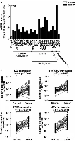
Four mRNAs of histone-modifying genes more than eightfold overexpressed in HCC tissues
To rate mRNA expression of histone-modifying genes in HCC, we performed RT-qPCR analysis
on total RNA extracted, classifying 24 histone-modifying genes as {1} acetylation of lysine, {2} methylation of lysine, {3} methylation of argi
-nine, or {4} phosphorylation of serine/threo -nine. Table 2 shows an overview of these his
-tone-modifying genes. RT-qPCR results show all 24 genes significantly up-regulated in HCC tissues (tumors) from 50 patients as compared
to adjacent non-cancerous tissues (normal) (P
< 0.05; Figure 1A). Since all these genes
showed statistically significant increase in tumors, we set eightfold up-regulation as threshold of mRNA overexpression. We found
G9a (↑8.85 folds; methylation of lysine),
SUV39H2 (↑9.78 folds; methylation of lysine), EZH2 (↑8.52 folds; methylation of lysine), and ARK2 (↑15.53 folds; phosphorylation of serine/ threonine) were more than eightfold overex -pressed in tumor (P < 0.0001; n = 50; Figure 1B). Figure 1B shows gene expression patterns in 50 paired tissues.
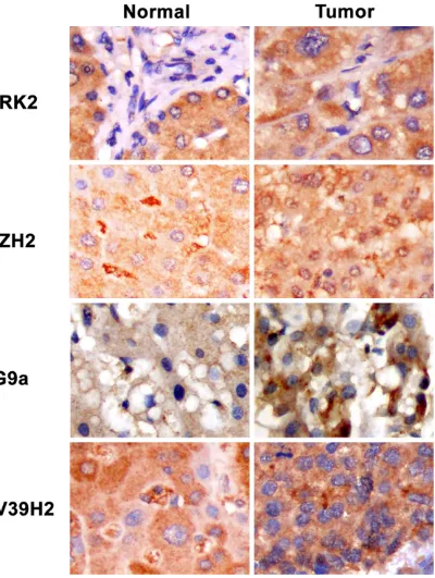
Increase protein expressions of ARK2, EZH2, G9a, and SUV39H2 in tumors
Since mRNAs of ARK2, EZH2, G9a, and
SUV39H2 were more than eightfold overex
[image:6.612.91.523.97.358.2]-pressed in tumor tissues of 50 HCC patients.
Table 3. Correction of ARK2 and EZH2 protein intensity with clinicopathological parameters in 50
Taiwanese patients with HCC
Characteristics
ARK2 EZH2
Tumor ≥
Normal Tumor < Normal Total P-value Tumor ≥ Normal Tumor < Normal Total p-value
Tumor Size ≤ 2 cm 1 5 6 1.000 0 6 6 1.000
> 2 cm 4 28 32 1 30 31
Grade Well 0 4 4 0.309 0 3 3 0.827
Moderate 5 22 27 1 26 27
Poor 0 7 7 0 7 7
Stage I, II 4 17 21 0.349 1 20 21 0.474
III, IV 1 17 18 1 17 18
Survival ≤ 2 years 1 16 17 0.363 1 16 17 0.447
> 2 years 4 18 22 0 21 21
HBV infection No 2 10 12 0.594 0 12 12 0.647
Yes 2 21 23 1 21 22
HCV infection No 1 19 20 0.303 0 19 19 0.458
Yes 3 13 16 1 15 16
Virus infection No 0 2 2 1.000 0 2 2 1.000
Yes 4 31 35 1 33 34
Cirrhosis No 1 15 16 0.618 1 15 16 0.444
We then studied protein expressions of ARK2, EZH2, G9a and SUV39H2 in 50 HCC patients by
IHC staining on tissue microarray. Figure 2
shows the typical staining patterns of ARK2, EZH2, G9a, and SUV39H2 in respective normal and tumor tissues from HCC patients. We found that protein levels of ARK2, EZH2, G9a, and
SUV39H2 were increase only in tumor but not
normal tissues. These results concur with our mRNA expression data.
Association of ARK2, EZH2, G9a, and SU-V39H2 protein levels with clinicopathological parameters
Table 3 shows relationship between protein
intensity of ARK2 or EZH2 and clinicopathologi
-Table 4. Correction of ARK2 and EZH2 protein intensity in tumor tissues with clinicopathological pa -rameters in 50 Taiwanese patients with HCC
Characteristics ARK2 in tumor EZH2 in tumor
≤ 1+ ≥ 2+ Total P-value ≤ 1+ ≥ 2+ Total P-value
Tumor Size ≤ 2 cm 2 4 6 1.000 2 4 6 0.668
> 2 cm 11 24 35 17 18 35
Stage I 3 4 7 0.780 3 4 7 0.344
II 3 11 14 4 10 14
III 4 9 13 8 5 13
IV 3 6 9 5 4 9
Grade Well 2 2 4 0.578 1 3 4 0.635
Moderate 8 22 30 15 15 30
Poor 3 5 8 4 4 8
Survival ≤ 2 years 7 14 21 0.747 14 7 21 0.015*
> 2 years 6 16 22 6 16 22
[image:7.612.91.523.97.269.2]*, p < 0.05.
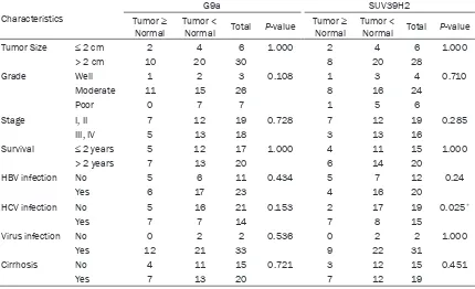
Table 5. Correction of G9a and SUV39H2 protein intensity with clinicopathological parameters in 50
Taiwanese patients with HCC
Characteristics
G9a SUV39H2
Tumor ≥
Normal Tumor < Normal Total P-value Tumor ≥ Normal Tumor < Normal Total P-value
Tumor Size ≤ 2 cm 2 4 6 1.000 2 4 6 1.000
> 2 cm 10 20 30 8 20 28
Grade Well 1 2 3 0.108 1 3 4 0.710
Moderate 11 15 26 8 16 24
Poor 0 7 7 1 5 6
Stage I, II 7 12 19 0.728 7 12 19 0.285
III, IV 5 13 18 3 13 16
Survival ≤ 2 years 5 12 17 1.000 4 11 15 1.000
> 2 years 7 13 20 6 14 20
HBV infection No 5 6 11 0.434 5 7 12 0.24
Yes 6 17 23 4 16 20
HCV infection No 5 16 21 0.153 2 17 19 0.025*
Yes 7 7 14 7 8 15
Virus infection No 0 2 2 0.536 0 2 2 1.000
Yes 12 21 33 9 22 31
Cirrhosis No 4 11 15 0.721 3 12 15 0.451
Yes 7 13 20 7 12 19
[image:7.612.92.522.330.592.2]cal parameters of 50 HCC patients. ARK2 and EZH2 intensity in normal and tumor liver tis
-sues were not significant associated with any clinicopathological parameters: i.e., tumor size, grade, stage, survival, HBV infection, HCV infec
-tion, virus infec-tion, and cirrhosis. We then restricted the analysis to ARK2 and EZH2 inten
-sity in tumor tissues and found EZH2 but not
ARK2 intensity in tumor tissues associated with survival (P = 0.015; Table 4). Table 5 shows the relationships between G9a and SUV39H2 intensity and clinicopathological parameters in 50 HCC patients; SUV39H2 intensity was
asso-ciated with HCV infection (P = 0.025); G9
inten-sity was not linked with any clinicopathological parameters: e.g., tumor size, grade, stage, sur
-vival, virus infection, and cirrhosis. If we further
restricted the analysis to G9a and SUV39H2
intensity in tumors, we found SUV39H2 but not
G9a intensity associated with tumor stage (P = 0.044; Table 6).
Discussion
In 2012, HCC ranked as second leading cause of cancer-related deaths in Taiwan (Department of Health, Executive Yuan). The hepatitis B vac
-cination program launched in 1984 has suc
-cessfully reduced HCC incidence in children,
yet incidence and mortality in the general Taiwanese population remain high (overall
5-year survival rate was 15% during 1987-1992) and lack diagnostic markers for early
detection [30-32]. Histones originally believed
lation and methylation of specific lysine resi -dues on histone H3 and H4 [34]. Kondo et al. showed that multiple epigenetic silencing mechanisms inappropriately active in HCC cells
and mRNAs of histone
methyltransferases-EZH2 and G9a (but not SUV39H1) higher in
malignant liver tissues of 23 HCC patients of
Japan [25]. Suggesting histone methylations contribute to gene silencing in HCC. In this
report, we have studied four groups of histone modifying genes including lysine acetylation,
lysine methylation, arginine methylation,
ser-ine/threonine phosphorylation. This is the first report discussing expressions of 24 histone-modifying genes in hepatocarcinogenesis. To assess histone-modifying genes from mRNA
to protein levels, both mRNAs and proteins were measured in tissue pairs by RT-qPCR and tissue microarray-based immunohistochemis-try (TMA-based IHC) staining. Since RT-qPCR accurately monitors mRNA expressions and immunohistochemistry staining allowed us to
generate staining profiles for various antibod
-ies specific to the same fixation and staining procedures for 40 cores normal and tumor tis
-sues from HCC patients on the same slide.
Although immunohistochemistry staining is
[image:8.612.90.401.108.282.2]essential for diagnostic pathology, it exhibits inconsistency of quality assurance in data interpretation. Despite widespread use of immunohistochemistry, significant problems remain with regard to variability in tissue fixa -tion, processing, staining methodologies and
Table 6. Correction of G9a and SUV39H2 proteins expression levels
in tumor tissues with clinicopathological parameters in 50 Taiwanese patients with HCC
Characteristics G9a in tumor SUV39H2 in tumor
≤ 1+ ≥ 2+ Total P-value No Yes Total P-value
Tumor Size ≤ 2 cm 1 5 6 1.000 2 4 6 0.206
> 2 cm 6 29 35 4 31 35
Stage I 2 5 7 0.143 3 4 7 0.044*
II 0 14 14 0 14 14
III 2 11 13 1 12 13
IV 3 6 9 2 7 9
Grade Well 0 4 4 0.170 0 4 4 0.098
Moderate 4 26 30 3 27 30
Poor 3 5 8 3 5 8
Survival ≤ 2 years 5 16 21 0.240 3 18 21 1.000
> 2 years 2 20 22 3 19 22
*, p < 0.05.
to function as a scaffold for DNA packing, have
now emerged in
post-translational
modificati-ons that regulated chro-matin condensation and DNA accessibility. His-
tone modifications affect chromatin structure fur -ther gene expression, and play a vital role in
establishment of gene
silencing during tumori-genesis [33]. N-terminal
tails of histone proteins
are subject to a wide
range of covalent modifi -cation at many sites. Over 60 residues on
his-tones are modified; the
acety-reagents, and interpretation of staining results persists between different slides [35].
Com-bining RT-qPCR and immunohistochemistry staining in this study, we monitor
histone-modi-fying gene’s mRNA expression and its’ coding protein level to glean more information to inter -pret our data.
Besides the well-known alteration of histone
acetylation patterns, cancer cells also display widespread changes in histone
phosphoryla-tion and methylaphosphoryla-tion patterns [24, 36-39]. We
selected genes that showed more than
eight-fold overexpression in tumor tissues for further
immunohistochemistry staining: histone phos-phorylation protein (ARK2) and methylation
proteins (G9a, EZH2 and SUV39H2). We previ
-ously showed that overexpression of histone
methylation- and phosphorylation-related
pro-teins-ARK2, G9a, EZH2 (but not SUV39H2) associated with prognosis of oral squamous cell carcinoma in 215 male of Taiwan [40]. ARK2 may serve as both an effective prognos
-tic factor and a biomarker for predicting various clinical outcomes of oral cancer [40]. In the present report, our data shows all mRNAs of 24 histone-modifying genes increasing in tumor tissues, suggesting that histone modification (includes acetylation of lysine, methylation of lysine, methylation of arginine, and phosphory
-lation of serine/threonine) plays a lead role in HCC carcinogenesis. Within these 24 genes, mRNAs of G9a, SUV39H2, EZH2, and ARK2
were more than eightfold overexpressed in tumor tissues. In spite of mRNAs expression, we found proteins of ARK2, EZH2, G9a, and
SUV39H2 consistently up-regulated in tumors
of 50 HCC patients.
ARK2 (a kinase for H3S10) overexpression proves associated with stages of malignant
progression in thyroid carcinomas and with poor prognosis in endometrial carcinomas [13, 41]. Lin et al. showed that ARK2 mRNA fre -quent overexpressed in 160 Taiwanese HCC cases, which can be used as an important
molecular marker of early recurrence and poor prognosis [18]. In addition, they suggest that ARK2 selected inhibitors-AZD1152-HQPA and VE-465 had anticancer effects in HCC cells [18, 19]. Sorafenib (Nexavar®) is an orally active
multikinase inhibitor approved in Europe for treatment of HCC, showing greater survival
duration in combination therapy with
doxorubi-cin in advanced HCC [42]. We found mRNA and protein of ARK2 were both overexpressed in 50
HCCs, yet staining intensity of ARK2 not corre -lated with any clinicopathologic characteristics.
Possible reasons of this variation may be differ
-ences in selected antibody, sample size, genet
-ic background, and/or risk factors between
selected populations.
The polycomb group protein-EZH2 functions as a histone methyltransferase for H3K27 (his
-tone H3 lysine 27) as catalytic subunit of poly -comb repressive complex 2 (PRC2) and medi-ates transcription silencing [43]. Increased
expression of EZH2 is frequently detected in
HCC tissues and it is associated with the aggressiveness and/or poor prognosis [44-46].
Knockdown of EZH2 expression in HCC cells
could reverse tumorigenicity in mice, which
suggests that EZH2 inhibition has therapeutic value in HCC [47]. In addition, EZH2 expression,
as examined by IHC, has been correlated with
aggressiveness and poor prognosis of HCC in the China population, indicating EZH2 as a diagnostic biomarker for HCC [23]. Our results prove EZH2 intensity of tumor tissues also
associated with survival in the 50 HCC patients,
such that EZH2 might serve as an efficient prognostic biomarker.
Histone H3K9 di-methylation (H3K9me2) and tri-methylation (H3K9me3) can be mediated by
histone methyltransferase (HMT)-G9a and
SUV39H1, respectively. G9a promotes lung cancer cell invasion and may play an early role
in metastasis cascade [48]. Nuclear intensity of G9a protein correlates with reduced overall survival and disease-free interval in lung can
-cer [48]. Kondo et al. showed mRNA expres
-sions of EZH2 and G9a but not SUV39H1 were
higher in cancerous tissues of 23 HCC patients in Japan [25]. We found both mRNA and protein expressions of ARK2, EZH2, G9a, and SUV39H2 in tumor tissues significantly enhanced, sug -gesting these histone methylation- and phos-phorylation-related genes involves in
hepato-carcinogenesis. But G9a protein intensity from immunohistochemistry staining shows no link
with clinicopathological parameters in 50 HCC cases. On the other hand, SUV39H2 intensity
This article focuses on markers of histone
methylation and phosphorylation proteins:
ARK2, G9a, EZH2, and SUV39H2. Findings sug
-gest EZH2 and SUV39H2 as highly correlated
with survival and tumor stage, respectively, and
may yield important biomarkers that predict
patient survival and prognosis in HCC patients
of Taiwan.
Acknowledgements
This work was supported by the grant from
CMU Hospital (DMR-103-047).
Disclosure of conflict of interest
None.
Address correspondence to: Dr. Jan-Gowth Chang, Epigenome Research Center, China Medical University Hospital, 2 Yude Road, Taichung 40447, Taiwan. Tel: +886-4-22052121 ext. 2008; Fax: +886-4-22037690; E-mail: d6781@mail.cmuh.org. tw
References
[1] Siegel R, Ward E, Brawley O and Jemal A. Can-cer statistics, 2011: the impact of eliminating socioeconomic and racial disparities on pre-mature cancer deaths. CA Cancer J Clin 2011; 61: 212-236.
[2] Siegel R, Naishadham D and Jemal A. Cancer statistics, 2012. CA Cancer J Clin 2012; 62: 10-29.
[3] Maluccio M and Covey A. Recent progress in understanding, diagnosing, and treating hepa-tocellular carcinoma. CA Cancer J Clin 2012; 62: 394-399.
[4] El-Serag HB. Hepatocellular carcinoma: an epi-demiologic view. J Clin Gastroenterol 2002; 35: S72-78.
[5] Song do S and Bae SH. Changes of guidelines diagnosing hepatocellular carcinoma during the last ten-year period. Clin Mol Hepatol 2012; 18: 258-267.
[6] Kornberg RD and Lorch Y. Twenty-five years of the nucleosome, fundamental particle of the eukaryote chromosome. Cell 1999; 98: 285-294.
[7] Bird A. Perceptions of epigenetics. Nature 2007; 447: 396-398.
[8] Herceg Z and Paliwal A. Epigenetic mecha-nisms in hepatocellular carcinoma: how envi-ronmental factors influence the epigenome. Mutat Res 2011; 727: 55-61.
[9] Dawson MA and Kouzarides T. Cancer epi-genetics: from mechanism to therapy. Cell 2012; 150: 12-27.
[10] Arrowsmith CH, Bountra C, Fish PV, Lee K and Schapira M. Epigenetic protein families: a new frontier for drug discovery. Nat Rev Drug Dis-cov 2012; 11: 384-400.
[11] Jones PA and Baylin SB. The epigenomics of cancer. Cell 2007; 128: 683-692.
[12] Feinberg AP. Phenotypic plasticity and the epi-genetics of human disease. Nature 2007; 447: 433-440.
[13] Sorrentino R, Libertini S, Pallante PL, Troncone G, Palombini L, Bavetsias V, Spalletti-Cernia D, Laccetti P, Linardopoulos S, Chieffi P, Fusco A and Portella G. Aurora B overexpression asso-ciates with the thyroid carcinoma undifferenti-ated phenotype and is required for thyroid car-cinoma cell proliferation. J Clin Endocrinol Metab 2005; 90: 928-935.
[14] Taby R and Issa JP. Cancer epigenetics. CA Cancer J Clin 2010; 60: 376-392.
[15] Sharma S, Kelly TK and Jones PA. Epigenetics in cancer. Carcinogenesis 2010; 31: 27-36. [16] Keen N and Taylor S. Aurora-kinase inhibitors
as anticancer agents. Nat Rev Cancer 2004; 4: 927-936.
[17] Jeng YM, Peng SY, Lin CY and Hsu HC. Overex-pression and amplification of Aurora-A in hepa-tocellular carcinoma. Clin Cancer Res 2004; 10: 2065-2071.
[18] Lin ZZ, Jeng YM, Hu FC, Pan HW, Tsao HW, Lai PL, Lee PH, Cheng AL and Hsu HC. Significance of Aurora B overexpression in hepatocellular carcinoma. Aurora B Overexpression in HCC. BMC Cancer 2010; 10: 461.
[19] Lin ZZ, Hsu HC, Hsu CH, Yeh PY, Huang CY, Huang YF, Chen TJ, Kuo SH, Hsu C, Hu FC, Jeng YM, Chung Y and Cheng AL. The Aurora kinase inhibitor VE-465 has anticancer effects in pre-clinical studies of human hepatocellular carci-noma. J Hepatol 2009; 50: 518-527.
[20] Rice JC, Briggs SD, Ueberheide B, Barber CM, Shabanowitz J, Hunt DF, Shinkai Y and Allis CD. Histone methyltransferases direct different de-grees of methylation to define distinct chroma-tin domains. Mol Cell 2003; 12: 1591-1598. [21] Tachibana M, Sugimoto K, Nozaki M, Ueda J,
Ohta T, Ohki M, Fukuda M, Takeda N, Niida H, Kato H and Shinkai Y. G9a histone methyl-transferase plays a dominant role in euchro-matic histone H3 lysine 9 methylation and is essential for early embryogenesis. Genes Dev 2002; 16: 1779-1791.
[22] Simon JA and Tamkun JW. Programming off and on states in chromatin: mechanisms of Polycomb and trithorax group complexes. Curr Opin Genet Dev 2002; 12: 210-218.
hepatocellular carcinomas in liver needle biop-sies. Gut 2011; 60: 967-976.
[24] Peters AH, O’Carroll D, Scherthan H, Mechtler K, Sauer S, Schofer C, Weipoltshammer K, Pa-gani M, Lachner M, Kohlmaier A, Opravil S, Doyle M, Sibilia M and Jenuwein T. Loss of the Suv39h histone methyltransferases impairs mammalian heterochromatin and genome sta-bility. Cell 2001; 107: 323-337.
[25] Kondo Y, Shen L, Suzuki S, Kurokawa T, Masu-ko K, Tanaka Y, Kato H, Mizuno Y, YoMasu-koe M, Sugauchi F, Hirashima N, Orito E, Osada H, Ueda R, Guo Y, Chen X, Issa JP and Sekido Y. Alterations of DNA methylation and histone modifications contribute to gene silencing in hepatocellular carcinomas. Hepatol Res 2007; 37: 974-983.
[26] Fan DN, Tsang FH, Tam AH, Au SL, Wong CC, Wei L, Lee JM, He X, Ng IO and Wong CM. His-tone lysine methyltransferase, suppressor of variegation 3-9 homolog 1, promotes hepato-cellular carcinoma progression and is nega-tively regulated by microRNA-125b. Hepatolo-gy 2013; 57: 637-647.
[27] Edge SB and Compton CC. The American Joint Committee on Cancer: the 7th edition of the AJCC cancer staging manual and the future of TNM. Ann Surg Oncol 2010; 17: 1471-1474. [28] Liu TC, Lin SF, Chang JG, Yang MY, Hung SY and
Chang CS. Epigenetic alteration of the SOCS1 gene in chronic myeloid leukaemia. Br J Hae-matol 2003; 123: 654-661.
[29] Hofmann M, Stoss O, Shi D, Buttner R, van de Vijver M, Kim W, Ochiai A, Ruschoff J and Hen-kel T. Assessment of a HER2 scoring system for gastric cancer: results from a validation study. Histopathology 2008; 52: 797-805. [30] Lee CL, Ko YC and Choong CS. Survival rate for
liver cancer in Taiwan. Zhonghua Yi Xue Za Zhi (Taipei) 2000; 63: 16-20.
[31] Shyu HJ, Lung CC, Ho CC, Sun YH, Ko PC, Huang JY, Pan CC, Chiang YC, Chen SC and Liaw YP. Geographic patterns of hepatocellular carcinoma mortality with exposure to iron in groundwater in Taiwanese population: an eco-logical study. BMC Public Health 2013; 13: 352.
[32] Chang MH, Chen CJ, Lai MS, Hsu HM, Wu TC, Kong MS, Liang DC, Shau WY and Chen DS. Universal hepatitis B vaccination in Taiwan and the incidence of hepatocellular carcinoma in children. Taiwan Childhood Hepatoma Study Group. N Engl J Med 1997; 336: 1855-1859. [33] Jones PA and Baylin SB. The fundamental role
of epigenetic events in cancer. Nat Rev Genet 2002; 3: 415-428.
[34] Kouzarides T. Chromatin modifications and their function. Cell 2007; 128: 693-705. [35] Hsu FD, Nielsen TO, Alkushi A, Dupuis B,
Huntsman D, Liu CL, van de Rijn M and Gilks
CB. Tissue microarrays are an effective quality assurance tool for diagnostic immunohisto-chemistry. Mod Pathol 2002; 15: 1374-1380. [36] Nguyen CT, Weisenberger DJ, Velicescu M,
Gonzales FA, Lin JC, Liang G and Jones PA. His-tone H3-lysine 9 methylation is associated with aberrant gene silencing in cancer cells and is rapidly reversed by 5-aza-2’-deoxycyti-dine. Cancer Res 2002; 62: 6456-6461. [37] Kondo Y, Shen L, Ahmed S, Boumber Y, Sekido
Y, Haddad BR and Issa JP. Downregulation of histone H3 lysine 9 methyltransferase G9a in-duces centrosome disruption and chromo-some instability in cancer cells. PLoS One 2008; 3: e2037.
[38] Valk-Lingbeek ME, Bruggeman SW and van Lo-huizen M. Stem cells and cancer; the polycomb connection. Cell 2004; 118: 409-418. [39] Kondo Y, Shen L, Cheng AS, Ahmed S,
Boum-ber Y, Charo C, Yamochi T, Urano T, Furukawa K, Kwabi-Addo B, Gold DL, Sekido Y, Huang TH and Issa JP. Gene silencing in cancer by his-tone H3 lysine 27 trimethylation independent of promoter DNA methylation. Nat Genet 2008; 40: 741-750.
[40] Chen JH, Yeh KT, Yang YM, Chang JG, Lee HE and Hung SY. High expressions of histone methylation- and phosphorylation-related pro-teins are associated with prognosis of oral squamous cell carcinoma in male population of Taiwan. Med Oncol 2013; 30: 513.
[41] Kurai M, Shiozawa T, Shih HC, Miyamoto T, Feng YZ, Kashima H, Suzuki A and Konishi I. Expression of Aurora kinases A and B in nor-mal, hyperplastic, and malignant human endo-metrium: Aurora B as a predictor for poor prog-nosis in endometrial carcinoma. Hum Pathol 2005; 36: 1281-1288.
[42] Keating GM and Santoro A. Sorafenib: a review of its use in advanced hepatocellular carcino-ma. Drugs 2009; 69: 223-240.
[43] Cao R, Wang L, Wang H, Xia L, Erdjument-Bro-mage H, Tempst P, Jones RS and Zhang Y. Role of histone H3 lysine 27 methylation in Poly-comb-group silencing. Science 2002; 298: 1039-1043.
[44] Sudo T, Utsunomiya T, Mimori K, Nagahara H, Ogawa K, Inoue H, Wakiyama S, Fujita H, Shirouzu K and Mori M. Clinicopathological sig-nificance of EZH2 mRNA expression in patients with hepatocellular carcinoma. Br J Cancer 2005; 92: 1754-1758.
[46] Yonemitsu Y, Imazeki F, Chiba T, Fukai K, Nagai Y, Miyagi S, Arai M, Aoki R, Miyazaki M, Naka-tani Y, Iwama A and Yokosuka O. Distinct ex-pression of polycomb group proteins EZH2 and BMI1 in hepatocellular carcinoma. Hum Pathol 2009; 40: 1304-1311.
[47] Chen Y, Lin MC, Yao H, Wang H, Zhang AQ, Yu J, Hui CK, Lau GK, He ML, Sung J and Kung HF. Lentivirus-mediated RNA interference target-ing enhancer of zeste homolog 2 inhibits hepa-tocellular carcinoma growth through down-reg-ulation of stathmin. Hepatology 2007; 46: 200-208.